Osteoarthritis (OA) is a debilitating joint disorder with high incidence. Therapies for OA are currently lacking due to limited understanding of its pathogenesis. Eucommia ulmoides (EU) is a traditional herbal medicine used to treat OA, but the mechanisms underlying its disease-controlling effects are poorly understood. This study reports the anti-OA effects of an EU extract as well as underlying mechanisms in a rat model of OA. In rats, EU diminished articular cartilage destruction. Furthermore, MMP3, MMP13, and FPR2 levels were decreased in the rat articular cartilage and primary chondrocytes treated with EU extract. Moreover, EU treatment regulated the levels of inflammatory cytokines, prostaglandin E2 (PGE2), and lipoxin A4 (LXA4) in intra-articular lavage fluid. These results demonstrate that EU inhibits the progression of OA by suppressing the FPR2-LXA4 pathway to reduce inflammatory cytokines levels and block cartilage degeneration. Therefore, EU is a potential therapeutic agent for OA.
Osteoarthritis (OA) is the most common degenerative joint disease, causing pain mainly in the weight-supporting joints, such as the knees, and is associated with many identified risk factors (for example, mechanical stress, metabolic or genetic factors) 1, 2, 3. OA is characterized by articular cartilage degeneration, and synovial inflammation 4, 5. A variety of latent OA-causing mechanisms have been included as biochemical factors, such as pro-inflammatory cytokines, which reduce the ability of chondrocytes to produce extracellular matrix and disrupt extracellular matrix molecules through catabolic matrix-degrading enzymes 6, 7, 8.
Previous studies have shown that inflammatory molecules, including cytokines and adipokines, can damage joints 9. Increased joint inflammation through synovitis in the joint cavity plays an important role in the pathology of OA 10, 11. Thus, studies on inflammation are an effective approach to understand the pathological response of OA, whereas the inflammation that occurs in OA is not serious. Regarding low-grade inflammation, the role of adipokines produced in adipose tissue and released into the blood has been widely studied in recent years 12. The lipid mediator lipoxin A4 (LXA4) is an endogenous bioactive product of arachidonic acid that has been reported to regulate a variety of effects in multiple tissues, including anti-inflammation, neutrophil infiltration, and macrophage polarization, and its therapeutic effect on OA has been confirmed by previous studies 13, 14, 15, 16, 17. LXA4 is formed by activation of the oxylipin pathway in the fat pad below the articular cartilage and the action of 12-lipoxygenase in the capillaries 14, 18. These activities of LXA4 are regulated by the G protein-coupled FPR2/ALX receptor, and recent studies have shown that FPR2 is expressed in human articular chondrocytes 19, 20, 21. LXA4 exerts antagonistic effects via FPR2/ALX, as it induces the accumulation of presqualene diphosphate, leading to the inhibition of NF-κB activation during the inflammatory response 22. NF-κB signaling plays a central role in regulating the inflammatory response during OA pathogenesis 23.
Eucommia ulmoides Oliver Bark (EU), also known as Eucommiae Cortex or Du Zhung, has been traditionally used in oriental medicine for its antiviral, antihypertensive, anti-inflammatory, and renal- and hepatoprotective effects (without side effects) 24, 25, 26, 27. In addition, it strengthens muscles and lungs, lowers blood pressure, prevents miscarriage, and strengthens tendons and bones. EU prevents OA by protecting cartilage through improved metabolism and the inhibition of extracellular matrix breakdown 28. Therefore, EU is the main herbal ingredient used in “Jaseng SHINBARO,” which has been used clinically for the treatment of arthritis, discs, and ankylosing spondylitis and was approved as a New Herbal Medicine for the treatment of osteoarthritis by the Korean FDA in 2011 29. SHINBARO is a purified extract from a mixture of six oriental herbs (Ledebouriellae Radix, Achyranthis Radix, Acanthopanacis Cortex, Cibotii Rhizoma, Glycine Semen, and Eucommiae Cortex). Studies have shown that SHINBARO reduces inflammation and cartilage degradation as a primary herbal prescription for degenerative arthritis 30. According to a recent study, EU extract was found to affect OA by inhibiting inflammatory mediators, but the precise mechanism is still not known. Therefore, we investigated the effects of EU extract on inflammation-mediated signaling in a rat model of OA, as well as the potential mechanisms.
EU was purchased from Green M.P. Pharm. Co., Ltd (Gyeonggido, Korea). The aqueous extract of EU was prepared with distilled water, then heated to boiling using the reflux apparatus at 105°C for 3 h, and filtered with filter paper at 20-22°C. The filtrate was cooled at −20°C and lyophilized with a freeze dryer (Ilshin BioBase Co., Ltd, Gyeonggido, Korea) to obtain EU dry extract, which was then stored at −20°C until use.
2.2. Experimental OA in RatsSprague-Dawley (SD) male rats were used for experimental OA studies. We purchased 5–6-week-old SD male rats from Orient Bio (Seongnam, Korea). Experimental OA was induced via the intra-articular (IA) injection of monoiodoacetate (MIA; 2 mg dissolved in 30 μl of phosphate-buffered saline) at the right knee joint space through the suprapatellar ligament 31. The MIA was purchased from Sigma-Aldrich (St Louis, MO, USA). To assess the recovery of articular cartilage from OA induction, the oral administration of EU extract dissolved in distilled water (50, 100, and 250 mg/kg body weight) was performed daily for 4 weeks, beginning 1 week after IA injection. Rats were euthanized at 5 weeks after IA injection and subjected to biochemical and histological analyses. All rats were maintained in separate cages with a constant temperature (23–25°C) and humidity (45–50%) and a 12 h light/dark cycle. The rats had free access to food and water. All animal experiments were approved by the Institutional Animal Care and Use Committees (IACUC) (Protocol No: JSR-2019-09-001-001) of Jaseng Spine and Joint Research Institute and conformed with the allowed National Research Council Guidelines.
2.3. Behavioral Test (Von Frey Withdrawal Threshold)Mechanical allodynia was measured using a dynamic plantar series (Plantar Test, Ugo Basile, Italy). The rat was first acclimated to a wire mesh cage (28 28 10 cm). Once the rat had acclimatized to the apparatus and ceased exploratory behavior, the operator placed the touch simulator below the rat’s paw. The unit then automatically raised the filament at a preset force until a signal was received that the rat had either moved its paw or the greatest preset force had been met for 20 min. Measurements were carried out for 4 weeks, were performed at intervals to reduce sensitization, and were repeated five times per measurement. Pain behavior was assessed as mechanical hyperalgesia, measured with a dynamic plantar esthesiometer, and was quantified as paw withdrawal latency.
2.4. Micro-CT AnalysisTo analyze the degree of OA, knee joints were isolated from rats at the end of experiments. The knee joints were fixed in 10% formaldehyde and scanned using a Viva CT 80 in vivo μCT scanner (Scanco Medical AG, Brüttisellen, Switzerland) at 55 kV and 72 μA. Total 360° views were acquired at 0.7° angle increments, each for an exposure time of 400 ms, to give a resolution of 32 mm. From the acquired two-dimensional images, structural parameters of the femur and tibial trabecular bone were evaluated with a μCT Evaluation Program V6.6 (Scanco Medical AG, Brüttisellen, Switzerland), including the BMD (mm3), BV/TV (%), and Tb.Th (1/μm).
2.5. Histology and ImmunohistochemistryAt the end of the experiments, the knee joints were fixed with 10% formaldehyde over 48 h at 4°C, decalcified in 0.5 M EDTA in phosphate-buffered saline (PH 7.4) for 2 weeks, and embedded in paraffin. Next, the paraffin blocks were cut into 5-μm sections and stained with safranin-O and fast green. Cartilage destruction was scored by the OARSI scores method (grade 0–6), a standard OA-grading system 32. Cartilage destruction were identified by safranin-O staining and measured using NIS-Elements BR 5.01 software (Nikon Instruments Inc., Seoul, Korea) with an Eclipse Ti2 (Nikon). Immunostaining was conducted using a standard protocol. Sectioned knee joints were incubated with a primary antibody against FPRL1/FPR2 (NLS1878; Novus Biologicals, LLC, CO, USA) overnight at 4 °C. For immunohistochemical staining, a DAB peroxidase substrate detection kit (Vector Laboratories, Inc, USA) was used to detect the immunoactivity, and knee joints were stained with hematoxylin by counter-staining. Additional immunostaining was performed using antibodies against MMP13 (ab53015; Abcam) and COL2A1 (SC 52658; Santa Cruz Biotechnology).
2.6. Enzyme-linked Immunosorbent Assays (ELISAs)To determine the level of LXA4, the knee IALF was collected at the end of the experiments and assessed using a commercial quantitative ELISA kit (MBS755644, MyBioSource, CA, USA) according to the manufacturer’s instructions. Commercial quantitative ELISA kits were as follows: TNF-α (558535, BD Biosciences Pharmingen, CA, USA), IL-6 (550319, BD Biosciences Pharmingen), IL-1β (DY501, R & D Systems Inc., MN, USA), PGE2 (ab133021, Abcam), and CTXII (NBP2-75851, Novus Biologicals).
2.7. Primary Culture of Articular ChondrocytesRat articular chondrocytes were isolated from femoral condyles and tibial plateaus of 3–4-day-old rats by digestion with 0.2% collagenase type II 33. The cells were maintained in Dulbecco’s modified Eagle’s medium (HyClone, Logan, UT, USA) containing 10% fetal bovine serum. Cells were treated with EU at various doses and treated with IL-1β (10 ng/ml) for 24 h.
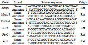
2.8. RNA Isolation and Real-time PCRTotal RNA was isolated from primary chondrocytes using TRIzol reagent (Life Technologies, Carlsbad, CA, USA) or knee joint tissues using a RNeasy Mini Kit (Qiagen, Hilden, Germany). It was then reverse transcribed to complementary DNA using AccuPower RT PreMix (Bioneer, Daejeon, Korea) following the manufacturer’s instructions. Real-time PCR analysis was performed using SYBR green supermix (Bio-Rad, Hercules, CA) on an iCycler iQ Real-Time PCR Detection System (Bio-Rad). Samples were analyzed in triplicate and normalized to β-actin mRNA expression. Primer sequences are listed in Table 1.
Chondrocytes were extracted with RIPA lysis buffer (Biosesang, Sungnam, Korea) containing protease and phosphatase inhibitors. The following antibodies were used for western blotting: anti-Col2a1 from Santa Cruz, anti-MMP3 and anti-MMP13 from Abcam, and anti-IL-1β and anti-FPR2 from Novus Biologicals. Glyceraldehyde-3-phosphate dehydrogenase (GAPDH; Santa Cruz Biotechnologies, Santa Cruz, CA) and β-actin (Santa Cruz) antibodies were used as loading controls.
2.10. StatisticsAll data are expressed as the means ± SDs from at least three independent experiments. Statistical analyses were performed using one-way analysis of variance to analyze differences between groups. Dunnett’s multiple comparisons tests were performed to determine significant differences among the groups. P values less than 0.05 were considered statistically significant.
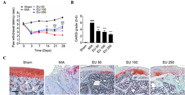
To elucidate the effect of EU extract in OA pathogenesis, we examined a rat model of OA induced by MIA injection. EU extract slightly mitigated the weight loss due to MIA-induced OA and resulted in a behavioral recovery effect based on the Von Frey observation experiment. Moreover, histological analysis revealed that EU extract resulted in gradually less cartilage destruction in a dose-dependent manner, as determined by Osteoarthritis Research Society International (OARSI) scoring with safranin-O staining (Figure 1).
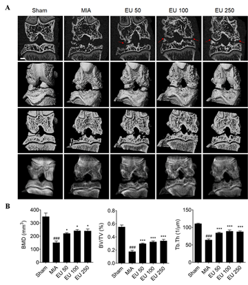
To assess morphological changes in the knee articular bone, the right femur and tibia of rats were analyzed by micro-CT. The EU extract not only suppressed cartilage destruction but also recovered the reduction of bone mineral density (BMD), bone volume/total volume (BV/TV), and trabecular thickness (Tb.Th) due to MIA. These results suggested that EU extract affects OA pathogenesis in rats (Figure 2).
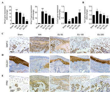
To determine the mechanism underlying the effect of the EU extract, the mRNA of articular cartilage tissue was analyzed by qRT-PCR analysis. Therefore, EU extract was found to regulate OA markers such as Mmp3, Mmp13, and Col2a1. In addition, EU decreased the expression of Fpr2, the LXA4 receptor, which was increased in the MIA-induced OA group. Moreover, immunohistochemical-analysis revealed that the EU extract regulated OA-related proteins including MMP13, COL2A1, and FPR2 (Figure 3).
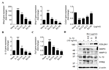
FPR2, the receptor for LXA4, is endogenously expressed in articular cartilage which regulates NF-κB activation in inflammatory response 22. To confirmed the inhibitory effects of EU extract on rat articular chondrocytes, these cells were incubated with multiple doses of EU extract for 24 h with IL-1β (10 ng/ml)-treated media. We found that EU extract regulated all OA-induced markers, including Mmp3, Mmp13, and Col2A1, by qRT-PCR analysis. EU extract also inhibited the expression of IL-1β, encoding an inflammatory cytokine, and Fpr2 in rat articular chondrocytes. These results were confirmed through western blot analysis (Figure 4).
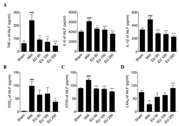
To investigate the inhibition of osteoarthritis through FPR2, we measured intra-articular lavage fluid (IALF) in the joint cavity. The levels of TNF-α, IL-6, and IL-1β significantly increased in the MIA-injected group, and that of these inflammatory cytokines was reduced in the EU extract-treated group. Moreover, level of PGE2, a pain marker, was attenuated in the EU extract (250 mg/kg)-treated group. CTXII is a fragment of collagen that increases in the IALF when cartilage is destroyed by OA. The level of CTXII was decreased in the EU groups. LXA4 binds to FPR2 and regulates inflammation caused by OA 15. Under normal conditions, LXA4 is present in the IALF of the joint cavity; however, when OA is triggered, it becomes attached to FPR2, a receptor present in the cartilage, and its concentration in the IALF decreases 15, 16, 17, 34. In the MIA group, the level of LXA4 was reduced compared with that in the sham group, whereas it was increased in the EU extract (250 mg/kg)-treated group. Our study suggests that EU extract suppresses the inflammatory response of OA and reduces pain through FPR2-LXA4, which inhibits OA markers such as MMP3 and MMP13 and prevents the suppression of COl2A1, helping to prevent and treat OA (Figure 5).
Degenerative diseases are on the rise as the elderly population increases worldwide. OA is a common degenerative disease, and its prevalence is steadily increasing. OA is a general degenerative arthritis that affects the joints of the knee, hip, lumbar region, shoulder, hand, and foot, in which joint cartilage is destroyed and becomes thinner 2, 35. People with OA suffer from movement limitations and pain. There are no OA therapeutic drugs, and only non-steroidal anti-inflammatory drugs (NSAIDs) and COX-2 inhibitors are used to reduce inflammation and pain. However, many of these pain relievers have side effects, such as gastrointestinal problems, and the best way to treat OA is joint replacement surgery 36, 37. Therefore, many researchers are looking for new therapeutics to reduce side effects, focusing on herbal extract that are generally more tolerable than synthetic medications 38, 39. OA is a debilitating disease affecting articular cartilage. Inflammation is closely related to all stages of OA progression, and the inflammatory factors TNF-α and IL-1β ultimately cause cartilage damage, increasing MMP3 and MMP13 and decreasing COL2A1 levels. The inflammatory response results in swelling of the tissues around the joints, associated with cartilage destruction 40, 41, 42, 43, 44.
EU extract is a herbal product traditionally known to be effective for bone and joint health, anti-inflammatory effects, and pain relief, with fewer side effects. Therefore, we studied the efficacy and mechanism of EU extract in OA. In this study, we found that EU extract showed inhibitory effects in the behavioral test to measure pain. In addition, EU extract reduced cartilage destruction and helped with the regeneration of bone based on micro-CT results. The levels of TNF-α, IL-6, and IL-1β in IALF were increased in OA-induced rats, but all decreased in a dose-dependent manner in EU extract-treated groups. PGE2 is a molecule that increases the sensitivity of neurons to pain. Like inflammatory cytokines, its expression was increased due to OA but was inhibited by the extract of EU, especially at 250 mg/kg. These results support the anti-inflammatory and analgesic effects of EU extract.
Type II collagen is primarily found in cartilage and is a major structural component 45. Collagen type II content is decreased during OA development; therefore, the level of CTXII, a fragment of type II collagen, is increased in IALF. Increased CTXII level is a biomarker of early OA 46, 47. EU extract was also effective in early OA and functioned by decreasing the content of CTXII.
OA is usually associated with the early development of mild inflammation, rather than severe inflammation. 14, 15, 16, 17 We, thus, assessed the level of LXA4, aside from the commonly known TNF-α and IL-1β markers, to determine the effects of EU extract on early inflammation. Specifically, 250 mg/kg EU extract increased LXA4 and showed an effective inhibitory effect on the initial inflammatory response.
LXA4 exists endogenously in IALF, and when inflammation occurs, it binds to the cell membrane receptor FPR2/ALX, after which its level in synovial fluid decreases, whereas FPR2 level increases 20, 22. EU extract decreased the increased level of FPR2 in MIA-induced OA and specifically attenuated the expression of MMP3, MMP13, and COL2A1 in the 250 mg/kg treatment group. These results were also verified in rat primary articular chondrocytes. Our studies showed that EU extract inhibits OA by regulating the initial inflammatory response, but further studies on specific binding molecules and their sub-signals should be performed.
Our findings suggest that EU extract reduces OA pathogenesis by inhibiting early inflammation via LXA4-FPR2. These results indicate that EU extract could be a potential therapeutic agent for OA treatment, with fewer side effects than conventional anti-inflammatory analgesics such as NSAIDs.
None.
The authors declare that there are no conflict of interests.
| [1] | Hootman, J. M., Helmick, C. G., “Projections of US prevalence of arthritis and associated activity limitations,” Arthritis. Rheum., 54, 226-229, 2006. | ||
| In article | View Article PubMed | ||
| [2] | Goldring, M. B., Goldring, S. R., “Osteoarthritis,” J. Cell. Physiol., 213, 626-634, 2007. | ||
| In article | View Article PubMed | ||
| [3] | Loeser, R. F., “Molecular mechanisms of cartilage destruction: mechanics, inflammatory mediators and aging collide,” Arthritis. Rheum., 54, 1357-1360, 2006. | ||
| In article | View Article PubMed | ||
| [4] | Suri, S., Walsh, D. A., “Osteochondral alterations in osteoarthritis,” Bone, 51, 204-211, 2012. | ||
| In article | View Article PubMed | ||
| [5] | Zhang, X., Xu, X., Xu, T., Qin, S., “β-Ecdysterone suppresses interleukin-1β-induced apoptosis and inflammation in rat chondrocytes via inhibition of NF-κB signaling pathway,” Drug. Dev. Res., 75, 195-201, 2014. | ||
| In article | View Article PubMed | ||
| [6] | Latourte, A., Cherifi, C., Maillet, J., Ea, H., Bouaziz, W., Funck-Brentano, T., Cohen-Solal, M., Hay, E., Richette, P., “Systemic inhibition of IL-6/Stat3 signalling protects against experimental osteoarthritis,” Ann. Rheum. Dis., 76, 748-755, 2017. | ||
| In article | View Article PubMed | ||
| [7] | Loeser, R. F., Goldring, S. R., Scanzello, C. R., Goldring, M. B., “Osteoarthritis: a disease of the joint as an organ,” Arthritis. Rheum., 64, 1697-1707, 2012. | ||
| In article | View Article PubMed | ||
| [8] | Mobasheri, A., Rayman, M. P., Gualillo, O., Sellam, J., van der Kraan, P., Fearon, U., “The role of metabolism in the pathogenesis of osteoarthritis,” Nat. Rev. Rheumatol., 13, 302-311, 2017. | ||
| In article | View Article PubMed | ||
| [9] | Cicuttini, F. M., Wluka, A. E., “Not just loading and age: the dynamics of osteoarthritis, obesity and inflammation,” Med. J. Aust., 204, 47, 2016. | ||
| In article | View Article PubMed | ||
| [10] | Malfait, A. M., “Osteoarthritis year in review 2015: biology,” Osteoarthritis Cartilage, 24, 21-26, 2016. | ||
| In article | View Article PubMed | ||
| [11] | Liu-Bryan, R., “Inflammation and intracellular metabolism: new targets in OA,” Osteoarthritis Cartilage, 23, 1835-1842, 2015. | ||
| In article | View Article PubMed | ||
| [12] | Berenbaum, F., van den Berg, W. B., “Inflammation in osteoarthritis: changing views,” Osteoarthritis Cartilage, 23, 182-1824, 2015. | ||
| In article | View Article PubMed | ||
| [13] | Serhan, C. N., Chiang, N., “Endogenous pro-resolving and anti-inflammatory lipid mediators: a new pharmacologic genus,” Br. J. Pharmacol., 153, S200-S215, 2008. | ||
| In article | View Article PubMed | ||
| [14] | McMahon, B., Mitchell, S., Brady, H. R., Godson, C., “Lipoxins: revelations on resolution,” Trends. Pharmacol. Sci., 22, 391-395, 2001. | ||
| In article | View Article | ||
| [15] | Sodin-Semrl, S., Taddeo, B., Tseng, D., Varga, J., Fiore, S., “Lipoxin A4 inhibits IL-1β-induced IL-6, IL-8, and matrix metalloproteinase-3 production in human synovial fibroblasts and enhances synthesis of tissue inhibitors of metalloproteinases,” J. Immunol., 164, 2660-2666, 2000. | ||
| In article | View Article PubMed | ||
| [16] | Chan, M. M., Moore, A. R., “Resolution of Inflammation in Murine Autoimmune Arthritis Is Disrupted by Cyclooxygenase-2 Inhibition and Restored by Prostaglandin E 2-Mediated Lipoxin A4 Production,” J. Immunol. 184, 6418-6426, 2010. | ||
| In article | View Article PubMed | ||
| [17] | Conte, F. P., Menezes-de-Lima, O. Jr., Verri, WA. Jr., Cunha, F. Q., Penido, C., Henriques, M. G., “Lipoxin A4 attenuates zymosan-induced arthritis by modulating endothelin-1 and its effects,” Br. J. Pharmacol., 161, 911-924, 2010. | ||
| In article | View Article PubMed | ||
| [18] | Gierman, L. M., Wopereis, S., van El, B., Verheij, E. R., Werff-van der Vat, B. J. C., Bastiaan-sen-Jenniskens, Y. M., van Osch, G. J. V. M., Kloppenburg, M., Stojanovic-Susulic, V., Huizinga, T. W. J., et al., “Metabolic Profiling Reveals Differences in Concentrations of Oxylipins and Fatty Acids Secreted by the Infrapatellar Fat Pad of Donors With End-Stage Osteoarthritis and Normal Donors,” Arthritis. Rheum., 65, 2606-2614, 2013. | ||
| In article | View Article PubMed | ||
| [19] | Krishnamoorthy, S., Recchiuti, A., Chiang, N., Yacoubian, S., Lee, C. H., Yang, R., Petasis, N. A., Serhan, C. N., “Resolvin D1 binds human phagocytes with evidence for proresolbing receptors,” Proc. Natl. Acad. Sci. U. S. A., 107, 1660-1665, 2010. | ||
| In article | View Article PubMed | ||
| [20] | Brink, C., Dahlén, S., Drazen, J., Evans, J. F., Hay, D. W. P., Nicosia, S., Serhan, C. N., Shimizu, T., Yokomizo, T., “International Union of Pharmacology XXXVII. Nomenclature for leukotriene and lipoxin receptors,” Pharmacol. Rev., 55, 195-227, 2003. | ||
| In article | View Article PubMed | ||
| [21] | Holmertz, A. S., Jonsson, C. A., Mohaddes, M., Lundqvist, C., Forsman, H., Gjertsson, I., Önn-heim, K., “Data describing expression of formyl peptide receptor 2 in human articular chondrocytes,” Data. Brief., 31, 105866, 2020. | ||
| In article | View Article PubMed | ||
| [22] | József, L., Zouki, C., Petasis, N. A., Serhan, C. N., Filep, J. G., “Lipoxin A4 and aspirin-triggered 15-epi-lipoxin A4 inhibit peroxynitrite formation, NF-κB and AP-1 activation, and IL-8 gene expression in human leukocytes,” Proc. Natl. Acad. Sci. U. S. A., 99, 13266-13271, 2002. | ||
| In article | View Article PubMed | ||
| [23] | Marcu, K. B., Otero, M., Olivotto, E., Borzi, R. M., Goldring, M. B., “NF-κB signaling: multiple angles to target OA,” Curr. Drug. Targets., 11, 599-613, 2010. | ||
| In article | View Article PubMed | ||
| [24] | He, X., Wang, J., Li, M., Hao, D., Yang, Y., Zhang, C., He, R., Tao, R., “Eucommia ulmoides Oliv.: ethnopharmacology, phytochemistry and pharmacology of an important traditional Chinese medicine,” J. Ethnopharmacol., 151, 78-92, 2014. | ||
| In article | View Article PubMed | ||
| [25] | Hsieh, C. L., Yen, G. C., “Antioxidant actions of du-zhong (Eucommia ulmoides Oliv.) toward oxidative damage in biomolecules,” Life. Sci., 66, 1387-1400, 2000. | ||
| In article | View Article | ||
| [26] | Kwan, C. Y., Chen, C., Deyama, T., Nishibe, S., “Endothelium-dependent vasorelaxant effects of the aqueous extracts of the Eucommia ulmoides Oliv. Leaf and bark: Implications on their antihypertensive action,” Vasc. Pharmacol., 40, 229-235, 2003. | ||
| In article | View Article PubMed | ||
| [27] | Zhao, Y., Li, Y., Wang, X., Sun, W., “The experimental study of Cortex Eucommiae on meridian tropsim: The distribution study of aucubin in rat tissues,” J. Pharm. Biomed. Anal., 46, 368-373, 2008. | ||
| In article | View Article PubMed | ||
| [28] | Lu, H., Jiang, J., Xie, G., Liu, W., Yan, G., “Effects of an aqueous extract of Eucommiae on articular cartilage in a rat model of osteoarthritis of the knee,” Exp. Ther. Med., 684-688, 2013. | ||
| In article | View Article PubMed | ||
| [29] | Lee, S., Kwon, H., Lee, S., “SHINBARO, a new herbal medicine with multifunctional mechanism for joint disease: First therapeutic application for the treatment of osteoarthriti,” Arch. Pharm. Res., 34, 73-1777, 2011. | ||
| In article | View Article PubMed | ||
| [30] | Kim, W. K., Chung, H., Pyee, Y., Choi, T. J., Park, H. J., Hong, J., Shin, J., Lee, J. H., Ha, I., Lee, S. K., “Effects of intra-articular SHINBARO treatment on monosodium iodoacetate-induced osteoarthritis in rats,” Chin. Med., 11, 17, 2016. | ||
| In article | View Article PubMed | ||
| [31] | Lee, S. W., Song, Y. S., Shin, S. H., Kim, K. T., Park, Y. C., Park, B. S., Yun, I., Kim, K., Lee, S. Y., Chung, W. T., et al., “Cilostazol protects rat chondrocytes against nitric oxide-induced apoptosis in vitro and prevents cartilage destruction in a rat model of osteoarthritis,” Arthritis. Rheum., 58, 790-800, 2008. | ||
| In article | View Article PubMed | ||
| [32] | Glasson, S. S., Chambers, M. G., Van Den Berg, W. B., Little, C. B., “The OARSI histopathology initiative–recommendations for histological assessments of osteoarthritis in the mouse,” Osteoarthritis Cartilage, 18, S17-S23, 2010. | ||
| In article | View Article PubMed | ||
| [33] | Gosset, M., Berenbaum, F., Thirion, S., Jacques, C., “Primary culture and phenotyping of murine chondrocytes,” Nat. Protoc., 3, 1253-1260, 2008. | ||
| In article | View Article PubMed | ||
| [34] | Yang, Y., Wang, Y., Kong, Y., Zhan, X., Bai, L., “The effects of different frequency treadmill exercise on lipoxin A4 and articular cartilage degeneration in an experimental model of monosodium iodoacetate-induced osteoarthritis in rats. Plos One, 12, e0179162, 2017. | ||
| In article | View Article PubMed | ||
| [35] | Felson, D. T., “Developments in the clinical understanding of osteoarthritis,” Arthitis. Res. Ther., 11, 203, 2009. | ||
| In article | View Article PubMed | ||
| [36] | Rannou, F., Pelletier, J. P., Martel-Pelletier, J., “Efficacy and safety of topical NSAIDs in the management of osteoarthritis: evidence from real-life setting trials and surveys,” Semin. Atrhtitis. Rheum., 45, S18-21, 2016. | ||
| In article | View Article PubMed | ||
| [37] | Brizuela, N. Y., Montrull, H. L., Demurtas, S. L., Meirovich, C. I., “Articular cartilage in osteoarthritic patients: effects of declofenac, celecoxib and glucosamine sulfate on inflammatory markers,” Rev. Fac. Cien. Med. Univ. Nac. Cordoba., 64, 9-15, 2007. | ||
| In article | |||
| [38] | Da Costa, B. R., Reichenbach, S., Keller, N., Nartey, L., Wandel, S., Jüni, P., Trelle, S., “Effectiveness of non-steroidal anti-inflammatory drugs for the treatment of pain in knee and hip osteoarthritis: A network meta-analysis,” Lancet, 390, e21-e33, 2017. | ||
| In article | View Article | ||
| [39] | Izzo, A. A., Kim, S. H., Radhakrishnan, R., Williamson, E. M., “A critical approach to evaluating clinical efficacy, adverse events and drug interactions of herbal remedies,” Phytother. Res., 30, 691-700, 2016. | ||
| In article | |||
| [40] | Pfander, D., Heinz, N., Rothe, P., Carl, H., Swoboda, B., “Tenascin and aggrecan expression by articular chondrocytes is influenced by interleukin 1β: A possible explanation for the changes in matrix synthesis during osteoarthritis,” Ann. Rheum. Dis., 63, 240-244, 2004. | ||
| In article | View Article PubMed | ||
| [41] | Meulenbelt, I., Seymour, A. B., Nieuwland, M., Huizinga, T. W. J., van Duijin, C. M., Slagboom, P. E., “Association of the interleukin-1 gene cluster with radiographic signs of osteoarthritis of the hip,” Arthritis. Rheum., 50, 1179-1186, 2004. | ||
| In article | View Article PubMed | ||
| [42] | Sakkas, L. I., Johanson, N. A., Scanzello, C. R., Platsoucas, C. D., “Interleukin-12 is expressed by infiltrating macrophages and synovial lining cells in rheumatoid arthritis and osteoarthritis,” Cell. Immunol., 188, 105-110, 1998. | ||
| In article | View Article PubMed | ||
| [43] | Scanzello, C. R., Umoh, E., Pessler, F., Diaz-Torne, C., Dicarlo, E., Potter, H. G., Mandl, L., Marx, R., Rodeo, S., Goldring, S. R., et al., “Local cytokine profiles in knee osteoarthritis: Elevated synovial fluid interleukin-15 differentiates early from end-stage disease,” Osteoarthritis Cartilage, 17, 1040-1048, 2009. | ||
| In article | View Article PubMed | ||
| [44] | Campo, G. M., Avenoso, A., D’Ascola, A., Scuruchi, M., Prestipino, V., Calatroni, A., Campo, S., “Hyaluronan in part mediates IL-1β-induced inflammation in mouse chondrocytes by up-regulating CD44 receptors,” Gene, 494, 24-35, 2012. | ||
| In article | View Article PubMed | ||
| [45] | Prockop, D. J., Kivirikko, K. I., “Collagens: Molecular biology, diseases, and potentials for therapy,” Annu. Rev. Biochem., 64, 403-434, 1995. | ||
| In article | View Article PubMed | ||
| [46] | Chu, X. Q., Wang, J. J., Dou, L. D., Zhao, G., “Cartilage oligomeric matrix protein and matrix metalloproteinase-3 expression in the serum and joint fluid of a reversible osteoarthritis rabbit model,” Genet. Mol. Res., 14, 14207-14215, 2015. | ||
| In article | View Article PubMed | ||
| [47] | Jiao, Q., Wei, L., Chen, C., Li, P., Wang, X., Li, Y., Guo, L., Zhang, C., Wei, X., “Cartilage oligomeric matrix protein and hyaluronic acid are sensitive serum biomarkers for early cartilage lesions in the knee joint,” Biomarkers, 21, 146-151, 2016. | ||
| In article | View Article PubMed | ||
Published with license by Science and Education Publishing, Copyright © 2022 Doo ri Park, Chang hwan Yeo, Jee Eun Yoon, Wan-Jin Jeon, Woo-Jae Choung and In-Hyuk Ha
![]() This work is licensed under a Creative Commons Attribution 4.0 International License. To view a copy of this license, visit
http://creativecommons.org/licenses/by/4.0/
This work is licensed under a Creative Commons Attribution 4.0 International License. To view a copy of this license, visit
http://creativecommons.org/licenses/by/4.0/
| [1] | Hootman, J. M., Helmick, C. G., “Projections of US prevalence of arthritis and associated activity limitations,” Arthritis. Rheum., 54, 226-229, 2006. | ||
| In article | View Article PubMed | ||
| [2] | Goldring, M. B., Goldring, S. R., “Osteoarthritis,” J. Cell. Physiol., 213, 626-634, 2007. | ||
| In article | View Article PubMed | ||
| [3] | Loeser, R. F., “Molecular mechanisms of cartilage destruction: mechanics, inflammatory mediators and aging collide,” Arthritis. Rheum., 54, 1357-1360, 2006. | ||
| In article | View Article PubMed | ||
| [4] | Suri, S., Walsh, D. A., “Osteochondral alterations in osteoarthritis,” Bone, 51, 204-211, 2012. | ||
| In article | View Article PubMed | ||
| [5] | Zhang, X., Xu, X., Xu, T., Qin, S., “β-Ecdysterone suppresses interleukin-1β-induced apoptosis and inflammation in rat chondrocytes via inhibition of NF-κB signaling pathway,” Drug. Dev. Res., 75, 195-201, 2014. | ||
| In article | View Article PubMed | ||
| [6] | Latourte, A., Cherifi, C., Maillet, J., Ea, H., Bouaziz, W., Funck-Brentano, T., Cohen-Solal, M., Hay, E., Richette, P., “Systemic inhibition of IL-6/Stat3 signalling protects against experimental osteoarthritis,” Ann. Rheum. Dis., 76, 748-755, 2017. | ||
| In article | View Article PubMed | ||
| [7] | Loeser, R. F., Goldring, S. R., Scanzello, C. R., Goldring, M. B., “Osteoarthritis: a disease of the joint as an organ,” Arthritis. Rheum., 64, 1697-1707, 2012. | ||
| In article | View Article PubMed | ||
| [8] | Mobasheri, A., Rayman, M. P., Gualillo, O., Sellam, J., van der Kraan, P., Fearon, U., “The role of metabolism in the pathogenesis of osteoarthritis,” Nat. Rev. Rheumatol., 13, 302-311, 2017. | ||
| In article | View Article PubMed | ||
| [9] | Cicuttini, F. M., Wluka, A. E., “Not just loading and age: the dynamics of osteoarthritis, obesity and inflammation,” Med. J. Aust., 204, 47, 2016. | ||
| In article | View Article PubMed | ||
| [10] | Malfait, A. M., “Osteoarthritis year in review 2015: biology,” Osteoarthritis Cartilage, 24, 21-26, 2016. | ||
| In article | View Article PubMed | ||
| [11] | Liu-Bryan, R., “Inflammation and intracellular metabolism: new targets in OA,” Osteoarthritis Cartilage, 23, 1835-1842, 2015. | ||
| In article | View Article PubMed | ||
| [12] | Berenbaum, F., van den Berg, W. B., “Inflammation in osteoarthritis: changing views,” Osteoarthritis Cartilage, 23, 182-1824, 2015. | ||
| In article | View Article PubMed | ||
| [13] | Serhan, C. N., Chiang, N., “Endogenous pro-resolving and anti-inflammatory lipid mediators: a new pharmacologic genus,” Br. J. Pharmacol., 153, S200-S215, 2008. | ||
| In article | View Article PubMed | ||
| [14] | McMahon, B., Mitchell, S., Brady, H. R., Godson, C., “Lipoxins: revelations on resolution,” Trends. Pharmacol. Sci., 22, 391-395, 2001. | ||
| In article | View Article | ||
| [15] | Sodin-Semrl, S., Taddeo, B., Tseng, D., Varga, J., Fiore, S., “Lipoxin A4 inhibits IL-1β-induced IL-6, IL-8, and matrix metalloproteinase-3 production in human synovial fibroblasts and enhances synthesis of tissue inhibitors of metalloproteinases,” J. Immunol., 164, 2660-2666, 2000. | ||
| In article | View Article PubMed | ||
| [16] | Chan, M. M., Moore, A. R., “Resolution of Inflammation in Murine Autoimmune Arthritis Is Disrupted by Cyclooxygenase-2 Inhibition and Restored by Prostaglandin E 2-Mediated Lipoxin A4 Production,” J. Immunol. 184, 6418-6426, 2010. | ||
| In article | View Article PubMed | ||
| [17] | Conte, F. P., Menezes-de-Lima, O. Jr., Verri, WA. Jr., Cunha, F. Q., Penido, C., Henriques, M. G., “Lipoxin A4 attenuates zymosan-induced arthritis by modulating endothelin-1 and its effects,” Br. J. Pharmacol., 161, 911-924, 2010. | ||
| In article | View Article PubMed | ||
| [18] | Gierman, L. M., Wopereis, S., van El, B., Verheij, E. R., Werff-van der Vat, B. J. C., Bastiaan-sen-Jenniskens, Y. M., van Osch, G. J. V. M., Kloppenburg, M., Stojanovic-Susulic, V., Huizinga, T. W. J., et al., “Metabolic Profiling Reveals Differences in Concentrations of Oxylipins and Fatty Acids Secreted by the Infrapatellar Fat Pad of Donors With End-Stage Osteoarthritis and Normal Donors,” Arthritis. Rheum., 65, 2606-2614, 2013. | ||
| In article | View Article PubMed | ||
| [19] | Krishnamoorthy, S., Recchiuti, A., Chiang, N., Yacoubian, S., Lee, C. H., Yang, R., Petasis, N. A., Serhan, C. N., “Resolvin D1 binds human phagocytes with evidence for proresolbing receptors,” Proc. Natl. Acad. Sci. U. S. A., 107, 1660-1665, 2010. | ||
| In article | View Article PubMed | ||
| [20] | Brink, C., Dahlén, S., Drazen, J., Evans, J. F., Hay, D. W. P., Nicosia, S., Serhan, C. N., Shimizu, T., Yokomizo, T., “International Union of Pharmacology XXXVII. Nomenclature for leukotriene and lipoxin receptors,” Pharmacol. Rev., 55, 195-227, 2003. | ||
| In article | View Article PubMed | ||
| [21] | Holmertz, A. S., Jonsson, C. A., Mohaddes, M., Lundqvist, C., Forsman, H., Gjertsson, I., Önn-heim, K., “Data describing expression of formyl peptide receptor 2 in human articular chondrocytes,” Data. Brief., 31, 105866, 2020. | ||
| In article | View Article PubMed | ||
| [22] | József, L., Zouki, C., Petasis, N. A., Serhan, C. N., Filep, J. G., “Lipoxin A4 and aspirin-triggered 15-epi-lipoxin A4 inhibit peroxynitrite formation, NF-κB and AP-1 activation, and IL-8 gene expression in human leukocytes,” Proc. Natl. Acad. Sci. U. S. A., 99, 13266-13271, 2002. | ||
| In article | View Article PubMed | ||
| [23] | Marcu, K. B., Otero, M., Olivotto, E., Borzi, R. M., Goldring, M. B., “NF-κB signaling: multiple angles to target OA,” Curr. Drug. Targets., 11, 599-613, 2010. | ||
| In article | View Article PubMed | ||
| [24] | He, X., Wang, J., Li, M., Hao, D., Yang, Y., Zhang, C., He, R., Tao, R., “Eucommia ulmoides Oliv.: ethnopharmacology, phytochemistry and pharmacology of an important traditional Chinese medicine,” J. Ethnopharmacol., 151, 78-92, 2014. | ||
| In article | View Article PubMed | ||
| [25] | Hsieh, C. L., Yen, G. C., “Antioxidant actions of du-zhong (Eucommia ulmoides Oliv.) toward oxidative damage in biomolecules,” Life. Sci., 66, 1387-1400, 2000. | ||
| In article | View Article | ||
| [26] | Kwan, C. Y., Chen, C., Deyama, T., Nishibe, S., “Endothelium-dependent vasorelaxant effects of the aqueous extracts of the Eucommia ulmoides Oliv. Leaf and bark: Implications on their antihypertensive action,” Vasc. Pharmacol., 40, 229-235, 2003. | ||
| In article | View Article PubMed | ||
| [27] | Zhao, Y., Li, Y., Wang, X., Sun, W., “The experimental study of Cortex Eucommiae on meridian tropsim: The distribution study of aucubin in rat tissues,” J. Pharm. Biomed. Anal., 46, 368-373, 2008. | ||
| In article | View Article PubMed | ||
| [28] | Lu, H., Jiang, J., Xie, G., Liu, W., Yan, G., “Effects of an aqueous extract of Eucommiae on articular cartilage in a rat model of osteoarthritis of the knee,” Exp. Ther. Med., 684-688, 2013. | ||
| In article | View Article PubMed | ||
| [29] | Lee, S., Kwon, H., Lee, S., “SHINBARO, a new herbal medicine with multifunctional mechanism for joint disease: First therapeutic application for the treatment of osteoarthriti,” Arch. Pharm. Res., 34, 73-1777, 2011. | ||
| In article | View Article PubMed | ||
| [30] | Kim, W. K., Chung, H., Pyee, Y., Choi, T. J., Park, H. J., Hong, J., Shin, J., Lee, J. H., Ha, I., Lee, S. K., “Effects of intra-articular SHINBARO treatment on monosodium iodoacetate-induced osteoarthritis in rats,” Chin. Med., 11, 17, 2016. | ||
| In article | View Article PubMed | ||
| [31] | Lee, S. W., Song, Y. S., Shin, S. H., Kim, K. T., Park, Y. C., Park, B. S., Yun, I., Kim, K., Lee, S. Y., Chung, W. T., et al., “Cilostazol protects rat chondrocytes against nitric oxide-induced apoptosis in vitro and prevents cartilage destruction in a rat model of osteoarthritis,” Arthritis. Rheum., 58, 790-800, 2008. | ||
| In article | View Article PubMed | ||
| [32] | Glasson, S. S., Chambers, M. G., Van Den Berg, W. B., Little, C. B., “The OARSI histopathology initiative–recommendations for histological assessments of osteoarthritis in the mouse,” Osteoarthritis Cartilage, 18, S17-S23, 2010. | ||
| In article | View Article PubMed | ||
| [33] | Gosset, M., Berenbaum, F., Thirion, S., Jacques, C., “Primary culture and phenotyping of murine chondrocytes,” Nat. Protoc., 3, 1253-1260, 2008. | ||
| In article | View Article PubMed | ||
| [34] | Yang, Y., Wang, Y., Kong, Y., Zhan, X., Bai, L., “The effects of different frequency treadmill exercise on lipoxin A4 and articular cartilage degeneration in an experimental model of monosodium iodoacetate-induced osteoarthritis in rats. Plos One, 12, e0179162, 2017. | ||
| In article | View Article PubMed | ||
| [35] | Felson, D. T., “Developments in the clinical understanding of osteoarthritis,” Arthitis. Res. Ther., 11, 203, 2009. | ||
| In article | View Article PubMed | ||
| [36] | Rannou, F., Pelletier, J. P., Martel-Pelletier, J., “Efficacy and safety of topical NSAIDs in the management of osteoarthritis: evidence from real-life setting trials and surveys,” Semin. Atrhtitis. Rheum., 45, S18-21, 2016. | ||
| In article | View Article PubMed | ||
| [37] | Brizuela, N. Y., Montrull, H. L., Demurtas, S. L., Meirovich, C. I., “Articular cartilage in osteoarthritic patients: effects of declofenac, celecoxib and glucosamine sulfate on inflammatory markers,” Rev. Fac. Cien. Med. Univ. Nac. Cordoba., 64, 9-15, 2007. | ||
| In article | |||
| [38] | Da Costa, B. R., Reichenbach, S., Keller, N., Nartey, L., Wandel, S., Jüni, P., Trelle, S., “Effectiveness of non-steroidal anti-inflammatory drugs for the treatment of pain in knee and hip osteoarthritis: A network meta-analysis,” Lancet, 390, e21-e33, 2017. | ||
| In article | View Article | ||
| [39] | Izzo, A. A., Kim, S. H., Radhakrishnan, R., Williamson, E. M., “A critical approach to evaluating clinical efficacy, adverse events and drug interactions of herbal remedies,” Phytother. Res., 30, 691-700, 2016. | ||
| In article | |||
| [40] | Pfander, D., Heinz, N., Rothe, P., Carl, H., Swoboda, B., “Tenascin and aggrecan expression by articular chondrocytes is influenced by interleukin 1β: A possible explanation for the changes in matrix synthesis during osteoarthritis,” Ann. Rheum. Dis., 63, 240-244, 2004. | ||
| In article | View Article PubMed | ||
| [41] | Meulenbelt, I., Seymour, A. B., Nieuwland, M., Huizinga, T. W. J., van Duijin, C. M., Slagboom, P. E., “Association of the interleukin-1 gene cluster with radiographic signs of osteoarthritis of the hip,” Arthritis. Rheum., 50, 1179-1186, 2004. | ||
| In article | View Article PubMed | ||
| [42] | Sakkas, L. I., Johanson, N. A., Scanzello, C. R., Platsoucas, C. D., “Interleukin-12 is expressed by infiltrating macrophages and synovial lining cells in rheumatoid arthritis and osteoarthritis,” Cell. Immunol., 188, 105-110, 1998. | ||
| In article | View Article PubMed | ||
| [43] | Scanzello, C. R., Umoh, E., Pessler, F., Diaz-Torne, C., Dicarlo, E., Potter, H. G., Mandl, L., Marx, R., Rodeo, S., Goldring, S. R., et al., “Local cytokine profiles in knee osteoarthritis: Elevated synovial fluid interleukin-15 differentiates early from end-stage disease,” Osteoarthritis Cartilage, 17, 1040-1048, 2009. | ||
| In article | View Article PubMed | ||
| [44] | Campo, G. M., Avenoso, A., D’Ascola, A., Scuruchi, M., Prestipino, V., Calatroni, A., Campo, S., “Hyaluronan in part mediates IL-1β-induced inflammation in mouse chondrocytes by up-regulating CD44 receptors,” Gene, 494, 24-35, 2012. | ||
| In article | View Article PubMed | ||
| [45] | Prockop, D. J., Kivirikko, K. I., “Collagens: Molecular biology, diseases, and potentials for therapy,” Annu. Rev. Biochem., 64, 403-434, 1995. | ||
| In article | View Article PubMed | ||
| [46] | Chu, X. Q., Wang, J. J., Dou, L. D., Zhao, G., “Cartilage oligomeric matrix protein and matrix metalloproteinase-3 expression in the serum and joint fluid of a reversible osteoarthritis rabbit model,” Genet. Mol. Res., 14, 14207-14215, 2015. | ||
| In article | View Article PubMed | ||
| [47] | Jiao, Q., Wei, L., Chen, C., Li, P., Wang, X., Li, Y., Guo, L., Zhang, C., Wei, X., “Cartilage oligomeric matrix protein and hyaluronic acid are sensitive serum biomarkers for early cartilage lesions in the knee joint,” Biomarkers, 21, 146-151, 2016. | ||
| In article | View Article PubMed | ||


