FSG-CA is a mixture of Centella asiatica extract and Portulaca oleracea extract. In previous in vitro studies, FSG-CA was shown to enhance osteoblastic activity. In this study, we investigated the effect of FSG-CA on longitudinal bone growth in vivo. Male adolescent SD rats were divided into four groups and administered FSG-CA (50 or 100 mg/kg BW/day), recombinant human growth hormone (rhGH, positive control), or vehicle (control) for 5 weeks. Longitudinal bone growth rate was measured using tetracycline labeling. Growth plate (GP) height was measured using hematoxylin and eosin staining. Serum GH, insulin-like growth factor (IGF)-1, and IGF-binding protein (IGFBP)-3 concentrations were measured by enzyme-linked immunosorbent assay. In the liver, mRNA expression of IGF-1 and IGFBP-3 was analyzed by RT-PCR. IGF-1 and bone morphogenetic protein (BMP)-2 in the tibial GP were analyzed by immunofluorescence staining. Oral administration of FSG-CA significantly increased endochondral bone formation and proliferation and tibial GP height. FSG-CA increased IGF-1 and IGFBP-3 mRNA expression in the liver and increased IGF-1 concentration in serum. FSG-CA also upregulated IGF-1 and BMP-2 expression in the tibial GP. These results suggest that FSG-CA may help increase longitudinal bone growth during puberty by utilizing the GH - IGF-1 pathway.
Short stature is characterized by a height greater than two standard deviations below the average for an individual’s age, sex, and population group. The causes of short stature are complex, including endocrine and wasting diseases 1. Children with short stature might have a high frequency of behavioral and psychosocial issues, including low self-esteem, social immaturity, or bullying 2. Individuals with shorter height are reported to have a lower health-related quality of life, even if they do not meet the criteria for short stature 3. There is an increasing interest in overcoming short stature because stature is considered one of the critical factors for healthy mental and physical growth in children and adolescents.
Bone grows in two ways: endochondral ossification and intramembranous ossification 4. The former is the primary mechanism of longitudinal bone growth and occurs within the GP. In the GP, chondrocytes proliferate and differentiate into mature hypertrophic chondrocytes, which then secrete extracellular matrix to form cartilage. Next, freshly formed cartilage is calcified and transformed into trabecular bone by osteoblasts and osteoclasts, resulting in bone growth 5. This process is mainly regulated by the growth hormone (GH) - insulin-like growth factor (IGF)-1 pathway 6. GH affects final height after birth and plays an important role during the growth spurt of adolescence.
GH therapy involves subcutaneous injection of recombinant human GH (rhGH), is the most common clinical practice for treating short stature. This therapy is advantageous for short stature caused by GH deficiency 7, 8. However, its effectiveness for idiopathic short stature (ISS), which accounts for 80% of all short stature, remains controversial because the estimated cost of GH therapy for adult height gain is very high 9. Furthermore, the long-term use of GH therapy, the pain due to daily injection, the abuse of GH to increase the height of children who are already of standard height, and the several adverse side effects when using it in healthy individuals are also considered controversial 8, 10, 11. Therefore, there is a growing need to develop effective alternatives that are effective and safe in all short statures. Traditional herbal medicines promoting longitudinal bone growth could be a promising option for developing safe and effective alternatives for treating short stature.
Centella asiatica, a perennial herbaceous creeper belonging to the Apiceae family, is found in swampy areas in most tropical and subtropical countries. It is an important medicinal herb used in the Orient and is also becoming popular in the West 12. C. asiatica exhibits various pharmacological activities, including wound healing 13, 14, antidepressant 15, cognition improvement 16, and antioxidant activity 17. The main active constituents responsible for its pharmacological activity are triterpenes, which include asiaticoside, asiatic acid, madecassoside, and madecassic acid 12, 18.
Portulaca oleracea, belonging to the Portulacaceae family, is a succulent annual plant that thrives in warm climates and is found worldwide. P. oleracea has been used as a herbal folk medicine in many countries 19. It exhibits a wide range of pharmacological effects, including hepatoprotective 20, antidiabetic 21, antioxidant 22, anti-inflammatory 23, and antiulcer 24 effects. P. oleracea contains a variety of active constituents such as flavonoids, alkaloids, fatty acids, terpenoids, and sterols, which contribute to its pharmacological effects 19.
In a preliminary study, we screened various traditional medicinal herbs for their ability to promote longitudinal bone growth in vitro. C. asiatica and P. oleracea extracts stimulated insulin-like growth factor (IGF)-1 and IGF-binding protein (IGFBP)-3 production in HepG2 hepatocytes and increased alkaline phosphatase (ALP) activity in MC3T3-E1 osteoblastic cells. Moreover, the mixture of C. asiatica extract and P. oleracea extract showed a synergistic effect 25. We hypothesized that the standardized extract mixing C. asiatica and P. oleracea (FSG-CA) exerts a growth-promoting effect. In this study, we investigated the effects of oral administration of FSG-CA on longitudinal bone growth in adolescent male Sprague-Dawley (SD) rats.
FSG-CA was prepared by Frombio Co. Ltd. (Yongin, Korea). To prepare C. asiatica extract (CAE), the dried leaves of C. asiatica were extracted with a 30-fold volume of 70% edible ethanol at 50°C for 6 h. The extract was then filtered, concentrated by vacuum evaporation, and freeze-dried. For the P. oleracea extract (POE), the dried whole P. oleracea plant was extracted with a 20-fold volume of 70% edible ethanol at 50°C for 6 h, followed by filtration, concentration, and freeze-drying. CAE and POE were homogenously mixed to prepare the standardized mixture at a 1:1 ratio. The resulting mixture was used as FSG-CA. Asiatic acid and adenosine content, the indicative compound, were 13.28 mg/g and 1.1 mg/g in FSG-CA, respectively, as determined by high-performance liquid chromatography analysis.
2.2. Ethical Statement and Animal CareAll animal experimental protocols were approved by the Institutional Animal Care and Use Committee of Hallym University (approved number: Hallym 2023-45). The care and use of the animals followed established guidelines for the care and use of laboratory animals. Three-week-old male SD rats were procured from Dooyeol Biotech Co., Ltd. (Seoul, Korea). The rats were maintained under controlled conditions (23 ± 3°C, 50 ± 10%) relative humidity, and 12 h light/dark cycles) and were provided with a commercial rodent diet and tap water ad libitum.
2.3. Experimental Design and TreatmentAfter 7-day acclimatization, rats were assigned into 4 groups: vehicle-treated group (CON), 50 mg/kg body weight (BW)/day FSG-CA-treated group (F50), 100 mg/kg BW/day FSG-CA -treated group (F100), or 200 μg/kg BW/day rhGH-treated group (GH). The rats in the F50 and F100 groups received FSG-CA dissolved in sterile water orally once daily for five weeks. The rats in the GH group received a daily subcutaneous injection of 200 μg/kg BW of rhGH for five weeks. The animal’s initial and final BW and lengths (from nose to tail, NT) were measured. After the final treatment, the rats were anesthetized with 2-3% isoflurane/N2O/O2 mixture, and blood was then collected from the orbital vein. The rats were then euthanized by carbon dioxide asphyxiation, after which the livers were excised and weighed. The tibiae were excised and dissected to remove soft tissue, and their weight and length were measured.
2.4. Measurement of Serum Biochemical IndicatorsSerum alanine aminotransferase (ALT) and aspartate aminotransferase (AST) activities were measured using a blood chemistry autoanalyzer (Indiko Plus, Thermo Fisher Scientific, Vantaa, Finland). Serum GH, IGF-1, and IGFBP-3 were measured using the relevant enzyme-linked immunosorbent assay (ELISA) kits (MyBioSource, San Diego, CA, USA).
2.5. Measurement of Longitudinal Bone GrowthTetracycline hydrochloride was used as a fluorescent marker to level the bone line on the surface of the tibia. To assess longitudinal bone growth, all rats received an intraperitoneal injection of tetracycline hydrochloride (20 mg/kg BW, Sigma-Aldrich) 72 h before sacrifice. The dissected tibia was fixed in 4% paraformaldehyde (PFA), decalcified in Calci-Clear Rapid (National Diagnostics, Atlanta, GA, USA) for 72 h, and embedded in OCT compound (Sakura Finetek, Torrance, CA, USA), and then sectioned longitudinally at a thickness of 30 μm using a cryostat (MICROM HM520, Thermo Scientific, Walldorf, Germany). The sections were observed under a microscope (AxioImager, Carl Zeiss, Jena, Germany). Longitudinal bone growth was estimated by measuring the distance between the fluorescent line and the epiphyseal end line of the GP at three different sites using Image M1 Software AxioVision 4.8 (Carl Zeiss).
2.6. Measurement of Epiphyseal Plate HeightThe dissected tibia was fixed, decalcified, and embedded in paraffin. Subsequently, the tissues were sectioned longitudinally at a thickness of 5 μm and stained with hematoxylin and eosin (H&E, Sigma-Aldrich) following the manufacturer’s protocol. The stained tissues were observed and photographed under a microscope (AxioImager, Carl Zeiss) in a blind manner. The height of the epiphyseal GP measured at three different sites using Image M1 Software AxioVision 4.8 (Carl Zeiss).
2.7. Immunofluorescence (IF) StainingSections of paraffin-embedded tibia tissues were deparaffinized using xylene, followed by rehydration through graded alcohol. Subsequently, the sections were blocked with 5% bovine serum albumin. IF staining was conducted with antibodies against IGF-1 (Santa Cruz Biotechnology, Santa Cruz, CA, USA) and bone morphogenic protein (BMP)-2 (Santa Cruz Biotechnology), followed by fluorochrome-conjugated secondary antibodies (Alexa-488 or 564). The nuclei were counterstained with 4′,6-diamidino-2-phenylindole (DAPI). The stained tissues were blindly observed and photographed under a microscope (AxioImager, Carl Zeiss).
2.8. Assessment of IGF-1 and IGFBP-3 mRNA expression in the LiversTotal RNA from the liver tissue was extracted using TRIzol reagent (Invitrogen Life Technologies, Carlsbad, CA, USA). Real-time reverse transcription-polymerase chain reaction (RT-PCR) conducted using a HyperScriptTM RT Master Mix kit (GeneAll Biotechnology, Seoul, Korea) and a QuantiNova SYBR Green PCR kit (Qiagen), as described previously 26. The primer sequences used in this study were as follows: IGF-1, forward, 5’-CCGCTGCAAGCCTACAAAGT-3’, reverse, 5’-TGAGTCTTGGGCATGTCAGTGT-3’; GFBP-3, forward, 5’-GGAAAGACGACGTGCATTG-3’, reverse, 5’-GCGTATTTGAGCTCCACGTT-3’; glyceraldehyde 3-phosphate dehydrogenase (GAPDH), forward, 5’-CTCAACTACATGGTCTACATGTTCCA-3’, reverse, 5’-CTTCCCATTCTCAGCCTTGACT-3’. The relative mRNA expression level of the target genes was normalized to that of GAPDH.
2.9. Statistical AnalysisStatistical analyses were performed utilizing the Statistical Analysis System for Windows version 9.4 (SAS Institute, Cary, NC, USA). All data are presented as the mean ± standard errors (SEM). Differences between groups were analyzed by one-way analysis of variation, followed by Duncan’s multiple comparisons test. P < 0.05 was considered to indicate a statistically significant difference.
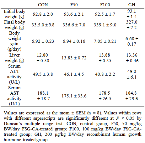
We measured BW, organ weight, and serum ALT and AST activities to determine whether FSG-CA administration led to any adverse effects in vivo. Oral administration of FSG-CA did not affect BW gain, liver weight, and activities of ALT and AST in serum (Table 1). These results indicate that FSG-CA administration did not cause noticeable adverse effects in SD rats.
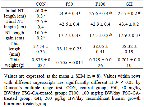
To examine the effect of FSG-CA on height growth, we measured the initial and final NT lengths and calculated the NT length gain. Initial NT length was significantly higher in the CON group than in the other experimental groups (p < 0.05). However, there was no significant difference in the final NT length among the experimental groups. NT length gain, which represents the change in NT length before and after FSG-CA administration, was significantly higher in the F50 and GH groups than in the CON group (p < 0.001). NT length gain increased by 7.3% and 8.5% in the F50 and GH groups, respectively, compared to the CON group (Table 2).
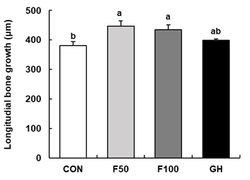
To evaluate the effect of FSG-CA on endochondral bone formation and its longitudinal bone growth, tetracycline fluorescent labeling was used to bind to a newly formed bone in the GP. As shown in Figure 1, longitudinal bone growth of the treatment of FSG-CA groups (F50 and F100 groups) significantly increased compared to the CON group. It reached the same level as the positive control group, the GH group. There was no significant difference between the FSA-CA administration concentrations of 50 mg/kg BW/day (F50) and 100 mg/kg BW/day (F100). These results suggest that oral administration of FSA-CA at 50 mg/kg BW/day is as effective as injection of rhGH 200 µg/kg BW/day in promoting longitudinal bone growth in adolescent male rats.
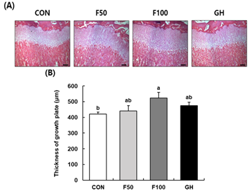
The GP height of the proximal tibia was measured using H&E staining. The microscopic picture of the H&E-stained section is shown in Figure 2A. As a result of quantifying this (Figure 2B), the F50 and F100 groups showed statistically significant differences, with increases of 6% and 25.3%, respectively, compared to the CON group. This suggests that oral administration of FSG-CA has the effect of increasing the GP height in a dose-dependent manner.
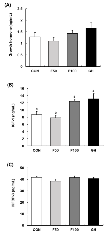
3.5. Effects of FS-CA on the Level of GH, IGF-1, and IGFBP-3 in Adolescent SD RatsThe levels of GH, IGF-1, and IGFBP-3 in the serum were analyzed and presented in Figure 3. There was no significant difference in the level of GH among the experimental groups. However, the GH level of the F100 group increased by about 11.5% compared to the CON group. The level of IGF-1 in the serum was significantly higher in the F100 and GH groups than in the CON group. The level of IGFBP-3 in the serum did not differ significantly among the experimental groups (Figure 3). These results suggest that oral administration of FSG-CA at 100 mg/kg BW/day increases the concentration of IGF-1 in the blood as effectively as injection of rhGH 200 µg/kg BW/day.
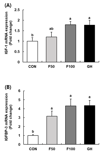
3.6. Effects of FS-CA on IGF-1 and IGFBP-3 mRNA expression in the liver of adolescent SD ratsCirculatory IGF-1 is mainly synthesized in the liver by GH stimulation 27. Therefore, to determine whether the increased serum IGF-1 by FSG-CA was due to increased IGF-1 secretion in the liver, we analyzed IGF-1 and IGFBP-3 mRNA expression in the liver by quantitative RT-PCR. Hepatic IGF-1 mRNA expression tended to increase dose-dependent by FSG-CA administration. In particular, the F100 group showed IGF-1 mRNA expression approximately 1.8 times higher than that of the CON group, which was the same level as that of the GH group. Hepatic IGFBP-3 mRNA expression showed a greater effect by FSG-CA administration, and was approximately 3 and 4 times higher in the F50 and F100 groups, respectively, than that of the CON group (Figure 4).
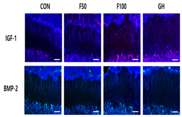
Protein expression of IGF-1 and BMP-2 was analyzed by antigen-specific IF staining in proximal tibial GP. Administration of FSG-CA or rhGH markedly increased the expressions of IGF-1 and BMP-2 in the GP compared to the control (Figure 5).
In this study, the results of serum analysis, bone length measurement, biomechanical evaluation, bone growth rate measurement, histological observation, and mRNA expression quantification showed that FSG-CA could promote longitudinal bone growth in adolescent male rats via the GH - IGF-1 pathway.
NT length gain increased by 10.7%, 10.5%, and 10.8% in the F50, F100, and GH groups, respectively, compared to the CON group (Table 2). The femur, tibia, and fibula are used to measure the growth of long bones. Among them, the tibia is the best indicator of stature 32. To investigate whether FSG-CA promotes longitudinal bone growth, we evaluated longitudinal bone growth by measuring the fluorescent line formed by tetracycline and the end-edge of the GP in the tibia. The FSG-CA-administered groups (F50 and F100) showed a significant increase in longitudinal bone growth compared to the CON group (P < 0.05) (Figure 1).
The GP is the cartilage between the metaphyseal and epiphyseal bones at the ends of the longitudinal long bones. In the GP, chondrocytes regulate longitudinal growth through proliferation, hypertrophy, apoptosis, cartilage matrix synthesis, mineralization, and vascularization. GP height is a significant index reflecting endochondral bone formation 33. Therefore, the GP height was measured to explore the potential way in which FSG-CA promotes bone growth. In tibia, the GP height of the F100 group was significantly higher than that of the other experimental groups (P < 0.05), and increased by 25.3% compared to the CON group (Figure 2). These results suggest that FSG-CA enhances entochondrostosis to promote longitudinal bone growth by promoting chondrocyte proliferation and hypertrophy.
To explore the possible mechanism of the increase in GP height and proliferation rate observed in the FSG-CA-administered group, serum and gene expression were analyzed. The bone elongation process after birth is regulated by endocrine molecules that act on target cells of the GP systemically, such as hormones, and paracrine/autocrine factors expressed in chondrocytes or surrounding perichondrium. Major endocrine factors include GH, IGF-1, thyroid hormones, and sex hormones, among which GH plays the most important role 34. Autocrine/paracrine molecules include BMPs, fibroblast growth factors, vascular endothelial growth factor, IGF-1, Wnt, indian hedgehog, and parathyroid hormone related protein 35. Bone lengthening results from coordinated signaling of endocrine and paracrine/autocrine factors, which interact to promote chondrocyte proliferation and GP hypertrophy 35. In particular, the GH - IGF-1 pathway is important for longitudinal bone growth 36. GH-IGF-1 regulation is achieved through circulating IGF-1 secreted from the liver by GH or through local IGF-1 production by direct stimulation of GH 37. The liver is a major source of circulating IGF-1 and IGFBP-3 in response to GH. Approximately 75% of circulating IGF-1 in serum comes from the liver 38. In our study, hepatic IGF-1 mRNA expression was dose-dependently increased by FSG-CA administration, and IGFBP-3 mRNA expression was increased by 3-fold and 4-fold in the F50 and F100 groups, respectively, compared with CON. The serum IGF-1 levels were more than 50% higher in the F100 and GH groups than in the CON group (Figure 3). IGF-1 plays a very important role in longitudinal bone growth after birth 39. It has been reported that locally produced IGF-1 may be more important than GH for longitudinal bone growth, as IGF-1-deficient mutant animals showed a much more severe impairment in height growth than mutant animals with impaired GH function 40. Our results suggest that FSG-CA may enhance local IGF-1 expression in bone by promoting systemic IGF-1 expression regulated by the liver.
BMP belongs to the transforming growth factor-β family and functions as both a growth and differentiation factor 29. Of the various forms of BMP, BMP-2 plays a role in developing the GP and enhancing longitudinal bone growth by promoting the proliferation and hypertrophy of GP chondrocytes 30. In the present study, FSG-CA was shown to increase the expression of not only IGF-1 but also BMP-2 in tibia (Figure 5). These results indicate that FSG-CA promotes the proliferation and differentiation of chondrocytes through the GH - IGF-1 pathway and upregulation of BMP-2 expression, thereby promoting endochondral ossification, which can promote longitudinal bone growth.
This study has some limitations that should be addressed in future studies. First, further research is needed on the components of FSG-CA related to longitudinal bone growth. Second, more detailed bone turnover biomarkers and molecular pathway analysis should be performed to elucidate the mechanism by which FSG-CA promotes longitudinal bone growth. Third, further research is needed on the effect of FSG-CA on osteoclasts. Osteoclast activity regulates the mineralization of chondrocytes and thus affects the GP height. Fourth, further research is needed on the appropriate concentration for applying FSG-CA to humans.
Oral administration of FSG-CA significantly increased endochondral bone formation, proliferation, and tibial GP height. FSG-CA upregulated the mRNA expressions of IGF-1 and IGFBP-3 in the liver and IGF-1 and BMP-2 expression in GP. FSG-CA also increased circulatory IGF-1 levels. Considering immunohistochemical studies, the effect of FSG-CA on longitudinal bone growth may be due to the increased local IGF-1 and BMP-2 expression in GP, which may be considered as growth hormone (GH) -dependent endocrine and autocrine/adrenal pathways. These results suggest that FSG-CA may help increase longitudinal bone growth in adolescence by utilizing the GH-IGF-1 pathway.
The authors thank the Frombio Co., Ltd. for providing the mixture of Centella asiatica extract and Portulaca oleracea extract (FSG-CA).
The authors have no competing interests.
| [1] | Cohen,P., Rogol, A.D., Deal, C.L., Saenger, P., Reiter, E.O., Ross, J.L., Chernausek, S.D., Savage, M.O. and Wit, M.O, “Consensus statement on the diagnosis and treatment of children with idiopathic short stature: a summary of the Growth Hormone Research Society, the Lawson Wilkins Pediatric Endocrine Society, and the European Society for Paediatric Endocrinology Workshop,” J Clin Endocrinol Metab, 93(11). 4210-4217. Nov.2008. | ||
| In article | View Article PubMed | ||
| [2] | Voss, L.D., Sandberg, D.E, “The psychological burden of short stature: evidence against,” Eur J Endocrinol, 151 Suppl 1. S29-33. Aug.2004. | ||
| In article | View Article PubMed | ||
| [3] | Christensen, T.L., Djurhuus, C.B., Clayton, P., Christiansen, J.S, “An evaluation of the relationship between adult height and health-related quality of life in the general UK population,” Clin Endocrinol (Oxf), 67(3). 407-412. Sep.2007. | ||
| In article | View Article PubMed | ||
| [4] | Zhan, Q., Tian, Y., Dai, Y., Li, Y., Li, Y., Liu, Y., Liu, Y., Xue, C., Wang, J, “Antarctic krill oil promotes longitudinal bone growth in adolescent male mice,” Food Biosc, 28. 170–176. Apr.2019. | ||
| In article | View Article | ||
| [5] | Burdan, F., Szumiło, J., Korobowicz, A., Farooquee, R., Patel, S., Patel, A., Dave, A., Szumilo, M., Solecki, M., Klepacz, R., Dudka, J, “Morphology and physiology of the epiphyseal growth plate,” Folia Histochem Cytobiol, 47(1). 5–16. May.2009. | ||
| In article | View Article | ||
| [6] | Nilsson, O., Marino, R., De Luca, F., Phillip, M., Baron, J, “Endocrine regulation of the growth plate,” Horm Res, 64(4). 157–165. Nov.2005. | ||
| In article | View Article PubMed | ||
| [7] | Wit, J.M., Kamp, G.A., Rikken, B, “Spontaneous growth and response to growth hormone treatment in children with growth hormone deficiency and idiopathic short stature,” Pediatr Res, 39(2). 295–302. Feb.1996. | ||
| In article | View Article PubMed | ||
| [8] | Barake, M., Klibanski, A., Tritos, N.A, “Effects of recombinant human growth hormone therapy on bone mineral density in adults with growth hormone deficiency: a meta-analysis,” J Clin Endocrinol Metab, 99(3). 852–860. Mar.2014. | ||
| In article | View Article PubMed | ||
| [9] | Bryant, J., Baxter, L., Cave, C.B., Milne, R, “Recombinant growth hormone for idiopathic short stature in children and adolescents,” Cochrane Database Syst Rev, 18(3). CD004440. Jul.2007. | ||
| In article | View Article | ||
| [10] | Hintz R.L, “Growth hormone: uses and abuses,” BMJ, 328(7445). 907–908. Apr.2004. | ||
| In article | View Article PubMed | ||
| [11] | Silverman, B.L., Blethen, S.L., Reiter, E.O., Attie, K.M., Neuwirth, R.B., Ford, K.M, “A long-acting human growth hormone (Nutropin Depot): efficacy and safety following two years of treatment in children with growth hormone deficiency,” J Pediatr Endocrinol Metab, 15 Suppl 2. 715–722. May.2002. | ||
| In article | View Article PubMed | ||
| [12] | Gohil, K.J., Patel, J.A., Gajjar, A.K, “Pharmacological review on Centella asiatica: a potential herbal cure-all,” Indian J Pharm Sci, 72(5). 546–556. Sep.2010. | ||
| In article | View Article PubMed | ||
| [13] | Brinkhaus, B., Lindner, M., Schuppan, D., Hahn, E.G, “Chemical, pharmacological and clinical profile of the East Asian medical plant Centella asiatica,” Phytomedicine, 7(5). 427-448. Oct.2000. | ||
| In article | View Article PubMed | ||
| [14] | Sunilkumar, Parameshwaraiah, S., Shivakumar, H.G, “Evaluation of topical formulations of aqueous extract of Centella asiatica on open wounds in rats,” Indian J Exp Biol, 36. 569–572. Jun.1998. | ||
| In article | |||
| [15] | Chen, Y., Han, T., Rui, Y., Yin, M., Qin, L., Zheng, H, “Effects of total triterpenes of Centella asiatica on the corticosterone levels in serum and contents of monoamine in depression rat brain,” Zhong Yao Cai, 28(6). 492–496. Jun.2005. | ||
| In article | |||
| [16] | Veerendra Kumar, M.H., Gupta, Y.K, “Effect of different extracts of Centella asiatica on cognition and markers of oxidative stress in rats,” J Ethnopharmaco, 79(2). 253–260. Feb.2002. | ||
| In article | View Article PubMed | ||
| [17] | Jayashree, G. Kurup Muraleedhara, G., Sudarslal, S., Jacob, V.B, “Anti-oxidant activity of Centella asiatica on lymphoma-bearing mice,” Fitoterapia, 74(5). 431–434. Jul.2003. | ||
| In article | View Article PubMed | ||
| [18] | Sun, B., Wu, L., Wu, Y., Zhang, C., Qin, L., Hayashi, M., Kudo, M., Gao, M., Liu, T, “Therapeutic potential of Centella asiatica and its triterpenes: a review,” Front Pharmacol, 11. 568032. Sep.2020. | ||
| In article | View Article PubMed | ||
| [19] | Zhou, Y.-X., Xin, H.-L., Rahman, K., Wang, S.-J., Peng, C., Zhang, H, “Portulaca oleracea L.: a review of phytochemistry and pharmacological effects,” Biomed Res Int, 2015. 925631. Jan.2015. | ||
| In article | View Article PubMed | ||
| [20] | Eidi, A., Mortazavi, P., Moghadam, J.Z., Mardani, P.M, “Hepatoprotective effects of Portulaca oleracea extract against CCl4-induced damage in rats,” Pharm Biol, 53(7). 1042–1051. Jul.2015. | ||
| In article | View Article PubMed | ||
| [21] | El-Sayed, M.-I.K, “Effects of Portulaca oleracea L. seeds in treatment of type-2 diabetes mellitus patients as adjunctive and alternative therapy,” J Ethnopharmacol, 137(1). 643–651. Sep.2011. | ||
| In article | View Article PubMed | ||
| [22] | Lim, Y.Y., Quah, E.P.L, “Antioxidant properties of different cultivars of Portulaca oleracea,” Food Chem, 103(3). 734–740. Mar.2007. | ||
| In article | View Article | ||
| [23] | Hwang, J.T., Kim, Y., Jang, H.-J., Oh, H.-M., Lim, C.-H., Lee, S.W. Rho, M.-C, “Study of the UV light conversion of feruloyl amides from Portulaca oleracea and their inhibitory effect on IL-6-induced STAT3 activation,” Molecules, 21(7). 865. Jun.2016. | ||
| In article | View Article PubMed | ||
| [24] | Karimi, G., Hosseinzadeh, H., Ettehad, N, “Evaluation of the gastric antiulcerogenic effects of Portulaca oleracea L. extracts in mice,” Phytother Res, 18(6). 484–487. Jun.2004. | ||
| In article | View Article PubMed | ||
| [25] | Shim, T.J., Kim, J.H., Hong, I.K., Jang, Y.S., Min, S.Y., Kim, E.R., Kim, H.B., Kim, J.I., Lee, E.J., Park, C.W., Kim, E.J., Jung, J.I, “Composition for promoting height growth using complex of natural extract,” Korea Patent, 10–2700325. Aug.2024. | ||
| In article | |||
| [26] | Lim, S.-M., Lee, H.S., Jung, J.I., Kim, S.M., Kim, N.Y., Seo, T.S., Bae, J.-S., Kim, E.J, “Cyanidin-3-O-galactoside-enriched aronia melanocarpa extract attenuates weight gain and adipogenic pathways in high-fat diet-induced obese C57BL/6 mice,” Nutrients, 11(5). 1190. May.2019. | ||
| In article | View Article PubMed | ||
| [27] | Kaplan, S.A., Cohen, P, “The somatomedin hypothesis 2007: 50 years later,” J Clin Endocrinol Metab, 92(12). 4529–4535. Dec.2007. | ||
| In article | View Article PubMed | ||
| [28] | Lee, S.H., Kim, J.Y., Kim, H., Park, S.K., Kim, C.Y., Chung, S.Y., Chang, G.T, “Amomum villosum induces longitudinal bone growth in adolescent female rats,” J Tradit Chin Med, 32(3). 453–458. Sep.2012. | ||
| In article | View Article PubMed | ||
| [29] | Kim, J.Y., Lee, J.-I., Song, M., Lee, D., Song, J., Kim, S.Y., Park, J., Choi, H.-Y., Kim, H, “The actions of IGF-1 in the growth plateia ulmoides extract on longitudinal bone growth rate in adolescent female rats,” Phytother Res, 29(1). 148–153. Jan.2015. | ||
| In article | View Article PubMed | ||
| [30] | Lee, D., Kim, Y.-S., Song, J., Kim, H.S., Lee, H.J., Guo, H., Kim, H, “Effects of Phlomis umbrosa root on longitudinal bone growth rate in adolescent female rats,” Molecules, 21(4). 461. Apr.2016. | ||
| In article | View Article PubMed | ||
| [31] | Chung, Y.H., Lee, D.Y., Lee, H.S., Hong, S.A., Park, E.S., Nam, Y., Nam, Y., Kim, H.C., Lee, S.J., Sohn, U.D., Kim, H., Jeong, J.H, “Effects of aqueous extract of Phyllostachyos Caulis in Taeniam on longitudinal bone growth in adolescent rats,” Planta Med, 82(4). 330–336. Mar.2016. | ||
| In article | View Article PubMed | ||
| [32] | Petrovecki, V., Mayer, D., Slaus, M., Strinović, D., Skavić, J, “Prediction of stature based on radiographic measurements of cadaver long bones: a study of the Croatian population,” J Forensic Sci, 52(3). 547–552. May.2007. | ||
| In article | View Article PubMed | ||
| [33] | Lee, D., Lee, S.H., Lee, Y.H., Song, J., Kim, H, “Astragalus extract mixture HT042 increases longitudinal bone growth rate by upregulating circulatory IGF-1 in rats,” Evid Based Complement Alternat Med, 2017. 6935802. Jun.2017. | ||
| In article | View Article PubMed | ||
| [34] | Lui, J.C., Nilsson, O., Baron, J, “Recent research on the growth plate: recent insights into the regulation of the growth plate,” J Mol Endocrinol, 53(1). T1-9. Aug.2014. | ||
| In article | View Article PubMed | ||
| [35] | Racine, H.L., Serrat, M.A, “The actions of IGF-1 in the growth plate and its role in postnatal bone elongation,” Curr Osteoporos Rep, 18(3). 210–227. Jun.2020. | ||
| In article | View Article PubMed | ||
| [36] | Blair, J.C., Savage, M.O, “The GH-IGF-I axis in children with idiopathic short stature,” Trends Endocrinol Metab, 13(8). 325–330. Oct.2002. | ||
| In article | View Article PubMed | ||
| [37] | Yakar, S., Liu, J.L., Stannard, B., Butler, A., Accili, D., Sauer, B., LeRoithe, D, “Normal growth and development in the absence of hepatic insulin-like growth factor I,” Proc Natl Acad Sci U S A, 96(13). 7324–7329. Jun.1999. | ||
| In article | View Article PubMed | ||
| [38] | Yakar, S., Rosen, C.J., Beamer, W.G., Ackert-Bicknell, C.L., Wu, Y., Liu, J.-L., Ooi, G.T., Setser, J., Frystyk, J., Boisclair, Y.R., LeRoith, D, “Circulating levels of IGF-1 directly regulate bone growth and density,” J Clin Invest, 110(6). 771–781. Sep.2002. | ||
| In article | View Article PubMed | ||
| [39] | Baker, J., Liu, J.P., Robertson, E.J., Efstratiadis, A, “Role of insulin-like growth factors in embryonic and postnatal growth,” Cell, 75(1). 73–82. Oct.1993. | ||
| In article | View Article PubMed | ||
| [40] | Tahimic, C.G.T., Wang, Y., Bikle, D.D, “Anabolic effects of IGF-1 signaling on the skeleton,” Front Endocrinol (Lausanne), 4. 6. Feb.2013. | ||
| In article | View Article PubMed | ||
Published with license by Science and Education Publishing, Copyright © 2025 Hyun Sook Lee, Jae In Jung, Seungtae Lim, YoungSun Jang and Eun Ji Kim
![]() This work is licensed under a Creative Commons Attribution 4.0 International License. To view a copy of this license, visit
http://creativecommons.org/licenses/by/4.0/
This work is licensed under a Creative Commons Attribution 4.0 International License. To view a copy of this license, visit
http://creativecommons.org/licenses/by/4.0/
| [1] | Cohen,P., Rogol, A.D., Deal, C.L., Saenger, P., Reiter, E.O., Ross, J.L., Chernausek, S.D., Savage, M.O. and Wit, M.O, “Consensus statement on the diagnosis and treatment of children with idiopathic short stature: a summary of the Growth Hormone Research Society, the Lawson Wilkins Pediatric Endocrine Society, and the European Society for Paediatric Endocrinology Workshop,” J Clin Endocrinol Metab, 93(11). 4210-4217. Nov.2008. | ||
| In article | View Article PubMed | ||
| [2] | Voss, L.D., Sandberg, D.E, “The psychological burden of short stature: evidence against,” Eur J Endocrinol, 151 Suppl 1. S29-33. Aug.2004. | ||
| In article | View Article PubMed | ||
| [3] | Christensen, T.L., Djurhuus, C.B., Clayton, P., Christiansen, J.S, “An evaluation of the relationship between adult height and health-related quality of life in the general UK population,” Clin Endocrinol (Oxf), 67(3). 407-412. Sep.2007. | ||
| In article | View Article PubMed | ||
| [4] | Zhan, Q., Tian, Y., Dai, Y., Li, Y., Li, Y., Liu, Y., Liu, Y., Xue, C., Wang, J, “Antarctic krill oil promotes longitudinal bone growth in adolescent male mice,” Food Biosc, 28. 170–176. Apr.2019. | ||
| In article | View Article | ||
| [5] | Burdan, F., Szumiło, J., Korobowicz, A., Farooquee, R., Patel, S., Patel, A., Dave, A., Szumilo, M., Solecki, M., Klepacz, R., Dudka, J, “Morphology and physiology of the epiphyseal growth plate,” Folia Histochem Cytobiol, 47(1). 5–16. May.2009. | ||
| In article | View Article | ||
| [6] | Nilsson, O., Marino, R., De Luca, F., Phillip, M., Baron, J, “Endocrine regulation of the growth plate,” Horm Res, 64(4). 157–165. Nov.2005. | ||
| In article | View Article PubMed | ||
| [7] | Wit, J.M., Kamp, G.A., Rikken, B, “Spontaneous growth and response to growth hormone treatment in children with growth hormone deficiency and idiopathic short stature,” Pediatr Res, 39(2). 295–302. Feb.1996. | ||
| In article | View Article PubMed | ||
| [8] | Barake, M., Klibanski, A., Tritos, N.A, “Effects of recombinant human growth hormone therapy on bone mineral density in adults with growth hormone deficiency: a meta-analysis,” J Clin Endocrinol Metab, 99(3). 852–860. Mar.2014. | ||
| In article | View Article PubMed | ||
| [9] | Bryant, J., Baxter, L., Cave, C.B., Milne, R, “Recombinant growth hormone for idiopathic short stature in children and adolescents,” Cochrane Database Syst Rev, 18(3). CD004440. Jul.2007. | ||
| In article | View Article | ||
| [10] | Hintz R.L, “Growth hormone: uses and abuses,” BMJ, 328(7445). 907–908. Apr.2004. | ||
| In article | View Article PubMed | ||
| [11] | Silverman, B.L., Blethen, S.L., Reiter, E.O., Attie, K.M., Neuwirth, R.B., Ford, K.M, “A long-acting human growth hormone (Nutropin Depot): efficacy and safety following two years of treatment in children with growth hormone deficiency,” J Pediatr Endocrinol Metab, 15 Suppl 2. 715–722. May.2002. | ||
| In article | View Article PubMed | ||
| [12] | Gohil, K.J., Patel, J.A., Gajjar, A.K, “Pharmacological review on Centella asiatica: a potential herbal cure-all,” Indian J Pharm Sci, 72(5). 546–556. Sep.2010. | ||
| In article | View Article PubMed | ||
| [13] | Brinkhaus, B., Lindner, M., Schuppan, D., Hahn, E.G, “Chemical, pharmacological and clinical profile of the East Asian medical plant Centella asiatica,” Phytomedicine, 7(5). 427-448. Oct.2000. | ||
| In article | View Article PubMed | ||
| [14] | Sunilkumar, Parameshwaraiah, S., Shivakumar, H.G, “Evaluation of topical formulations of aqueous extract of Centella asiatica on open wounds in rats,” Indian J Exp Biol, 36. 569–572. Jun.1998. | ||
| In article | |||
| [15] | Chen, Y., Han, T., Rui, Y., Yin, M., Qin, L., Zheng, H, “Effects of total triterpenes of Centella asiatica on the corticosterone levels in serum and contents of monoamine in depression rat brain,” Zhong Yao Cai, 28(6). 492–496. Jun.2005. | ||
| In article | |||
| [16] | Veerendra Kumar, M.H., Gupta, Y.K, “Effect of different extracts of Centella asiatica on cognition and markers of oxidative stress in rats,” J Ethnopharmaco, 79(2). 253–260. Feb.2002. | ||
| In article | View Article PubMed | ||
| [17] | Jayashree, G. Kurup Muraleedhara, G., Sudarslal, S., Jacob, V.B, “Anti-oxidant activity of Centella asiatica on lymphoma-bearing mice,” Fitoterapia, 74(5). 431–434. Jul.2003. | ||
| In article | View Article PubMed | ||
| [18] | Sun, B., Wu, L., Wu, Y., Zhang, C., Qin, L., Hayashi, M., Kudo, M., Gao, M., Liu, T, “Therapeutic potential of Centella asiatica and its triterpenes: a review,” Front Pharmacol, 11. 568032. Sep.2020. | ||
| In article | View Article PubMed | ||
| [19] | Zhou, Y.-X., Xin, H.-L., Rahman, K., Wang, S.-J., Peng, C., Zhang, H, “Portulaca oleracea L.: a review of phytochemistry and pharmacological effects,” Biomed Res Int, 2015. 925631. Jan.2015. | ||
| In article | View Article PubMed | ||
| [20] | Eidi, A., Mortazavi, P., Moghadam, J.Z., Mardani, P.M, “Hepatoprotective effects of Portulaca oleracea extract against CCl4-induced damage in rats,” Pharm Biol, 53(7). 1042–1051. Jul.2015. | ||
| In article | View Article PubMed | ||
| [21] | El-Sayed, M.-I.K, “Effects of Portulaca oleracea L. seeds in treatment of type-2 diabetes mellitus patients as adjunctive and alternative therapy,” J Ethnopharmacol, 137(1). 643–651. Sep.2011. | ||
| In article | View Article PubMed | ||
| [22] | Lim, Y.Y., Quah, E.P.L, “Antioxidant properties of different cultivars of Portulaca oleracea,” Food Chem, 103(3). 734–740. Mar.2007. | ||
| In article | View Article | ||
| [23] | Hwang, J.T., Kim, Y., Jang, H.-J., Oh, H.-M., Lim, C.-H., Lee, S.W. Rho, M.-C, “Study of the UV light conversion of feruloyl amides from Portulaca oleracea and their inhibitory effect on IL-6-induced STAT3 activation,” Molecules, 21(7). 865. Jun.2016. | ||
| In article | View Article PubMed | ||
| [24] | Karimi, G., Hosseinzadeh, H., Ettehad, N, “Evaluation of the gastric antiulcerogenic effects of Portulaca oleracea L. extracts in mice,” Phytother Res, 18(6). 484–487. Jun.2004. | ||
| In article | View Article PubMed | ||
| [25] | Shim, T.J., Kim, J.H., Hong, I.K., Jang, Y.S., Min, S.Y., Kim, E.R., Kim, H.B., Kim, J.I., Lee, E.J., Park, C.W., Kim, E.J., Jung, J.I, “Composition for promoting height growth using complex of natural extract,” Korea Patent, 10–2700325. Aug.2024. | ||
| In article | |||
| [26] | Lim, S.-M., Lee, H.S., Jung, J.I., Kim, S.M., Kim, N.Y., Seo, T.S., Bae, J.-S., Kim, E.J, “Cyanidin-3-O-galactoside-enriched aronia melanocarpa extract attenuates weight gain and adipogenic pathways in high-fat diet-induced obese C57BL/6 mice,” Nutrients, 11(5). 1190. May.2019. | ||
| In article | View Article PubMed | ||
| [27] | Kaplan, S.A., Cohen, P, “The somatomedin hypothesis 2007: 50 years later,” J Clin Endocrinol Metab, 92(12). 4529–4535. Dec.2007. | ||
| In article | View Article PubMed | ||
| [28] | Lee, S.H., Kim, J.Y., Kim, H., Park, S.K., Kim, C.Y., Chung, S.Y., Chang, G.T, “Amomum villosum induces longitudinal bone growth in adolescent female rats,” J Tradit Chin Med, 32(3). 453–458. Sep.2012. | ||
| In article | View Article PubMed | ||
| [29] | Kim, J.Y., Lee, J.-I., Song, M., Lee, D., Song, J., Kim, S.Y., Park, J., Choi, H.-Y., Kim, H, “The actions of IGF-1 in the growth plateia ulmoides extract on longitudinal bone growth rate in adolescent female rats,” Phytother Res, 29(1). 148–153. Jan.2015. | ||
| In article | View Article PubMed | ||
| [30] | Lee, D., Kim, Y.-S., Song, J., Kim, H.S., Lee, H.J., Guo, H., Kim, H, “Effects of Phlomis umbrosa root on longitudinal bone growth rate in adolescent female rats,” Molecules, 21(4). 461. Apr.2016. | ||
| In article | View Article PubMed | ||
| [31] | Chung, Y.H., Lee, D.Y., Lee, H.S., Hong, S.A., Park, E.S., Nam, Y., Nam, Y., Kim, H.C., Lee, S.J., Sohn, U.D., Kim, H., Jeong, J.H, “Effects of aqueous extract of Phyllostachyos Caulis in Taeniam on longitudinal bone growth in adolescent rats,” Planta Med, 82(4). 330–336. Mar.2016. | ||
| In article | View Article PubMed | ||
| [32] | Petrovecki, V., Mayer, D., Slaus, M., Strinović, D., Skavić, J, “Prediction of stature based on radiographic measurements of cadaver long bones: a study of the Croatian population,” J Forensic Sci, 52(3). 547–552. May.2007. | ||
| In article | View Article PubMed | ||
| [33] | Lee, D., Lee, S.H., Lee, Y.H., Song, J., Kim, H, “Astragalus extract mixture HT042 increases longitudinal bone growth rate by upregulating circulatory IGF-1 in rats,” Evid Based Complement Alternat Med, 2017. 6935802. Jun.2017. | ||
| In article | View Article PubMed | ||
| [34] | Lui, J.C., Nilsson, O., Baron, J, “Recent research on the growth plate: recent insights into the regulation of the growth plate,” J Mol Endocrinol, 53(1). T1-9. Aug.2014. | ||
| In article | View Article PubMed | ||
| [35] | Racine, H.L., Serrat, M.A, “The actions of IGF-1 in the growth plate and its role in postnatal bone elongation,” Curr Osteoporos Rep, 18(3). 210–227. Jun.2020. | ||
| In article | View Article PubMed | ||
| [36] | Blair, J.C., Savage, M.O, “The GH-IGF-I axis in children with idiopathic short stature,” Trends Endocrinol Metab, 13(8). 325–330. Oct.2002. | ||
| In article | View Article PubMed | ||
| [37] | Yakar, S., Liu, J.L., Stannard, B., Butler, A., Accili, D., Sauer, B., LeRoithe, D, “Normal growth and development in the absence of hepatic insulin-like growth factor I,” Proc Natl Acad Sci U S A, 96(13). 7324–7329. Jun.1999. | ||
| In article | View Article PubMed | ||
| [38] | Yakar, S., Rosen, C.J., Beamer, W.G., Ackert-Bicknell, C.L., Wu, Y., Liu, J.-L., Ooi, G.T., Setser, J., Frystyk, J., Boisclair, Y.R., LeRoith, D, “Circulating levels of IGF-1 directly regulate bone growth and density,” J Clin Invest, 110(6). 771–781. Sep.2002. | ||
| In article | View Article PubMed | ||
| [39] | Baker, J., Liu, J.P., Robertson, E.J., Efstratiadis, A, “Role of insulin-like growth factors in embryonic and postnatal growth,” Cell, 75(1). 73–82. Oct.1993. | ||
| In article | View Article PubMed | ||
| [40] | Tahimic, C.G.T., Wang, Y., Bikle, D.D, “Anabolic effects of IGF-1 signaling on the skeleton,” Front Endocrinol (Lausanne), 4. 6. Feb.2013. | ||
| In article | View Article PubMed | ||


