Tooth dentin-derived autogenous bone graft material is very effective for dental bone defect repair but is limited by the need for sufficient material. A xenogeneic material with retained organic matrix, Ivory Dentin Graft, has thus been developed. The properties of this material in comparison to a bone-derived material were examined in a clinically relevant porcine model. Using a split-mouth design, two types of bone defects were created and grafted using either Ivory Dentin Graft or a bone-derived material with retained organic component. The extraction site of an extracted mandibular incisor modelled post-extraction socket preservation, while a mandibular sub-periosteal pouch modelled bone augmentation in procedures such as sinus lifting. At 10 weeks after grafting, when new bone growth and site remodeling is active, the graft sites were solid, dense and stable with no sign of loose particles with both materials. The dentin-derived material was distinguished by having a much higher mean percentage of intimate contact between the graft material and new bone growth (77.5% versus 45.5%) which was statistically different (p < 0.001, paired t-test, 2-tailed). This confirms that Ivory Dentin Graft retains the key property of autologous dentin, of forming an ankylosed network with new bone ingrowth which is key for early and maintained graft site stability.
Recently, autogenous tooth-derived dentin materials have demonstrated great promise for bone repair and implant integration in dental procedures 1. Such materials can be produced from the patient’s extracted teeth either chairside for immediate use 2, 3, 4, 5 or via a central facility 6, 7. In the simplest to prepare form, the cleaned extracted teeth are ground to produce particles within the size range of 250 to 1300 microns, which retain the mineral and organic components of the dentin and the dentin tubular microstructure. This material attracts host bone-forming cells, provides an excellent substrate for their growth and differentiation, and has physicochemical properties that promote rapid integration of the graft material into the bone.
The relatively dense dentin particles rapidly form a structurally solid structure by ankylosing with the ingrowing bone and are then only resorbed by the slow process of external replacement resorption in normal bone turnover 1. The early formation of a solid graft site, combined with a slow remodeling that replaces the graft material with host bone, while maintaining mechanical properties, are thus potential advantages for dentin-derived materials. In particular, the slow external replacement resorption allows for volume maintenance at the graft site with eventual complete replacement by host bone.
This differs from the mechanism for bone-derived graft materials. These are either rapidly resorbed, particularly when they retain their organic component or even cells (autologous bone) and thus often fail to maintain the required volume. Alternatively, bone-derived materials may persist almost indefinitely when the organic material has been removed by sintering. This preserves volume but hinders proper restoration of the normal bone structure during remodeling 8, 9, 10.
Partially or completely demineralized dentin has also been shown to be effective for bone grafting, although a high level of demineralization would not allow ankylosis, and the few comparative studies do not show major advantages over mineralized dentin 11. Optimization of the processing and use of dentin-derived materials is thus an area of active development.
A current limitation of autologous dentin-derived bone graft materials for more general use is the need for the patient to have sufficient teeth extracted to cover the amount of material required. It would therefore be desirable to have a high quality, sterile, dentin-derived material available in unlimited quantity for off-the-shelf use. Such material would not only offer convenience and reproducibility for dental procedures but may allow for expansion of the use into orthopedic procedures where autologous tooth-derived material is generally not available.
We have therefore developed a xenogeneic dentin-derived bone graft material, Ivory Dentin Graft, that can be manufactured in large quantities to high quality standards but retains the properties of autologous material because the production process retains the organic components of the dentin. It has recently been demonstrated that this material is clinically safe and effective for bone regeneration and implant placement following tooth extraction 12.
In the current study, to examine the properties of this new material in more detail, we have characterized the macroscopic, radiological and histological outcome at an early stage of regeneration in a clinically relevant large animal model.
A split-mouth comparison to a bone-derived material that also has retained organic material provided additional clinical validation. The comparator material, Osteobiol Gen-Os®, has a demonstrated excellent clinical performance for bone preservation or augmentation in a range of dental procedures 13. Both materials were grafted into third incisor extraction sites or sub-periosteal pouches in the mandibles of minipigs, which are a good clinically relevant animal model 14, 15, 16.
This study was a non-GLP efficacy study performed as a requirement for being able to place a new device on the market in accordance with DIN EN ISO 22794: 2009-11, which requires that a clinically relevant animal study is performed prior to use in humans. The study was conducted following Good Documentation Practices according to ISO 9001:2008 and meets the requirements of the Israeli ‘Prevention of Cruelty to Animals Law’ (1994) with Ethical Committee approval number IL-15-04-127.
The study was a multiple facility study run by Ivory Graft Ltd. Ivory Graft Ltd. provided the material to be tested, coordinated the study, and generated the overall protocol and study report documents. Lahav CRO (Kibbutz Lahav, D.N. Negev 85335 Israel) provided the animals, surgical facilities, animal holding facility, and pathology services. Histological processing and evaluation were performed by Pharmaseed Ltd (9 Hamazmera St. Ness Ziona 7404709 Israel).
This is a comparative study of the ability of the dentin-derived bone graft material, Ivory Dentin Graft, to produce solid bone regeneration in extraction sockets of incisors and bone augmentation in sub-periosteal defects, in the mandible of adult minipigs, in comparison to the bone-derived graft material, Osteobiol Gen-Os®. In order to minimize variability each animal was operated bilaterally with one side being treated with Ivory Dentin Graft and the other side with Gen-Os®. By performing multiple procedures in each animal and using a split-mouth comparison, the number of animals required could be minimized while having an adequate number of sites for comparison. A total of six animals were operated. The grafted sites were examined macroscopically and histologically at 10 weeks after the procedures.
2.2. Animals and Surgical ProceduresAdult Sinclair minipigs (2 years old, weight 60 – 80Kg), purposely bred for medical research at the controlled breeding facilities of Lahav CRO, were used. Following 3 days of acclimatization, during which the health and suitability of the animals was controlled, the animals were fasted on the day of surgery but allowed water ad libitum. Anesthesia was induced with inhalational isoflurane (1-3%) and intramuscular ketamine (10 mg/Kg) and xylazine (2 mg/Kg). Intravenous infusion of diazepam was then initiated via an ear vein and the animal intubated and maintained with an isoflurane, nitrous oxide and oxygen mixture.
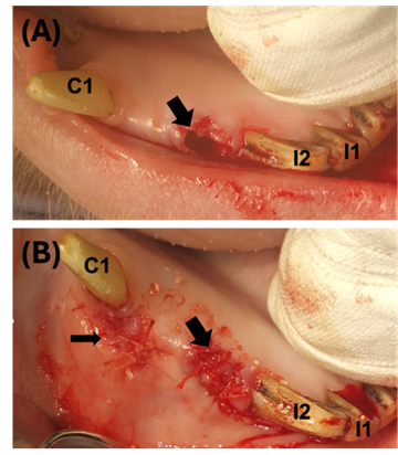
Atraumatic tooth extraction was performed by a certified dentist using the standard procedure for human patients (Figure 1A). Curettage of the socket was then performed and the socket filled with the graft material up to the alveolar gum ridge. The socket was then closed with Vicryl resorbable interrupted sutures (Figure 1B). In the first animal both the first and third incisors were extracted but it was found that extraction of the first incisor was difficult to control, and so in subsequent animals only the third incisor was extracted. Below the incisor a vertical incision 5 mm long was made, including attached and free gingiva, with full mucoperiosteal flap releasing. A periosteal elevator was then used to form a pouch in the mandibular bone with a depth of 10 mm. The sub-periosteal pouch thus formed was filled with graft material. The flap was then closed using Vicryl resorbable horizontal mattress sutures (Figure 1B).
At completion of the procedures, 1000 mg of Cefazolin was administered by IV injection and 5 mg/Kg Gentamicin by slow IV infusion. Following recovery from the surgery the animals received analgesics (Buprenorphine 0.02 mg/Kg IM) twice a day for three days. In the recovery period the antibiotic Vetrimoxin (7 mL) was administered intramuscularly every second day for four days. On the first post-operative day the animals were given liquid food (‘ENSURE’). On the second post-operative day and thereafter they received normal solid food. At 10 weeks after the procedures the animals were humanely sacrificed, and the graft sites examined.
2.3. Clinical and Radiographic EvaluationThe general condition of the animals was monitored twice daily by animal husbandry personnel and daily by a veterinary surgeon. Once weekly veterinary examination monitored surgical incision condition, mucous membrane color, overall condition, behavior, gait, appetite, and signs of pain. Feed and water consumption were also monitored.
Following animal sacrifice, the mandibles were removed and the macroscopic appearance of the implant sites described and photographed, both with the intact soft tissue and after removal of the overlying soft tissue. The mandible was then X-rayed and underwent a CT scan. The density of the grafted area was recorded for each graft site.
2.4. Histological EvaluationIndividual graft sites were then isolated and fixed in 4% paraformaldehyde in labelled vials. The vials were then sent to Pharmaseed Ltd for further processing. Here the graft sites were decalcified with EDTA, embedded in paraffin and then sectioned at approximately 5 microns. Sagittal sections through the center of each site were stained with hematoxylin and eosin (H&E) and examined microscopically.
The microscopic analysis involved a qualitative analysis of new bone formation using the following categories:
0.5 = Very Mild (up to 5 %) new bone formation per x10 magnification).
1 = Mild (up to 10 %) new bone formation per x10 magnification).
1.5 = Moderate (between 10-15 %) new bone formation per x10 magnification).
2 = Moderate (between 15-20 %) new bone formation per x10 magnification).
2.5 = Strong (between 20-25 %) new bone formation per x10 magnification).
3.0 = Strong (more than 25 %) new bone formation per x10 magnification).
In addition, the following parameters were quantified:
1. The area of mineralized tissue (host bone ingrowth plus graft material) as a percentage of overall bone tissue in the graft.
2. The area of non-mineralized connective tissue as a percentage of the overall tissue around the graft particles.
3. The percentage of new bone that was in close contact with the graft material to assess the degree of bone-graft contact.
Finally, a qualitative assessment of the presence of inflammatory cells or fibrosis was performed to examine the biocompatibility of the graft materials.
Six animals were successfully operated. All animals had uncomplicated bilateral I3 extraction with material grafting, and also bilateral sub-periosteal pouch production with material grafting, apart from the first animal for which the I1 was additionally extracted and filled and only one pouch was produced. Only the I3 extraction and pouch sites were included in the data collection because the I1 sites proved to be too difficult to extract atraumatically and resulted in very large defects. Depending on animal variation and surgical conditions there was some variation in the amount of material grafted. For the I3 sites between 0.25 and 1.5 grams of graft material were required, whereas for the sub-periosteal pouches the graft material required ranged from 0.4 to 2 grams.
Apart from the early surgical recovery period, the animals were without signs of pain or tenderness at the sites, ate normally and were generally healthy. A more detailed examination of the implant site is not possible in the un-anaesthetized Sinclair™ minipig. However, there were no signs of obvious discomfort or healing problems in any of the animals.
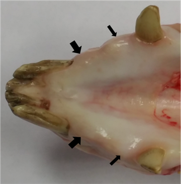
Post-mortem macroscopic examination of the mandibles showed that the soft tissue overlying the grafted sites had healed, with no signs of inflammation or any abnormalities, in all animals. As illustrated in a representative animal (Figure 2), both the incisor extraction sites, and sub-periosteal pouch sites had excellent healing of the overlying soft tissue.
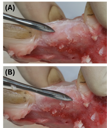
Removal of the overlying soft tissue revealed that the graft sites were filled with solid material having the same appearance as the surrounding bone (Figure 3).
On panoramic radiographs the bone density and height between the canine tooth and the I3 showed that the defects had been filled (Figure 4). The regions of the implants could be identified from the panoramic views and then examined in more detail in CT cross-section.
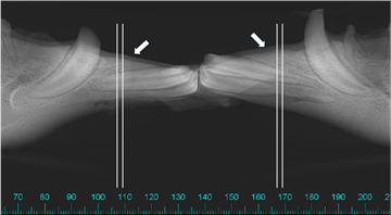
One-millimeter radiographic sections were examined through the grafted sites (Figure 5) and all the images showed homogenous radio-opacity consistently at every implantation site, in the range of 700-800 HU (Hounsfield Units), indicating similarity to values for cortical bone. There was no radiolucency, which indicates inflammation, at any of the implantation sites. No non-integrated graft particles could be distinguished separately from the host bone at either the Gen-Os® or Ivory Dentin Graft implant sites, either for the extraction sockets or the pouches.
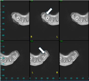
Thus, the radiographic examination confirmed uniform filling of the grafted sites with a radio-opacity approaching that of cortical bone. There was no radiolucency indicative of inflammation and no evidence for graft particles separated from the bone. There was no major difference between the Ivory Dentin Graft and Gen-Os® sites.
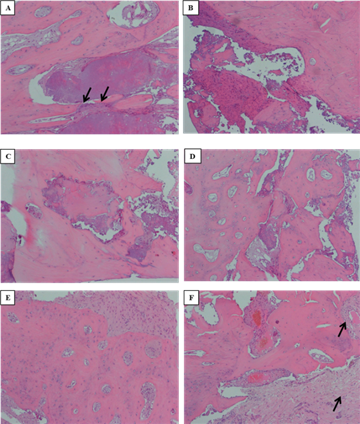
3.3. Histological FindingsTheoretically 12 Gen-Os® graft sites (6 incisor and 6 pouch) should have been available to compare with 12 Ivory Graft sites. Insufficient quality of the histological preparation in two of the cases for each group resulted in 10 versus 10 sites being evaluable in terms of the required parameters.
Qualitative classification of the H&E labeled sections showed that the degree of bone formation at all sites was at a relatively early stage with the maximal degree being moderate (grade 2.0 – 15-20% new bone formation), as may be expected at 10 weeks after grafting. There was no statistical difference in the qualitative degree of bone formation between Ivory Dentin Graft and Gen-Os® and no difference between the extraction and sub-periosteal pouch sites. As there was no difference in the degree of bone formation between extraction and pouch sites the results for the more detailed histological parameters were pooled to increase statistical power for the comparison of the Ivory Dentin Graft and Gen-Os® graft sites.
Detailed examination of the histology showed new bone ingrowth without any signs of inflammatory responses for both materials (Figure 6 a – d). New bone was often in very close contact with the graft material particles. In addition to proliferating bone tissue, fibrous, soft tissue ingrowth was also seen (Figure 6 e & f).
Quantification of histological parameters allowed for a direct comparison of the two materials. For a split mouth study, the correct procedure for assessing the statistical significance of differences is the paired t-test. Theoretically 12 Gen-Os® graft sites (6 incisor and 6 pouch) should have been available to compare with 12 Ivory Graft sites. Insufficient quality of the histological preparation for some sites, however, meant that some pairwise comparisons were not possible. For the strict pairwise comparison 10 versus 10 sites were evaluable in terms of the required parameters.
In terms of the percentage area of mineralized tissue there was no statistical difference (p > 0.05, paired t-test, two tailed) between Ivory Dentin Graft (mean 11.7%, SD 7.1) and Gen-Os® (mean 19.4%, SD 7.3). The percentage area of non-mineralized connective tissue was also not statistically different (p > 0.05, paired t-test, two tailed) between Ivory Dentin Graft (mean 48.9%, SD 30.2) and Gen-Os® (mean 60.0%, SD 16.4). There was a relatively high variability in these parameters which probably reflects differences in the degree of host tissue ingrowth in this early stage of repair. Addition of the non-paired assessments gave only marginally different mean values, showing the consistency of the data.
The percentage of new bone in close contact with Ivory Dentin Graft (mean 77.5%, SD 6.3) was, however, statistically significantly higher (p = 0.00001, paired t-test, two tailed; Figure 7) than for Gen-Os® (mean 45.5%, SD 9.3). Inclusion of the non-paired data resulted in only marginally different mean values, showing consistency of the data. This parameter was therefore clearly different despite the early stage of repair and variability. An over 30% difference in the degree of new bone to graft material contact is likely to impact the mechanical strength of the grafted region in light of the formation of ankylosis-like contacts between dentin and bone.
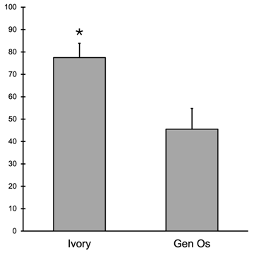
In summary, within the variability inherent in early repair parameters there was no statistical difference between Ivory Dentin Graft and Gen-Os® in terms of overall bone and soft tissue ingrowth, but there was a highly significant difference in the degree of new bone contact with the graft material, with the dentin material showing much higher contact.
In this study we have examined the ability of a novel xenogeneic dentin-derived bone graft material to repair bone defects in the mandible of the minipig in comparison with a clinically established bone-derived bone graft material.
The minipig is an accepted large animal, clinically relevant, model system for the examination of bone repair 14, 15, 16. Porcine bone has a similar mineral density and mineral concentration to human bone. The lamellar structure and remodeling process is also comparable to that in humans. Extraction of the third incisor creates a substantial defect of comparable size to human molar extraction sockets and is therefore a good model for extraction socket repair prior to implant placement. To examine bone augmentation comparable to that in sinus elevation procedures, a sub-periosteal pouch was created into which the graft material was placed. These procedures followed standard clinical practices for bone grafting but should best be interpreted as models for prediction of clinical effects because certain aspects, such as the use of membranes and the different anatomical arrangement in the minipig, preclude direct equivalence with the clinical situation. Thus, the use of a clinically validated positive control for comparison is important for interpretation.
Both types of bone defects were adequately repaired at 10 weeks after grafting using Ivory Dentin Graft as assessed by macroscopic appearance, radiographic density and histopathology. A direct comparison with other published studies is difficult due to differences in the type of bone defect examined, the use of membranes and different strains of minipig 17, 18. Use of an internal positive control was thus important for interpretation of the results. As an internal positive control Osteobiol® Gen-Os® was used in defects of the contralateral mandible in the same animals. Gen-Os® is a porcine bone-derived graft material which has retained the organic extracellular matrix, similarly to Ivory Dentin Graft. In clinical dental procedures it has been shown to provide excellent bone repair and augmentation but is relatively rapidly resorbed 13. Compared to this clinically well validated bone-derived material, Ivory Dentin Graft demonstrated similar early defect repair, in terms of host bone ingrowth and low inflammatory response, to create a solid graft potentially suitable for implant placement. It was, however, evident that Ivory Dentin Graft demonstrated a much higher degree of intimate contact with the in-growing host bone than the Gen-Os®. Although this is just a single timepoint assessment, this is consistent with the hypothesis that dentin-derived material forms a network of ankylosis-like connections with the host bone which, combined with high density and strength of dentin, gives structural stability and slows the resorption of the graft material 1. The xenograft material thus demonstrated the same favorable properties as chairside prepared autologous dentin material. In a clinical study of extraction socket grafting with implant placement using Ivory Dentin Graft a qualitative assessment of graft material to bone contact showed a similar effect 12.
This animal study therefore strengthens the overall evidence that this novel xenogeneic dentin material can be used for effective bone regeneration by confirming efficacy in extraction sockets and extending the evidence for use to unsupported augmentation in sub-periosteal pouches.
Beyond efficacy for dental procedures there is also the possibility to extend the use of this material to orthopedic situations because the material is available off-the-shelf in potentially unlimited quantity, and it provides early mechanical stability which is maintained by the ankylotic network. Future directions for study will therefore be to test its efficacy in models more relevant to orthopedic uses.
A novel xenogeneic dentin-derived bone graft material with retained organic extracellular matrix, demonstrated excellent bone defect repair and bone augmentation at 10 weeks after grafting in the mandible of minipigs. In a direct split-mouth comparison to a clinically validated bone-derived material the dentin-derived material showed much higher intimate contact with the ingrowing bone at this relatively early stage of repair. This is consistent with the material retaining the ability to form an ankylosis-like network, similar to that of autologous dentin, which produces early stability and slow resorption during the repair process. The off-the-shelf availability of this material potentially expands the uses of dentin derived bone graft material beyond the current chair-side prepared autologous material.
Dr. Sapoznikov is the owner and employee of Ivory Dentin Graft Ltd. which is developing Ivory Dentin Graft for clinical use. Martin Humphrey is an independent consultant to Ivory Dentin Graft Ltd.
| [1] | Sapoznikov, L., Humphrey, M. Progress in Dentin-Derived Bone Graft Materials: A New Xenogeneic Dentin-Derived Material with Retained Organic Component Allows for Broader and Easier Application. Cells, 13(21), 1806. Oct 2024. | ||
| In article | View Article PubMed | ||
| [2] | Binderman, I.; Hallel, G.; Nardy, C.; Yaffe, A.; Sapoznikov, L. A Novel Procedure to Process Extracted Teeth for Immediate Grafting of Autogenous Dentin. J. Interdiscipl. Med. Dent. Sci., 2, 154. Oct 2014. | ||
| In article | |||
| [3] | Murata, M.; Nezu, T.; Takebe, H.; Hirose, Y.; Okubo, N.; Saito, T.; Akazawa, T. Human dentin materials for minimally invasive bone regeneration: Animal studies and clinical cases. J. Oral Biosci., 65(1), 13–18. Nov 2023. | ||
| In article | View Article PubMed | ||
| [4] | Cervera-Maillo, J.M.; Morales-Schwarz, D.; Morales-Melendez, H.; Mahesh, L.; Calvo-Guirado, J.L. Autologous Tooth Dentin Graft: A Retrospective Study in Humans. Medicina, 58(1), 56. Dec 2021. | ||
| In article | View Article PubMed | ||
| [5] | Inchingolo, A.M.; Patano, A.; Di Pede, C.; Inchingolo, A.D.; Palmieri, G.; de Ruvo, E.; Campanelli, M.; Buongiorno, S.; Carpentiere, V.; Piras, F.; et al. Autologous Tooth Graft: Innovative Biomaterial for Bone Regeneration. Tooth Transformer® and the Role of Microbiota in Regenerative Dentistry. A Systematic Review. J. Funct. Biomater., 14(3), 132. Feb 2023. | ||
| In article | View Article PubMed | ||
| [6] | Kim, Y.K.; Kim, S.G.; Byeon, J.H.; Lee, H.J.; Um, I.U.; Lim, S.C.; Kim, S.Y. Development of a novel bone grafting material using autogenous teeth. Oral Surg. Oral Med. Oral Pathol. Oral Radiol. Endodontol. 109(4), 496–503. Apr 2010. | ||
| In article | View Article PubMed | ||
| [7] | Kim, Y.; Um, I.; Murata, M. Tooth Bank System for Bone Regeneration - Safety Report. J. Hard Tissue Biol., 23(3), 371–376. Jul 2014. | ||
| In article | View Article | ||
| [8] | Galindo-Moreno, P.; Hernández-Cortés, P.; Mesa, F.; Carranza, N.; Juodzbalys, G.; Aguilar, M.; O'Valle, F. Slow resorption of anorganic bovine bone by osteoclasts in maxillary sinus augmentation. Clin. Implant Dent. Relat. Res., 15(6), 858–866. Dec 2013. | ||
| In article | View Article PubMed | ||
| [9] | Trzaskowska, M.; Vivcharenko, V.; Przekora, A. The Impact of Hydroxyapatite Sintering Temperature on Its Microstructural, Mechanical, and Biological Properties. Int. J. Mol. Sci., 24(6), 5083. Mar 2023. | ||
| In article | View Article PubMed | ||
| [10] | Miron, R.J.; Fujioka-Kobayashi, M.; Pikos, M.A.; Nakamura, T.; Imafuji, T.; Zhang, Y.; Shinohara, Y.; Sculean, A.; Shirakata, Y. The development of non-resorbable bone allografts: Biological background and clinical perspectives. Periodontol 2000, 94(1), 161–179. Feb 2024. | ||
| In article | View Article PubMed | ||
| [11] | Dłucik, R.; Orzechowska-Wylęgała, B.; Dłucik, D.; Bogus, K. Histological examination of tooth-derived biomaterials obtained from different devices. Expert Rev. Med. Devices, 20(11), 979–988. Jul-Dec 2023. | ||
| In article | View Article PubMed | ||
| [12] | Sapoznikov, L.; Haim, D.; Zavan, B.; Scortecci, G.; Humphrey, M.F. A novel porcine dentin-derived bone graft material provides effective site stability for implant placement after tooth extraction: A randomized controlled clinical trial. Clin. Oral Investig., 27(6), 2899–2911. Jun 2023. | ||
| In article | View Article PubMed | ||
| [13] | Romasco, T.; Tumedei, M.; Inchingolo, F.; Pignatelli, P.; Montesani, L.; Iezzi, G.; Petrini, M.; Piattelli, A.; Di Pietro, N. A Narrative Review on the Effectiveness of Bone Regeneration Procedures with OsteoBiol® Collagenated Porcine Grafts: The Translational Research Experience over 20 Years. J. Funct. Biomater., 13(3), 121. Aug 2022. | ||
| In article | View Article PubMed | ||
| [14] | Wang, S.; Liu, Y.; Fang, D.; Shi, S. (2007). The miniature pig: a useful large animal model for dental and orofacial research. Oral diseases, 13(6), 530–537. Nov 2007. | ||
| In article | View Article PubMed | ||
| [15] | Li, Y.; Chen, S. K.; Li, L.; Qin, L.; Wang, X. L.; Lai, Y. X. (2015). Bone defect animal models for testing efficacy of bone substitute biomaterials. Journal of orthopaedic translation, 3(3), 95–104. Jun 2015. | ||
| In article | View Article PubMed | ||
| [16] | Zhang, Z.; Gan, Y.; Guo, Y.; Lu, X.; Li, X. Animal models of vertical bone augmentation (Review). Experimental and therapeutic medicine, 22(3), 919. Sep 2021. | ||
| In article | View Article PubMed | ||
| [17] | Buser, D.; Hoffmann, B.; Bernard, J. P.; Lussi, A.; Mettler, D.; Schenk, R. K. Evaluation of filling materials in membrane--protected bone defects. A comparative histomorphometric study in the mandible of miniature pigs. Clinical oral implants research, 9(3), 137–150. Jun 1998. | ||
| In article | View Article PubMed | ||
| [18] | Jensen, S. S.; Broggini, N.; Hjørting-Hansen, E.; Schenk, R.; Buser, D. Bone healing and graft resorption of autograft, anorganic bovine bone and beta-tricalcium phosphate. A histologic and histomorphometric study in the mandibles of minipigs., Clinical oral implants research, 2006, 17(3), 237–243. Jun 2006. | ||
| In article | View Article PubMed | ||
Published with license by Science and Education Publishing, Copyright © 2025 Lari Sapoznikov and Martin Humphrey
![]() This work is licensed under a Creative Commons Attribution 4.0 International License. To view a copy of this license, visit
http://creativecommons.org/licenses/by/4.0/
This work is licensed under a Creative Commons Attribution 4.0 International License. To view a copy of this license, visit
http://creativecommons.org/licenses/by/4.0/
| [1] | Sapoznikov, L., Humphrey, M. Progress in Dentin-Derived Bone Graft Materials: A New Xenogeneic Dentin-Derived Material with Retained Organic Component Allows for Broader and Easier Application. Cells, 13(21), 1806. Oct 2024. | ||
| In article | View Article PubMed | ||
| [2] | Binderman, I.; Hallel, G.; Nardy, C.; Yaffe, A.; Sapoznikov, L. A Novel Procedure to Process Extracted Teeth for Immediate Grafting of Autogenous Dentin. J. Interdiscipl. Med. Dent. Sci., 2, 154. Oct 2014. | ||
| In article | |||
| [3] | Murata, M.; Nezu, T.; Takebe, H.; Hirose, Y.; Okubo, N.; Saito, T.; Akazawa, T. Human dentin materials for minimally invasive bone regeneration: Animal studies and clinical cases. J. Oral Biosci., 65(1), 13–18. Nov 2023. | ||
| In article | View Article PubMed | ||
| [4] | Cervera-Maillo, J.M.; Morales-Schwarz, D.; Morales-Melendez, H.; Mahesh, L.; Calvo-Guirado, J.L. Autologous Tooth Dentin Graft: A Retrospective Study in Humans. Medicina, 58(1), 56. Dec 2021. | ||
| In article | View Article PubMed | ||
| [5] | Inchingolo, A.M.; Patano, A.; Di Pede, C.; Inchingolo, A.D.; Palmieri, G.; de Ruvo, E.; Campanelli, M.; Buongiorno, S.; Carpentiere, V.; Piras, F.; et al. Autologous Tooth Graft: Innovative Biomaterial for Bone Regeneration. Tooth Transformer® and the Role of Microbiota in Regenerative Dentistry. A Systematic Review. J. Funct. Biomater., 14(3), 132. Feb 2023. | ||
| In article | View Article PubMed | ||
| [6] | Kim, Y.K.; Kim, S.G.; Byeon, J.H.; Lee, H.J.; Um, I.U.; Lim, S.C.; Kim, S.Y. Development of a novel bone grafting material using autogenous teeth. Oral Surg. Oral Med. Oral Pathol. Oral Radiol. Endodontol. 109(4), 496–503. Apr 2010. | ||
| In article | View Article PubMed | ||
| [7] | Kim, Y.; Um, I.; Murata, M. Tooth Bank System for Bone Regeneration - Safety Report. J. Hard Tissue Biol., 23(3), 371–376. Jul 2014. | ||
| In article | View Article | ||
| [8] | Galindo-Moreno, P.; Hernández-Cortés, P.; Mesa, F.; Carranza, N.; Juodzbalys, G.; Aguilar, M.; O'Valle, F. Slow resorption of anorganic bovine bone by osteoclasts in maxillary sinus augmentation. Clin. Implant Dent. Relat. Res., 15(6), 858–866. Dec 2013. | ||
| In article | View Article PubMed | ||
| [9] | Trzaskowska, M.; Vivcharenko, V.; Przekora, A. The Impact of Hydroxyapatite Sintering Temperature on Its Microstructural, Mechanical, and Biological Properties. Int. J. Mol. Sci., 24(6), 5083. Mar 2023. | ||
| In article | View Article PubMed | ||
| [10] | Miron, R.J.; Fujioka-Kobayashi, M.; Pikos, M.A.; Nakamura, T.; Imafuji, T.; Zhang, Y.; Shinohara, Y.; Sculean, A.; Shirakata, Y. The development of non-resorbable bone allografts: Biological background and clinical perspectives. Periodontol 2000, 94(1), 161–179. Feb 2024. | ||
| In article | View Article PubMed | ||
| [11] | Dłucik, R.; Orzechowska-Wylęgała, B.; Dłucik, D.; Bogus, K. Histological examination of tooth-derived biomaterials obtained from different devices. Expert Rev. Med. Devices, 20(11), 979–988. Jul-Dec 2023. | ||
| In article | View Article PubMed | ||
| [12] | Sapoznikov, L.; Haim, D.; Zavan, B.; Scortecci, G.; Humphrey, M.F. A novel porcine dentin-derived bone graft material provides effective site stability for implant placement after tooth extraction: A randomized controlled clinical trial. Clin. Oral Investig., 27(6), 2899–2911. Jun 2023. | ||
| In article | View Article PubMed | ||
| [13] | Romasco, T.; Tumedei, M.; Inchingolo, F.; Pignatelli, P.; Montesani, L.; Iezzi, G.; Petrini, M.; Piattelli, A.; Di Pietro, N. A Narrative Review on the Effectiveness of Bone Regeneration Procedures with OsteoBiol® Collagenated Porcine Grafts: The Translational Research Experience over 20 Years. J. Funct. Biomater., 13(3), 121. Aug 2022. | ||
| In article | View Article PubMed | ||
| [14] | Wang, S.; Liu, Y.; Fang, D.; Shi, S. (2007). The miniature pig: a useful large animal model for dental and orofacial research. Oral diseases, 13(6), 530–537. Nov 2007. | ||
| In article | View Article PubMed | ||
| [15] | Li, Y.; Chen, S. K.; Li, L.; Qin, L.; Wang, X. L.; Lai, Y. X. (2015). Bone defect animal models for testing efficacy of bone substitute biomaterials. Journal of orthopaedic translation, 3(3), 95–104. Jun 2015. | ||
| In article | View Article PubMed | ||
| [16] | Zhang, Z.; Gan, Y.; Guo, Y.; Lu, X.; Li, X. Animal models of vertical bone augmentation (Review). Experimental and therapeutic medicine, 22(3), 919. Sep 2021. | ||
| In article | View Article PubMed | ||
| [17] | Buser, D.; Hoffmann, B.; Bernard, J. P.; Lussi, A.; Mettler, D.; Schenk, R. K. Evaluation of filling materials in membrane--protected bone defects. A comparative histomorphometric study in the mandible of miniature pigs. Clinical oral implants research, 9(3), 137–150. Jun 1998. | ||
| In article | View Article PubMed | ||
| [18] | Jensen, S. S.; Broggini, N.; Hjørting-Hansen, E.; Schenk, R.; Buser, D. Bone healing and graft resorption of autograft, anorganic bovine bone and beta-tricalcium phosphate. A histologic and histomorphometric study in the mandibles of minipigs., Clinical oral implants research, 2006, 17(3), 237–243. Jun 2006. | ||
| In article | View Article PubMed | ||


