Piebaldism is a rare autosomal dominant genetic disorder characterized by congenitalareas of depigmentation in a specific pattern. The condition is commonly marked by a white depigmented patchon the scalp and forehead, observed in 80-90% of cases. Depigmentation can also extend to the eyebrows, eyelashes, and nasal root. In rare instances, hypopigmented or depigmented patches may extend to atypical sites such as the chin, anterior neck, trunk, abdomen, and limbs. This case report presents an unusual manifestation of Piebaldism, highlighting the variability of clinical manifestations associated with this genetic condition.
Piebaldism is an uncommon autosomal dominant genodermatosis characterized by sharply demarcated congenital depigmented patches of skin and hair. The condition most frequently results from mutations in the KIT proto-oncogene, which encodes a transmembrane receptor critical for the development and survival of melanocytes, stem cells, and mast cells. Mutations in this gene impair the migration of melanoblasts from the neural crest and their subsequent differentiation into melanocytes, leading to stable depigmented areas that typically persist throughout life 1, 2.
The most recognizable clinical feature is a white forelock, present in approximately 80–90% of affected patients 2. Depigmented patches may also appear on the chin, neck, trunk, abdomen, and extremities 3. Although usually limited to cutaneous findings, piebaldism has occasionally been reported in association with developmental anomalies such as Hirschsprung’s disease and congenital deafness 4.
In this report, we present what we believe to be an unusual manifestation of piebaldism confined to the lateral forehead, without involvement of the frontal hairline. To the best of our knowledge, such a presentation has rarely been documented; highlighting these atypical features is crucial for achieving diagnostic accuracy and preventing misclassification.
A 5-month-old female infant was brought by her mother to the dermatology outpatient clinic at Al-Karak Governmental Teaching Hospital with concern about a white patch on the right side of the face. The lesion had been noted at birth and remained unchanged since then. The child was otherwise healthy, with no history of hearing problems, visual disturbances, or neurological symptoms.
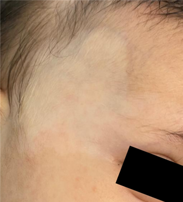
On physical examination, a well-defined, irregularly shaped depigmented patch was observed extending from the right temporal hairline toward the lateral aspect of the eyebrow (Figure 1). Within the lesion, fine vellus hairs displayed poliosis, whereas the surrounding eyebrows, eyelashes, and scalp hair retained normal pigmentation. Developmental milestones were appropriate for age, and there was no family history of similar pigmentary disorders.
Dermoscopy of the lesion revealed a distorted melanocytic network with reduced pigment intensity and absence of perifollicular pigmentation. Small islands of normally pigmented skin were visible within the depigmented patch. Under Wood’s light examination, the lesion appeared completely depigmented, confirming the loss of melanin.
Based on these clinical and dermoscopic findings, a diagnosis of piebaldism was established. Interestingly, the depigmentation was confined to the lateral forehead, sparing the frontal hairline, which made this a distinctly unusual presentation. The patient was advised to attend regular follow-up visits to monitor for any changes over time.
Piebaldism is an uncommon inherited disorder caused by mutations in the KIT proto-oncogene. These mutations interfere with the migration and differentiation of melanoblasts, which explains the absence of melanocytes in affected skin and hair 5. Most patients are recognized at birth with clearly demarcated, stable depigmented patches. The majority also develop the classic white forelock, a feature that remains one of the most recognizable signs of the condition 1. Depigmented patches can also be found on the chin, neck, trunk, abdomen, and limbs 3.
From a clinical perspective, our patient was unusual. The depigmentation was confined to the lateral forehead while sparing the frontal hairline. Such a distribution is rarely described in the literature and may be overlooked if clinicians rely only on the more typical features. Recognizing these atypical presentations is therefore essential to avoid misdiagnosis.
The main differentials considered were Waardenburg syndrome, nevus depigmentosus, and vitiligo 6, 7, 8, 9. Waardenburg syndrome was excluded as the child had no systemic abnormalities such as deafness or neurological deficits. Nevus depigmentosus usually shows faint hypopigmentation with serrated borders, perifollicular pigmentation, and eccrine white dots on dermoscopy. In our case, Wood’s light revealed complete depigmentation, which argued strongly against this diagnosis. Vitiligo was also excluded since it is typically acquired and progressive, whereas our patient’s lesion was congenital and stable.
Dermoscopy proved particularly helpful. The lesion showed a distorted melanocytic network, reduced pigment intensity, and islands of normally pigmented skin, findings that align well with piebaldism 10, 11, 12. These changes are different from the more uniform loss of pigment seen in vitiligo or the subtle reticular network described in nevus depigmentosus. As Al-Refu 12 has emphasized, dermoscopy can be a powerful non-invasive tool for distinguishing among hypopigmented macular disorders.
Interestingly, similar forehead involvement has been reported, though usually together with a frontal white forelock. The absence of such a forelock in our case makes the presentation even more distinctive and supports the idea that piebaldism can appear with wider clinical variability than generally assumed 13.
Overall, piebaldism remains a benign condition with no systemic consequences and no adverse effects on growth or cognition. Therefore, treatment is rarely needed except for cosmetic reasons. For this patient, a conservative plan with regular follow-up was chosen, mainly to document stability of the lesion over time.
| [1] | Spritz RA, Giebel LB, Holmes SA. Dominant negative and loss of function mutations of the c-kit (mast/stem cell growth factor receptor) proto-oncogene in human piebaldism. Am J Hum Genet. 1992 Feb; 50(2): 261-9. PMID: 1370874; PMCID: PMC1682440. | ||
| In article | |||
| [2] | Oiso N, Fukai K, Kawada A, Suzuki T. Piebaldism. J Dermatol. 2013 May; 40(5): 330-5. | ||
| In article | View Article PubMed | ||
| [3] | Paller AS, Mancini AJ. Hurwitz Clinical Pediatric Dermatology. 6th ed. Philadelphia: Elsevier; 2022. | ||
| In article | |||
| [4] | Treadwell PA. Systemic conditions in children associated with pigmentary changes. Clin Dermatol. 2015 May-Jun; 33(3): 362-7. | ||
| In article | View Article PubMed | ||
| [5] | Li X, Xing X, Liang X, Song C, Yang J, Ren D, Zhou Y. Piebaldism with café-au-lait macules resulting from a novel mutation of KIT gene in a three-generation Chinese family. Skin Res Technol. 2023 May; 29(6): e13352. | ||
| In article | View Article PubMed | ||
| [6] | Bolognia JL, Schaffer JV, Cerroni L. Dermatology. 5th ed. Philadelphia: Elsevier; 2021. | ||
| In article | |||
| [7] | Read AP, Newton VE. Waardenburg syndrome. J Med Genet. 1997 Aug; 34(8): 656-65. | ||
| In article | View Article PubMed | ||
| [8] | Ullah F, Schwartz RA. Nevus depigmentosus: review of a mark of distinction. Int J Dermatol. 2019 Dec; 58(12): 1366-1370. | ||
| In article | View Article PubMed | ||
| [9] | Taïeb A, Picardo M. Clinical practice. Vitiligo. N Engl J Med. 2009 Jan 8; 360(2): 160-9. | ||
| In article | View Article PubMed | ||
| [10] | Kaminska-Winciorek G, Spiewak R. Tips and tricks in the dermoscopy of pigmented lesions. BMC Dermatol. 2012 Aug 24; 12: 14. | ||
| In article | View Article PubMed | ||
| [11] | Ankad BS, Shah S. Dermoscopy of nevus depigmentosus. Pigment Int. 2017 Jul-Dec; 4(2): 121-123. | ||
| In article | View Article | ||
| [12] | Al-Refu K. Dermoscopy is a new diagnostic tool in diagnosis of common hypopigmented macular disease: A descriptive study. Dermatol Reports. 2018 Dec 21; 11(1): 7916. | ||
| In article | View Article PubMed | ||
| [13] | Grob A, Grekin S. Piebaldism in children. Cutis. 2016 Feb; 97(2): 90-2. PMID: 26919497. | ||
| In article | |||
Published with license by Science and Education Publishing, Copyright © 2025 Professor Khitam Al-Refu, Heba Al-lala and Hiba Harbi
![]() This work is licensed under a Creative Commons Attribution 4.0 International License. To view a copy of this license, visit
http://creativecommons.org/licenses/by/4.0/
This work is licensed under a Creative Commons Attribution 4.0 International License. To view a copy of this license, visit
http://creativecommons.org/licenses/by/4.0/
| [1] | Spritz RA, Giebel LB, Holmes SA. Dominant negative and loss of function mutations of the c-kit (mast/stem cell growth factor receptor) proto-oncogene in human piebaldism. Am J Hum Genet. 1992 Feb; 50(2): 261-9. PMID: 1370874; PMCID: PMC1682440. | ||
| In article | |||
| [2] | Oiso N, Fukai K, Kawada A, Suzuki T. Piebaldism. J Dermatol. 2013 May; 40(5): 330-5. | ||
| In article | View Article PubMed | ||
| [3] | Paller AS, Mancini AJ. Hurwitz Clinical Pediatric Dermatology. 6th ed. Philadelphia: Elsevier; 2022. | ||
| In article | |||
| [4] | Treadwell PA. Systemic conditions in children associated with pigmentary changes. Clin Dermatol. 2015 May-Jun; 33(3): 362-7. | ||
| In article | View Article PubMed | ||
| [5] | Li X, Xing X, Liang X, Song C, Yang J, Ren D, Zhou Y. Piebaldism with café-au-lait macules resulting from a novel mutation of KIT gene in a three-generation Chinese family. Skin Res Technol. 2023 May; 29(6): e13352. | ||
| In article | View Article PubMed | ||
| [6] | Bolognia JL, Schaffer JV, Cerroni L. Dermatology. 5th ed. Philadelphia: Elsevier; 2021. | ||
| In article | |||
| [7] | Read AP, Newton VE. Waardenburg syndrome. J Med Genet. 1997 Aug; 34(8): 656-65. | ||
| In article | View Article PubMed | ||
| [8] | Ullah F, Schwartz RA. Nevus depigmentosus: review of a mark of distinction. Int J Dermatol. 2019 Dec; 58(12): 1366-1370. | ||
| In article | View Article PubMed | ||
| [9] | Taïeb A, Picardo M. Clinical practice. Vitiligo. N Engl J Med. 2009 Jan 8; 360(2): 160-9. | ||
| In article | View Article PubMed | ||
| [10] | Kaminska-Winciorek G, Spiewak R. Tips and tricks in the dermoscopy of pigmented lesions. BMC Dermatol. 2012 Aug 24; 12: 14. | ||
| In article | View Article PubMed | ||
| [11] | Ankad BS, Shah S. Dermoscopy of nevus depigmentosus. Pigment Int. 2017 Jul-Dec; 4(2): 121-123. | ||
| In article | View Article | ||
| [12] | Al-Refu K. Dermoscopy is a new diagnostic tool in diagnosis of common hypopigmented macular disease: A descriptive study. Dermatol Reports. 2018 Dec 21; 11(1): 7916. | ||
| In article | View Article PubMed | ||
| [13] | Grob A, Grekin S. Piebaldism in children. Cutis. 2016 Feb; 97(2): 90-2. PMID: 26919497. | ||
| In article | |||


