Human health was affected strongly by diet, due to the composition and structure of intestinal microbiota mainly. This study aimed to characterize the intestinal mucosal bacteria of diarrheal mice caused by high-fat and high-protein diet. Ten specific pathogen free (SPF) Kunming male mice were chosen and randomly divided into control group and model group. The control group (FCM) mice were general feed diet. The model group (FMM) mice were high-fat and high-protein diet. After successful diarrhea, the mice's intestinal mucosa was collected for microbial analysis. Results showed that the number of OTUs (Operational Taxonomic Unit), the Chao 1 index and Shannon index (p<0.05) increased in high-fat and high-protein diet group. Compared to FCM, taxonomic composition indicated Bacteroidetes, Verrucomicrobia, Actinobacteria, Streptophyta, Deferribacteres were significantly increased and the Firmicutes was decreased in FMM. The genus of Helicobacter, Afipia., Methylobacterium, Pseudomonas, Clostridium, Phocaeicola and Faecalibaculum were higher in FMM, while Bradyrhizobium and Lactobacillus were lower than FCM. Bacterial species such as Helicobacter typhlonius, Methylobacterium sp., Afipia genosp. 1, Bacteroides vulgatus ATCC 8482, Faecalibaculum rodentium and Pseudomonas in FMM were significantly increased than FCM, but Lactobacillus johnsonii and Lactobacillus reuteri were decreased in FMM. To sum up, High-fat and high-protein diet results in intestinal mucosal microbiota dysbiosis, increases the number of conditionally pathogenic bacteria, reduces the number of beneficial bacteria, which trigger for diarrhea and may affect the Protein and fat catabolism and metabolism.
Diarrhea is a digestive system disease with the symptom featured by over three times of watery stools daily, and may be life-threatening, especially in children and adults who are immunosuppressed or malnourished 1. Depending on the duration there are two types of diarrhea in clinic: acute and chronic diarrhea. Normally, acute diarrhea is caused by microbial infections such as bacteria, viruses, and even fungi, however, chronic diarrhea is usually associated with disorders in the bowel or intestine, such as Crohn’s disease 2. Research indicated that diarrhea patients with the original microecological balance was disrupted, immunity reduced and bowel barrier function injured 3. Antibiotic-associated diarrhea (AAD) patient was characterized by a decrease of intestinal beneficial bacteria such as Bifidobacterium and Lactobacillus, and an increase of harmful bacteria such as Clostridium perfringens, Staphylococcus aureus and Clostridium difficile 4. Inflammatory bowel disease diarrhea patients have a lower relative abundance of Firmicutes phyla and an increase in Bacillus species, and the spatial distribution of Bacillus spp. and Bacteroides fragiles changed 5, 6. Therefore, treating diarrheal diseases by balancing intestinal microbiota is an important initiative.
It is well known that gut microbiota plays vital roles in maintaining human health and microecological balance and nutrient absorption (protein, fat, and fiber) 7, 8. Firmicutes and Proteobacteria have been identified as the most abundant phyla within the intestinal mucosa of mice, accounting for more than 90%, followed by Actinobacteria, however, which is different from the mouse small intestinal contents where the Firmicutes and Bacteroidetes are the main phylum 9, 10. The human body under healthy condition, the gut flora is in a relative balance, unfortunately, once inherent equilibrium is disturbed, which in turn increase risk of a variety of intestinal diseases 8, 11.
Since the dawn of the industrialized era, diet of people has changed dramatically, including a substantial reduction in the ingestion of dietary fiber, but the intake of foods rich in fat and protein has increased mutually 12. Numerous studies have shown that diet plays an important role in mediating alterations of gut flora. Research revealing that high-fat diet (HFD) inhibits the growth of Lactobacillus, Bifidobacterium 13, but enhances the growth of harmful bacteria (Escherichia coli), and promots choline catabolism which is further metabolized by intestinal flora into trimethylamine (TMA), then converts into trimethylamine oxide (TMAO), and ultimately leads to increas TMAO levels in the blood 14, 15. Dietary protein is the main source of intestinal flora amino acids, moreover, the undigested amino acids are usually fermented into many bacterial metabolites or end products, and different levels of ingested protein have different effects on the intestinal internal environment 16. Low protein diet increases beneficial bacteria Bifidobacterium and Lactobacillus, but reduces harmful bacteria in the intestinal, meanwhile, stimulated intestinal immunity and the formation of short chain fatty acid (SCFA) 17. Unfortunately, excessive protein intake by the organism, which causes an elevation of potential pathogens Coliforms, Streptococcus and Bacillus 8, 18, 19. Individuals who consumed more fiber had a higher abundance of Prevotella spp. compared to those who consumed high protein and fat, while the latter one had a higher abundance of anaphyla spp in the intestine, that suggested that Prevotella has a better fiber degradation capacity compared to anaphyla 20. Therefore, through a healthy and safe dietary interventions maintaining a healthy intestinal microbiota balance is important for the prevention and alleviation of intestinal diseases. Additionally, understanding the implications of different diet on intestinal microbiota is important. Based on people's current diet fat and protein intake are excessive 12, so this study aim to compile the available evidence on the contribution of dietary protein and fat to intestinal microbiota dysbiosis. We will describe the current knowledge on how dietary protein and fat affect the gut microbiota, how interactions between dietary protein and fat and the gut microbiota influence host physiology and health, and how the gut microbiota affects host protein and fat metabolism. Providing a fairly reliable basis for the treatment of various intestinal diseases especially associated with intestinal microbiota dysbiosis caused by high-fat and high-protein diet.
Ten 6-week-old SPF Kunming male mice, weighing 18~22 g, were purchased from Hunan Sleika Jingda Experimental Animal Co., Ltd (SCXK (xiang) 2016-0002), and the mice were housed under stable conditions at the Animal Experiment Center of Hunan University of Chinese Medicine (temperature 23°C~25°C, relative humidity 50%~70%), and the animal experimental procedures were performed in accordance with the Hunan University of Chinese Medicine Animal Ethics and Welfare Committee (LL2020062302).
2.2. Feeds PreparationThe normal feeds is breeding feed for rats and mice (5 kg/bag, protein: 20%, fat: 4%). The high-fat, high-protein group refers to the consisting of milk powder (Nestle, 300 g/bag, product execution standard number: GB 19644, 30% protein and 20% fat), soybean milk flour (Huiyi, 350 g/bag, product execution standard number: GB/T18738, 33% protein and 18% fat), low gluten flour (Huiyi, 1000 g/bag, Product Execution Standard No.: GB/T 8608, 13% protein and 2% fat), meat pine (Anhui Lizheng, 500 g/bag, Manufacturer: Anhui Lizheng Food Co., Ltd., Produ, 30% protein and 25% fat) are mixed 1:2:1:1.
2.3. Animal GroupsTen SPF KM male mice were randomly divided into control (FCM) and model groups (FMM). Five mice onecages in each group.
2.4. Modeling and Dosage [21,22]Mice in the FMM were fed high-fat and high-protein chow and gavaged with vegetable oil at 4 days, 0.4 mL/time, twice daily for 3 d, for a total duration of 6 d in the modeling phase. Mice in the FCM were fed with normal chow and gavaged with equal amounts of distilled water.
2.5. Extraction of Mice Intestinal Mucosa [23]After 9 days, mice in each group were sacrificed rapidly by cervical dislocation, placed on a super clean workbenchthe. Under aseptic environment, the whole section of the small intestine was removed, and the intestinal contents were extruded with ophthalmic forceps, followed by cutting open the whole intestine with surgical scissors. After rinsing in physiological saline, the intestinal contents adhering to the intestinal wall and the fat adhering to the top of the intestine were removed, and then placed on sterile filter paper to absorb the water. Afterwards, the intestinal mucosa was scraped on sterilized petri dishes using sterilized glass sheets and weighing paper, and the intestinal mucosa of the same group of mice was weighed and placed together in sterilized centrifuge tubes containing glass beads.
2.6. DNA ExtractionTotal bacterial DNA was extracted from the samples using CTAB method, and the quality of DNA extraction was detected by 0.8% agarose gel electrophoresis, while DNA was quantified by UV spectrophotometer.
2.7. PCR AmplificationThe full-length bacterial 16S rRNA gene sequence was amplified using the extracted DNA as a template. The amplification primers were 27F (5'-AGAGTTTGATCMTGGCTCAG-3') and 1492R (5'-ACCTTGTTACGACTT-3'), which were provided by Shanghai Paisano Biotechnology Co. Ltd. (http://www.personalbio.cn/). The amplification system was shown in Table 1: After the required components of PCR reaction were configured, the template DNA was pre-denatured at 98°C for 2 min on a PCR instrument to denature fully, and then entered the amplification cycle. In each cycle, at 98°C for 15s to denature the template, then lower the temperature to 55°C for 30s to fully anneal the primers to the template; at 72°C for 30s to extend the primers over the template, and synthesize DNA to complete a cycle. Repeat this cycle 25~30 times to make a large accumulation of amplified DNA fragments. Finally, hold at 72°C for 5 min to make the product extension complete and store at 10°C.
Sequences with similarity higher than 97% was assigned to one OTU, which include OTU division, annotation and streamlining, and finally get the OTU table for analysis 24.
In community ecology, the abundance and diversity of microbial communities can be reflected by analyzing sample diversity. Chao 1 (http://scikit-bio.org/docs/latest/generated/skbio.diversity.alpha.chao1.html) is an index that reflects the abundance of the community. The larger the Chao1 index, the higher abundance of the community. Simpson (http://scikit-bio.org/docs/latest/generated/skbio.diversity.alpha.simpson.html) and Shannon (http://scikit-bio.org/docs/latest/generated/skbio.diversity.alpha.Shannon.html) are reflecting community diversity, and hi-gher Shannon and Simpson indexes indicate higher community diversity.
The main purpose of Beta diversity analysis is to examine the similarity of community structure among different samples. Including three main methods, Principal component analysis (PCA), Multidimensional scaling (MDS), observing samples differences by natural decomposition of the community data structure and sorting the samples 25.
The specific composition of each sample at each classification level can be obtained based on the OTU division and classification status identification results.
IBM SPSS 21.0 statistical software was used for statistical analysis, and all measures were expressed as "mean ± standard deviation" (x ± s). Single sample t-test was used when multisample means were normally distributed and variances were uniform; Wilcoxon rank-sum test was used when variances were not uniform, p<0.01 or p<0.05 indicated that the differences were statistically significant, otherwise there was not statistical significance.
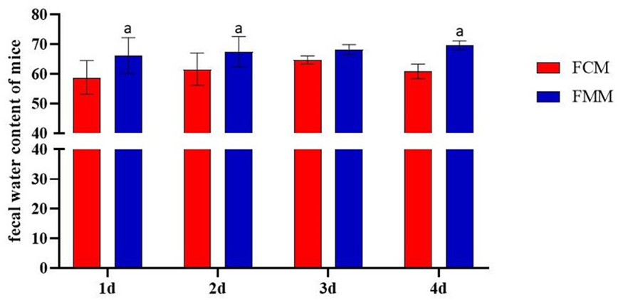
During the modeling period, compared with the FCM, the mice in the FMM gradually showed symptoms: darkened fur, dull eyes, easily provoked, fighting, increased frequency of pole climbing, filthy perianal area, and diluted stools, and on the 2nd day of modeling, soft feces, significant increase in fecal water content (t=-5.301, p=0.006) and half of the mice developed diarrhea after three days, while the the FCM were pellet.

The OTU Venn diagram analyzes the unique or common OTUs between the different sample groups and visualizes the similarity and uniqueness of the samples at the OTU level. From Figure 2, it can be seen that the common OTUs were 80. The number of OTUs exclusive to the FCM was 41, and the number of OTUs unique to the FMM was 175. The total number of OTUs in the FCM was 121, and the total number of OTUs in the FMM was 255. The analysis shows that after intervention with a high-fat and high-protein diet, the number of mucosal flora species and taxonomic units were significantly increased.

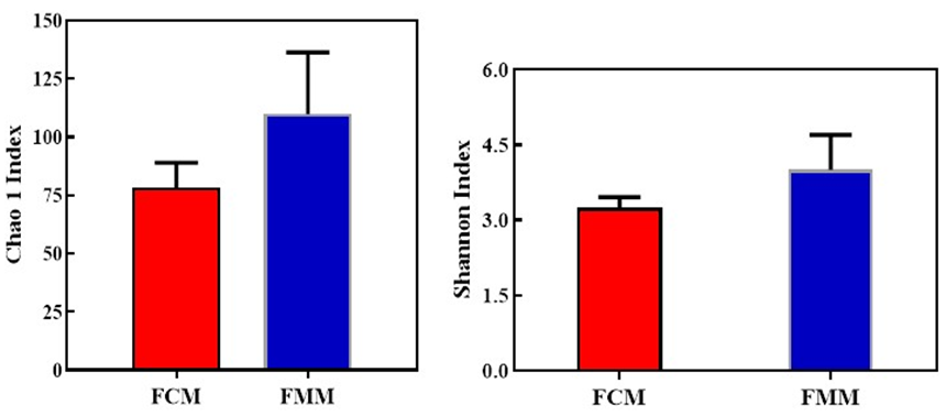
Alpha diversity index is a comprehensive index reflecting the richness and diversity of microbial communities, which includes species diversity, genetic diversity, physiological diversity, and ecological diversity. In this experiment, Chao 1 index was used to assess the abundance of intestinal mucosal flora, and Shannon index responded to community diversity. As shown in Figure 3, the Chao 1 index and Shannon index of FMM both were significantly higher than the FCM (t1= -2.472, p1=0.039; t2= -2.356, p2=0.046). This indicated that high-fat and high-protein diet increased the abundance and diversity of intestinal mucosal flora in mice, which was consistent with the OTU results.


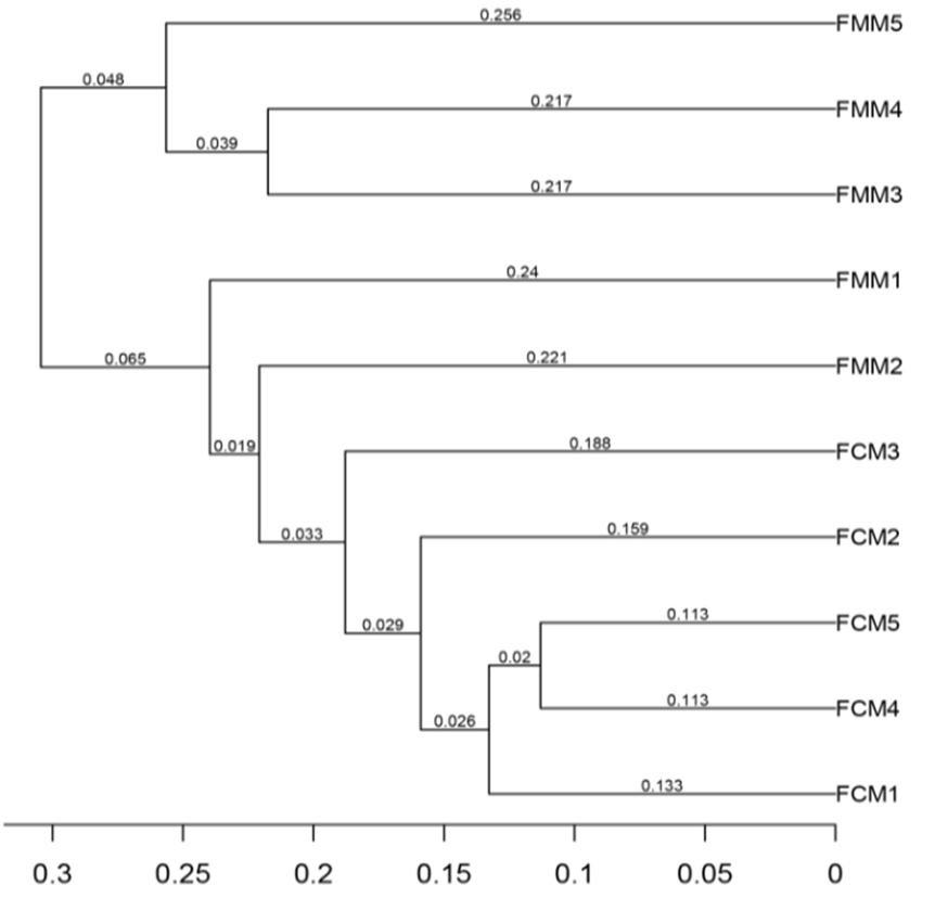
The main purpose of Beta diversity analysis is to examine the similarity of community structure among different samples. From Figure 5, the FCM samples were grouped at a distance of 0.033, which indicated the small variations in the FCM samples. In the FMM, except for the FMM1 and FMM2, the remaining samples can be well clustered into one group. Compared to the FCM, FMM1 and FMM2 are more likely to cluster with the rest of the model group samples. Indicating that the intra-group variation in FMM were larger than FCM, but still well separated from the FCM samples.

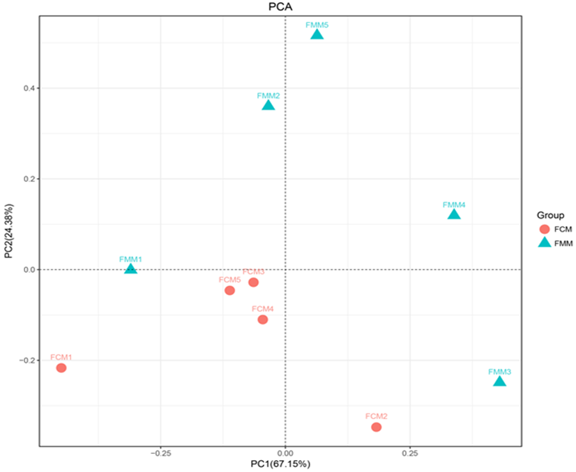
PCA was a visualization method used to study similarities or differences in data and to observe differences between individuals or communities. As shown in Figure 6, the FMM samples were mainly located in the first, third and fourth quadrants and were relatively dispersed, while the FCM samples were relatively concentrated in the second quadrant. It was suggested that the high-fat and high-protein diet changed the bacterial flora homogeneity.

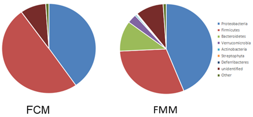
At the phylum level, there was a large difference in the flora composition between the FCM and FMM (Figure 7). The top three phyla in both groups were Proteobacteria, Firmicutes, and Bacteroidetes, respectively accounting for 30%, 49.77%, and 3.10% in the FCM, compared with 43.83%, 30.42%, and 10.89% in the FMM. The results suggested that except for Proteobacteria, the two groups differed more in Firmicutes and Bacteroidetes.

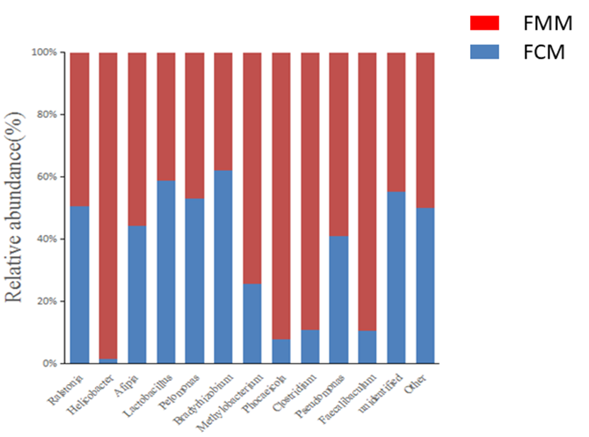
Total 63 genera were detected in the FCM and FMM, of which 25 genera were in the FCM and 56 genera in the FMM, again demonstrating the increased diversity gut mucosal flora diversity in mice treated with the special diet combined with lard gavage. The dominant genera in the FCM were Lactobacillus and Bradyrhizobium, and the dominant genera in the FMM were Helicobacter, Afipia, Methylobacterium, Phocaeicola, Clostridium, Pseudomonas, and Faecalibaculum (Figure 8).

Specifically to bacterial species, 98 species were detected in the FCM and FMM groups, of which 22 species were in the FCM and FMM (Table 2). Compared with the FCM, we found that the abundance of Lactobacillus johnsonii and Lactobacillus reuteri in the FMM was significantly decreased. Similarly, the dominant genera in the FMM were Helicobacter typhlonius, Methylobacterium sp., Afipia genosp. 1, Bacteroides vulgatus ATCC 8482, and Faecalibaculum rodentium, Pseudomonas. The above results suggested that high-fat and high-protein diet had a large effcct on the intestional microbiota of mice.
The gut flora that inhabits in the gastrointestinal tract has been co-evolving with us, which plays an important role in the gastrointestinal system and normal physiological function. In modern westernized diets, which contain higher fat and protein but are poor in fiber caused intestinal microecological dysbiosis, which promoted local inflammation and increased intestinal wall permeability and other hazards 26, 27. Studies found that an unbalanced diet altered the composition of the gut flora, causing a decrease in the dominant flora and an increase in non-dominant flora, at the same time, an increase of lipopolysaccharide (LPS) and endotoxin in the intestine 28, 29. Combined with the results of this experiment, we found that the proportion and abundance of intestinal flora in mice after high-fat and high-protein diet changed. Compared with the FCM, the number of OTU in the intestinal mucosa of the FMM increased remarkably, and the diversity and richness of its species changed. On the analysis of Alpha diversity results, the Chao 1 and Shannon indexes of FMM was significantly different with FCM (p<0.05). According to the results of Beta diversity analysis, PCA principal coordinate analysis and UPGMA analysis showed that the sample information of FCM was relatively concentrated, and there was a certain distance from the FMM. The above results suggested that the intestinal microbial community of mice has changed after high-fat and high-protein feeding.
Regarding human-based studies demonstrated that a longer term high-fat diet generated a positive association with an abundance of Bacteroides and Actinobacteria, but a negative association with Firmicutes and Proteobacteria 30. Chronic excess protein diet could also disrupt the intestinal microecological balance, mainly by increasing the abundance of the phylum Bacteroides, Proteobacteria, and Actinobacter, indicating that there were some differences in the variations between the two diets 31. In this experiment, we further analyzed the taxonomic components changes of intestinal mucosal microbial from phylum, genus, and species, found that high-fat and high-protein diet increased the abundance of Proteobacteria and Bacteroides, while the Firmicutes decreased. This is inconsistent with previous studies that HFD reduced the proportion of Bacteroides phylum and increased the proportion of Firmicutes and Proteobacteria phylum in the mouse intestinal mucosa, however, it was consistent with gut flora in diarrheic mice 6, 30. It may be due to high-protein diet increases the intestinal mucosal Bacteroidetes more than HFD decreases its abundance, but the exact mechanism remains to be explored 9. At the genus level, the relative abundance of Helicobacter, Afipia, Methylobacterium, Pseudomonas, Clostridium, Phocaeicola, and Faecalibaculum in the FMM was higher than the FCM, which is a specific embodiment of the increase of OTUs and the Alpha diversity index in the intestinal mucosa fed with the special diet combined with gavage of vegetable oil. Many studies have found that Helicobacter, Afipia, Pseudomonas, and Clostridium are conditional pathogens. Helicobacter has capable of causing a variety of intragastric and extragastric diseases, which severity is correlated with the virulence factors it expresses and these factors through involved in inflammatory responses to maintain chronic inflammation 32. Afipia is a genus of gram-negative bacteria, including Afipia gen. nov, Afipia broommeae sp. nov, Afipia clevelandensis sp. nov and other species, which is the main causative agent of cat claw disease 33. Pseudomonas is a conditionally pathogenic bacterium with weak pathogenicity and strong resistance to drugs, easily causing septic lesions 34. Some Clostridium can secrete exotoxins and invasive enzymes that cause disease 35. In addition, a study found that without affecting adaptive immune cells Faecalibaculum spp. could inhibit tumor cell growth by releasing SCFAs 36, which further demonstrated that the intestinal microenvironment of the FMM was damaged. Meanwhile, the abundance of Lactobacillus spp in the FMM was significantly lower than FCM. The genus Lactobacillus were long thought to be a common beneficial bacteria, and its members are the most abundant microorganisms in the human gastrointestinal (GI) tract, with animal studies and clinical results suggesting that they help prevent and treat various digestive disorders 37. At the species level, we found that the abundance of the beneficial bacteria Lactobacillus johnsonii and Lactobacillus reuteri was significantly decreased in the FMM. Reports have shown that Lactobacillus reuteri could inhibit the growth and colonization of various pathogenic bacteria and reshape the composition of the host's symbiotic flora by producing Reuterin, as well as promoting regulatory T cells to improve the immunity of the body 38. From the above, we hypothesized that the specially formulated high-fat, high-protein diets with gavage of vegetable oil increased the abundance of conditionally pathogenic bacteria, but the abundance of beneficial bacteria decreased, resulting in impaired intestinal microecological balance and causing diarrhea.
In the GI, some flora can break down fats and proteins. Cultivation analysis has identified at least Bacteroides and propionibacteria, as well as various bacilli, to be proteolytic bacteria, and several of these bacteria carry genes for serine and other proteases in their genomes 17. It has been reported that some dominant bacteria can secrete various proteases and peptidases to hydrolyze proteins and amino acids (AA) or metabolize AA directly, and the proteolytic activity has been mainly attributed to the genera of Bacteroides, Clostridium, and Lactobacillucan 39. The gut microbes are involved in lipid metabolic processes which may be mediated by the intestinal microbiota metabolites (SCFAs, secondary bile acids) or pro-inflammatory bacterial-derived factors (lipopolysaccharides). SCFAs regulate host metabolism by providing energy to the host, improving peripheral tissue metabolism and stimulating incretin hormone production; bile acids can emulsify lipids directly or binding to the bile acid receptor TGR5, and Bacillus spp., Eubacterium spp. and Clostridium spp. can promote the metabolism of dietary fat and cholesterol absorption by secreting bile acids 40; LPS interacts with lipids in multiple ways and increases blood triglyceride concentrations through a variety of mechanisms 41. In combination with the present experiment, compared with the FCM, the FMM abundance of Clostridium spp. and Lactobacillu spp. were significantly changed. Revealing that high-fat and high-protein diet may affect the breakdown and metabolism of protein and fat by some metabolism-related bacteria.
In summary, High-fat and high-protein diet is not conducive to human digestive function, leading to imbalance of intestinal mucosal microecological in mice, increased the abundance of Helicobacter typhlonius, Methylobacterium sp., Afipia genosp. 1. Faecalibaculum rodentium, Pseudomonas and other conditional pathogens , but suppressed the abundance of beneficial bacteria Lactobacillus johnsonii and Lactobacillus reuteri. Conversely, some increased flora (Clostridium, Lactobacillu) may affect the catabolism and absorption of proteins and fats in the organism, leading to an increased risk of intestinal diseases. Future research shall further focus on lifestyle strategies for wellbeing should integrate advice on the optimal establishment and maintenance of a healthy gut flora through regulating diet.
This work was supported by the National Natural Science Foundation of China (No. 81874460).
The authors declare that there is no confict of interests regarding the publication of this paper
Author contributions were as follows: study design (Zhoujin Tan), data collection (Tao Theng), statistical analysis (Jiayuan Zhu, Yawei Liu), data interpretation (Jiayuan Zhu, Yawei Liu), manuscript preparation (Jiayuan Zhu, Yawei Liu), and funds collection (Zhoujin Tan).
The study was approved by the Animal Ethics and Welfare Committee of Hunan University of Chinese Medicine (LL2020062302).
| [1] | Yu M, Jin X, Liang C, Bu F, Pan D, He Q et al. Berberine for diarrhea in children and adults: a systematic review and meta-analysis. Therap Adv Gastroenterol. 2020; 13: 1-19. | ||
| In article | View Article PubMed | ||
| [2] | He L, Long C, Liu Y, Guo Y, Xiao N, Tan Z. Effects of Debaryomyces hansenii treatment on intestinal microorganisms in mice with antibiotics-induced diarrhea. 3 Biotech. 2017; 7(5): 347. | ||
| In article | View Article PubMed | ||
| [3] | Wang W, Cui L.H. Advances in intestinal microecology and its therapeutic effect on diarrhea. Academic Journal of Chinese PLA Medical School. 2016; 37(7): 813-816+819. | ||
| In article | |||
| [4] | Silverman MA, Konnikova L, Gerber JS. Impact of antibiotics on necrotizing enterocolitis and antibiotic-associated diarrhea. Gastroenterol Clin North Am. 2017; 46(1): 61-76. | ||
| In article | View Article PubMed | ||
| [5] | Walker AW, Sanderson JD, Churcher C, Parkes GC et al. High-throughput clone library analysis of the mucosa-associated microbiota reveals dysbiosis and differences between inflamed and non-inflamed regions of the intestine in inflammatory bowel disease. BMC Microbiology. 2011; 11: 7. | ||
| In article | View Article PubMed | ||
| [6] | Lee JR, Magruder M, Zhang L, Westblade LF et al. Gut Microbiota Dysbiosis and Diarrhea in Kidney Transplant Recipients. Am J Transplant. 2020; 19(2): 488-500. | ||
| In article | View Article PubMed | ||
| [7] | Xi L, Song Y, Qin X, Han J, Chang YF. Microbiome Analysis Reveals the Dynamic Alternations in Gut Microbiota of Diarrheal Giraffa camelopardalis. Front Vet Sci. 2021; 8: 1-13. | ||
| In article | View Article PubMed | ||
| [8] | Meng X, Zhang G, Cao H, Yu D, Fang X et al. Gut dysbacteriosis and intestinal disease: mechanism and treatment. J Appl Microbiol. 2020; 129(4): 787-805. | ||
| In article | View Article PubMed | ||
| [9] | Meng Y, Li X., Zhang J, Wang C, Lu F. Effects of Different Diets on Microbiota in The Small Intestine Mucus and Weight Regulation in Rats. Sci Rep. 2019; 9(1):1-12. | ||
| In article | View Article PubMed | ||
| [10] | Zhu JY, Xiao NQ, Tan Z.J. Research progress on intestinal mucosal injury induced by traditional chinese medicine. World Chinese Journal of Digestology. 2021; 29:1-6. | ||
| In article | View Article | ||
| [11] | Weiss GA, Hennet T. Mechanisms and consequences of intestinal dysbiosis. Cell Mol Life Sci. 2017; 74(16): 2959-2977. | ||
| In article | View Article PubMed | ||
| [12] | Barber TM, Kabisch S, Pfeiffer A, Weickert M. The Health Benefits of Dietary Fibre Thomas. Nutrients. 2020; 12:3209. | ||
| In article | View Article PubMed | ||
| [13] | Zhang M, Yang XJ. Effects of a high fat diet on intestinal microbiota and gastrointestinal diseases. World J Gastroenterol. 2016; 22(40):8905-8909. | ||
| In article | View Article PubMed | ||
| [14] | Yoo W, Zieba JK, Foegeding NJ et al. High-fat diet–induced colonocyte dysfunction escalates microbiota-derived trimethylamine N-oxide. Science. 2021; 373(6556): 813-818. | ||
| In article | View Article PubMed | ||
| [15] | Rohr MW, Narasimhulu CA, Rudeski-Rohr TA, Parthasarathy S. Negative Effects of a High-Fat Diet on Intestinal Permeability: A Review. Adv Nut. 2020; 11(1):77-91. | ||
| In article | View Article PubMed | ||
| [16] | Zhao J, Zhang X, Liu H, Brown MA, Qiao S. Dietary Protein and Gut Microbiota Composition and Function. Curr Protein Pept Sci. 2019; 20(2):145-154. | ||
| In article | View Article PubMed | ||
| [17] | Ma N, Tian Y, Wu Y, Ma X. Contributions of the Interaction Between Dietary Protein and Gut Microbiota to Intestinal Health. Curr Protein Pept Sci. 2017; 18(8):795-808. | ||
| In article | View Article PubMed | ||
| [18] | Peng M, Bitsko E, Biswas D. Functional properties of peanut fractions on the growth of probiotics and foodborne bacterial pathogens. J Food Sci. 2015; 80(3):M635-M641. | ||
| In article | View Article PubMed | ||
| [19] | Cani PD. Human gut microbiome: Hopes, threats and promises. Gut. 2018; 67(9):1716-1725. | ||
| In article | View Article PubMed | ||
| [20] | Marietta E, Horwath I, Taneja V. Microbiome, Immunomodulation, and the Neuronal System. Neurotherapeutics. 2018; 159(1): 23-30. | ||
| In article | View Article PubMed | ||
| [21] | Windey K, De Preter V, Verbeke K Relevance of protein fermentation to gut health. Mol Nutr Food Res. 2012; 56(1):184-196. | ||
| In article | View Article PubMed | ||
| [22] | Yu T, Wang Y, Chen X, Xiong W, Tang Y, Lin L. Spirulina platensis alleviates chronic inflammation with modulation of gut microbiota and intestinal permeability in rats fed a high-fat diet. J Cell Mol Med. 2020; 24(15):8603-8613. | ||
| In article | View Article PubMed | ||
| [23] | Wu Y, Tang, Y, Liu YW, Hui HY, Tan ZJ.Effects of Tongxie Yaofang prescription on activity of digestive enzymes in intestinal mucosa and contents in Gan-qi-cheng diarrhea mice -pi diarrhea mice. Chinese Journal of Microecology. 2020; 32:745-749. | ||
| In article | |||
| [24] | Wang WY, Srivathsan A, Foo M, Yamane S.K, Meier R. Sorting specimen-rich invertebrate samples with cost-effective NGS barcodes: Validating a reverse workflow for specimen processing. Mol Ecol Resour.2018; 18(3):490-501. | ||
| In article | View Article PubMed | ||
| [25] | Turgut G. (2017). Board Diversity and Corporate Social Performance: A Review and Empirical Investigations. HEC Montreal (Canada). | ||
| In article | |||
| [26] | Chen Y, Shen T. Rarefaction and extrapolation of species richness using an based Fisher’s logseries. Ecol Evol.2017; 7(23): 10066-10078. | ||
| In article | View Article PubMed | ||
| [27] | Barber TM, Valsamakis G, Mastorakos G, Hanson P et al. Dietary influences on the microbiota-gut-brain axis. Int J Mol Sci. 2021; 22(7): 1-18. | ||
| In article | View Article PubMed | ||
| [28] | Bibbò S, Ianiro G, Giorgio V, Scaldaferri F et al.The role of diet on gut microbiota composition. Eur Rev Med Pharmacol Sci. 2016; 20(22): 4742-4749. | ||
| In article | |||
| [29] | Li X, Peng X, Guo K, Tan Z. Bacterial diversity in intestinal mucosa of mice fed with Dendrobium officinale and high-fat diet. 3 Biotech. 2021; 11(1): 1-12. | ||
| In article | View Article PubMed | ||
| [30] | Liu JP, Zou WL, Chen SJet al.Effects of different diets on intestinal microbiota and nonalcoholic fatty liver disease development. World J Gastroenterol. 2016; 22(32): 7353-7364. | ||
| In article | View Article PubMed | ||
| [31] | Dong TS, Luu K, Lagishetty V, Sedighian Fet al. A high protein calorie restriction diet alters the gut microbiome in obesity. Nutrients. 2020; 12(10): 1-16. | ||
| In article | View Article PubMed | ||
| [32] | Baj J, Forma A, Sitarz M, Portincasa P et al. Helicobacter pylori Virulence Factors-Mechanisms of Bacterial Pathogenicity in the Gastric Microenvironment. Cells. 2020; 10(1): 1-37. | ||
| In article | View Article PubMed | ||
| [33] | Feng ZS.The causative agent of cat-scratch disease and the genus Afipia. Acta Neuropharmacologica. 1993; 10: 97-98. | ||
| In article | |||
| [34] | Thi M, Wibowo D, Rehm B. Pseudomonas aeruginosa biofilms. Int J Mol Sci. 2020; 21(22): 1-25. | ||
| In article | View Article PubMed | ||
| [35] | Shen A, Edwards AN, Sarker MR, Paredes-Sabja D. Sporulation and Germination in Clostridial Pathogens Aimee. Microbiol Spectr. 2016; 7(6): 139-148. | ||
| In article | View Article PubMed | ||
| [36] | Zagato E, Pozzi C, Bertocchi A, Schioppa et al.Europe PMC Funders Group Endogenous murine microbiota member Faecalibaculum rodentium and its human homolog protect from intestinal tumor growth. Nat Microbiol. 2020; 5(3): 511-524. | ||
| In article | View Article PubMed | ||
| [37] | Heeney DD, Gareau MG, Marco ML. ntestinal Lactobacillus in health and disease, a driver or just along for the ride? Curr Opin Biotechnol. 2018; 49: 140-147. | ||
| In article | View Article PubMed | ||
| [38] | Mu Q, Tavella VJ, Luo XM. Role of Lactobacillus reuteri in human health and diseases. Front Microbiol. 2018; 9:1-17. | ||
| In article | View Article PubMed | ||
| [39] | Lin R, Liu W, Piao M, Zhu H. A review of the relationship between the gut microbiota and amino acid metabolism. Amino Acids. 2017; 49(12): 2083-2090. | ||
| In article | View Article PubMed | ||
| [40] | Sanmiguel C, Gupta A, Mayer EA. Gut Microbiome and Obesity: A Plausible Explanation for Obesity. Curr Obes Rep. 2015; 4(2): 250-261. | ||
| In article | View Article PubMed | ||
| [41] | Schoeler M, Caesar R.Dietary lipids, gut microbiota and lipid metabolism. Rev Endocr Metab Disord. 2019; 20(4): 461-472. | ||
| In article | View Article PubMed | ||
Published with license by Science and Education Publishing, Copyright © 2022 Jiayuan Zhu, Tao Zheng, Yawei Liu, Maijiao Peng and Zhoujin Tan
 This work is licensed under a Creative Commons Attribution 4.0 International License. To view a copy of this license, visit
http://creativecommons.org/licenses/by/4.0/
This work is licensed under a Creative Commons Attribution 4.0 International License. To view a copy of this license, visit
http://creativecommons.org/licenses/by/4.0/
| [1] | Yu M, Jin X, Liang C, Bu F, Pan D, He Q et al. Berberine for diarrhea in children and adults: a systematic review and meta-analysis. Therap Adv Gastroenterol. 2020; 13: 1-19. | ||
| In article | View Article PubMed | ||
| [2] | He L, Long C, Liu Y, Guo Y, Xiao N, Tan Z. Effects of Debaryomyces hansenii treatment on intestinal microorganisms in mice with antibiotics-induced diarrhea. 3 Biotech. 2017; 7(5): 347. | ||
| In article | View Article PubMed | ||
| [3] | Wang W, Cui L.H. Advances in intestinal microecology and its therapeutic effect on diarrhea. Academic Journal of Chinese PLA Medical School. 2016; 37(7): 813-816+819. | ||
| In article | |||
| [4] | Silverman MA, Konnikova L, Gerber JS. Impact of antibiotics on necrotizing enterocolitis and antibiotic-associated diarrhea. Gastroenterol Clin North Am. 2017; 46(1): 61-76. | ||
| In article | View Article PubMed | ||
| [5] | Walker AW, Sanderson JD, Churcher C, Parkes GC et al. High-throughput clone library analysis of the mucosa-associated microbiota reveals dysbiosis and differences between inflamed and non-inflamed regions of the intestine in inflammatory bowel disease. BMC Microbiology. 2011; 11: 7. | ||
| In article | View Article PubMed | ||
| [6] | Lee JR, Magruder M, Zhang L, Westblade LF et al. Gut Microbiota Dysbiosis and Diarrhea in Kidney Transplant Recipients. Am J Transplant. 2020; 19(2): 488-500. | ||
| In article | View Article PubMed | ||
| [7] | Xi L, Song Y, Qin X, Han J, Chang YF. Microbiome Analysis Reveals the Dynamic Alternations in Gut Microbiota of Diarrheal Giraffa camelopardalis. Front Vet Sci. 2021; 8: 1-13. | ||
| In article | View Article PubMed | ||
| [8] | Meng X, Zhang G, Cao H, Yu D, Fang X et al. Gut dysbacteriosis and intestinal disease: mechanism and treatment. J Appl Microbiol. 2020; 129(4): 787-805. | ||
| In article | View Article PubMed | ||
| [9] | Meng Y, Li X., Zhang J, Wang C, Lu F. Effects of Different Diets on Microbiota in The Small Intestine Mucus and Weight Regulation in Rats. Sci Rep. 2019; 9(1):1-12. | ||
| In article | View Article PubMed | ||
| [10] | Zhu JY, Xiao NQ, Tan Z.J. Research progress on intestinal mucosal injury induced by traditional chinese medicine. World Chinese Journal of Digestology. 2021; 29:1-6. | ||
| In article | View Article | ||
| [11] | Weiss GA, Hennet T. Mechanisms and consequences of intestinal dysbiosis. Cell Mol Life Sci. 2017; 74(16): 2959-2977. | ||
| In article | View Article PubMed | ||
| [12] | Barber TM, Kabisch S, Pfeiffer A, Weickert M. The Health Benefits of Dietary Fibre Thomas. Nutrients. 2020; 12:3209. | ||
| In article | View Article PubMed | ||
| [13] | Zhang M, Yang XJ. Effects of a high fat diet on intestinal microbiota and gastrointestinal diseases. World J Gastroenterol. 2016; 22(40):8905-8909. | ||
| In article | View Article PubMed | ||
| [14] | Yoo W, Zieba JK, Foegeding NJ et al. High-fat diet–induced colonocyte dysfunction escalates microbiota-derived trimethylamine N-oxide. Science. 2021; 373(6556): 813-818. | ||
| In article | View Article PubMed | ||
| [15] | Rohr MW, Narasimhulu CA, Rudeski-Rohr TA, Parthasarathy S. Negative Effects of a High-Fat Diet on Intestinal Permeability: A Review. Adv Nut. 2020; 11(1):77-91. | ||
| In article | View Article PubMed | ||
| [16] | Zhao J, Zhang X, Liu H, Brown MA, Qiao S. Dietary Protein and Gut Microbiota Composition and Function. Curr Protein Pept Sci. 2019; 20(2):145-154. | ||
| In article | View Article PubMed | ||
| [17] | Ma N, Tian Y, Wu Y, Ma X. Contributions of the Interaction Between Dietary Protein and Gut Microbiota to Intestinal Health. Curr Protein Pept Sci. 2017; 18(8):795-808. | ||
| In article | View Article PubMed | ||
| [18] | Peng M, Bitsko E, Biswas D. Functional properties of peanut fractions on the growth of probiotics and foodborne bacterial pathogens. J Food Sci. 2015; 80(3):M635-M641. | ||
| In article | View Article PubMed | ||
| [19] | Cani PD. Human gut microbiome: Hopes, threats and promises. Gut. 2018; 67(9):1716-1725. | ||
| In article | View Article PubMed | ||
| [20] | Marietta E, Horwath I, Taneja V. Microbiome, Immunomodulation, and the Neuronal System. Neurotherapeutics. 2018; 159(1): 23-30. | ||
| In article | View Article PubMed | ||
| [21] | Windey K, De Preter V, Verbeke K Relevance of protein fermentation to gut health. Mol Nutr Food Res. 2012; 56(1):184-196. | ||
| In article | View Article PubMed | ||
| [22] | Yu T, Wang Y, Chen X, Xiong W, Tang Y, Lin L. Spirulina platensis alleviates chronic inflammation with modulation of gut microbiota and intestinal permeability in rats fed a high-fat diet. J Cell Mol Med. 2020; 24(15):8603-8613. | ||
| In article | View Article PubMed | ||
| [23] | Wu Y, Tang, Y, Liu YW, Hui HY, Tan ZJ.Effects of Tongxie Yaofang prescription on activity of digestive enzymes in intestinal mucosa and contents in Gan-qi-cheng diarrhea mice -pi diarrhea mice. Chinese Journal of Microecology. 2020; 32:745-749. | ||
| In article | |||
| [24] | Wang WY, Srivathsan A, Foo M, Yamane S.K, Meier R. Sorting specimen-rich invertebrate samples with cost-effective NGS barcodes: Validating a reverse workflow for specimen processing. Mol Ecol Resour.2018; 18(3):490-501. | ||
| In article | View Article PubMed | ||
| [25] | Turgut G. (2017). Board Diversity and Corporate Social Performance: A Review and Empirical Investigations. HEC Montreal (Canada). | ||
| In article | |||
| [26] | Chen Y, Shen T. Rarefaction and extrapolation of species richness using an based Fisher’s logseries. Ecol Evol.2017; 7(23): 10066-10078. | ||
| In article | View Article PubMed | ||
| [27] | Barber TM, Valsamakis G, Mastorakos G, Hanson P et al. Dietary influences on the microbiota-gut-brain axis. Int J Mol Sci. 2021; 22(7): 1-18. | ||
| In article | View Article PubMed | ||
| [28] | Bibbò S, Ianiro G, Giorgio V, Scaldaferri F et al.The role of diet on gut microbiota composition. Eur Rev Med Pharmacol Sci. 2016; 20(22): 4742-4749. | ||
| In article | |||
| [29] | Li X, Peng X, Guo K, Tan Z. Bacterial diversity in intestinal mucosa of mice fed with Dendrobium officinale and high-fat diet. 3 Biotech. 2021; 11(1): 1-12. | ||
| In article | View Article PubMed | ||
| [30] | Liu JP, Zou WL, Chen SJet al.Effects of different diets on intestinal microbiota and nonalcoholic fatty liver disease development. World J Gastroenterol. 2016; 22(32): 7353-7364. | ||
| In article | View Article PubMed | ||
| [31] | Dong TS, Luu K, Lagishetty V, Sedighian Fet al. A high protein calorie restriction diet alters the gut microbiome in obesity. Nutrients. 2020; 12(10): 1-16. | ||
| In article | View Article PubMed | ||
| [32] | Baj J, Forma A, Sitarz M, Portincasa P et al. Helicobacter pylori Virulence Factors-Mechanisms of Bacterial Pathogenicity in the Gastric Microenvironment. Cells. 2020; 10(1): 1-37. | ||
| In article | View Article PubMed | ||
| [33] | Feng ZS.The causative agent of cat-scratch disease and the genus Afipia. Acta Neuropharmacologica. 1993; 10: 97-98. | ||
| In article | |||
| [34] | Thi M, Wibowo D, Rehm B. Pseudomonas aeruginosa biofilms. Int J Mol Sci. 2020; 21(22): 1-25. | ||
| In article | View Article PubMed | ||
| [35] | Shen A, Edwards AN, Sarker MR, Paredes-Sabja D. Sporulation and Germination in Clostridial Pathogens Aimee. Microbiol Spectr. 2016; 7(6): 139-148. | ||
| In article | View Article PubMed | ||
| [36] | Zagato E, Pozzi C, Bertocchi A, Schioppa et al.Europe PMC Funders Group Endogenous murine microbiota member Faecalibaculum rodentium and its human homolog protect from intestinal tumor growth. Nat Microbiol. 2020; 5(3): 511-524. | ||
| In article | View Article PubMed | ||
| [37] | Heeney DD, Gareau MG, Marco ML. ntestinal Lactobacillus in health and disease, a driver or just along for the ride? Curr Opin Biotechnol. 2018; 49: 140-147. | ||
| In article | View Article PubMed | ||
| [38] | Mu Q, Tavella VJ, Luo XM. Role of Lactobacillus reuteri in human health and diseases. Front Microbiol. 2018; 9:1-17. | ||
| In article | View Article PubMed | ||
| [39] | Lin R, Liu W, Piao M, Zhu H. A review of the relationship between the gut microbiota and amino acid metabolism. Amino Acids. 2017; 49(12): 2083-2090. | ||
| In article | View Article PubMed | ||
| [40] | Sanmiguel C, Gupta A, Mayer EA. Gut Microbiome and Obesity: A Plausible Explanation for Obesity. Curr Obes Rep. 2015; 4(2): 250-261. | ||
| In article | View Article PubMed | ||
| [41] | Schoeler M, Caesar R.Dietary lipids, gut microbiota and lipid metabolism. Rev Endocr Metab Disord. 2019; 20(4): 461-472. | ||
| In article | View Article PubMed | ||