Cutaneous blastomycosis is the second most common presentation of Blastomyces Dermatitidis infection. Commonly, skin involvement results from hematogenous spread of infection from a distant site but direct skin involvement can also occur from trauma and animal scratch or bite resulting in cutaneous inoculation blastomycosis. Clinical features of cutaneous blastomycosis are nonspecific and overlap with other fungal and bacterial infections and skin malignancies. A high degree of suspicion is required for diagnosis while evaluating a patient with recurrent skin lesions not responding to antibiotics. In these cases, fungal cultures and histology are of paramount importance for prompt diagnosis and treatment. We present a case of primary cutaneous blastomycosis with secondary lung involvement.
Blastomycosis is a fungal infection that can involve multiple organ systems of the body. Although once known as Chicago disease, the current incidence of Blastomycosis in Illinois lags other states with Wisconsin having the highest reported annual incidence 1. While it is common to see Blastomycosis as primary lung disease with secondary skin involvement, we present a case of primary cutaneous blastomycosis with secondary pulmonary involvement.
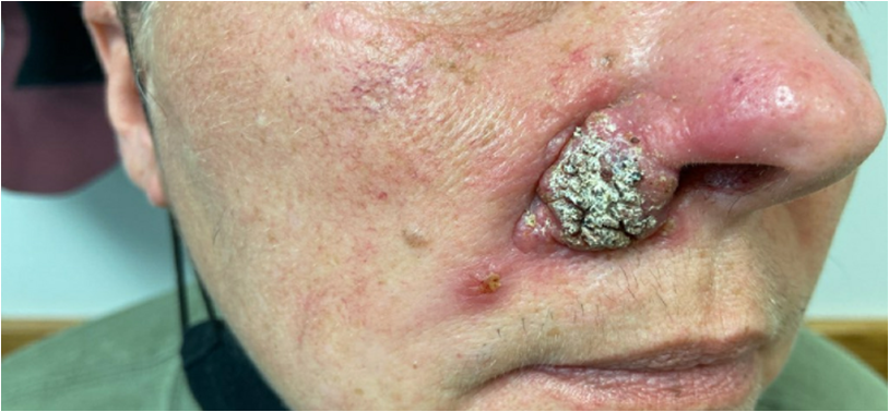
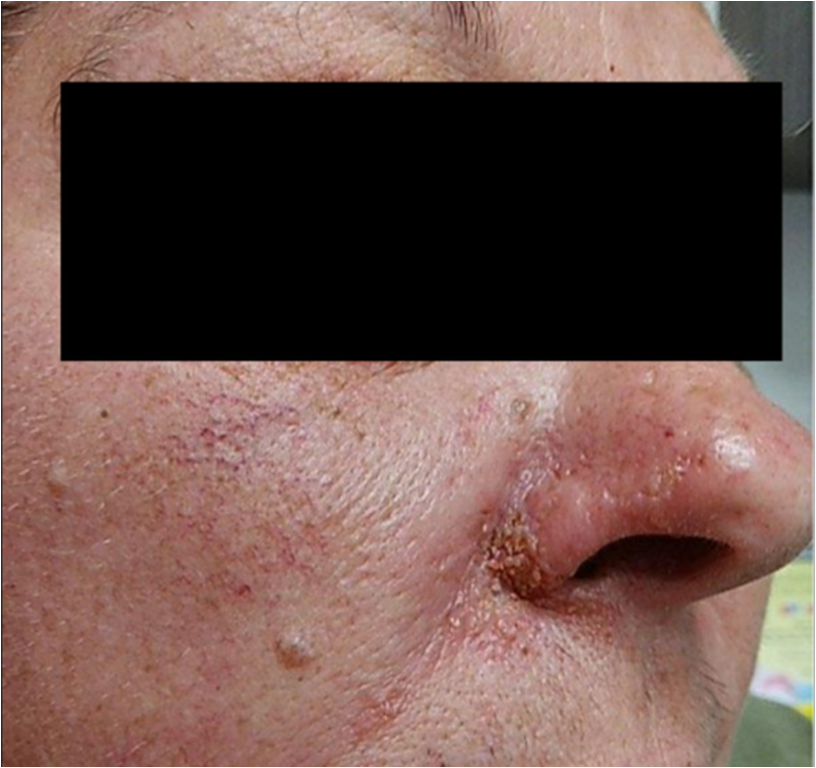
A 48-year-old Caucasian female presented in the Otolaryngology clinic for evaluation of right facial lesion that had appeared as a pimple over the right nasolabial fold 3 weeks ago and had been growing since with increased redness. She denied fever/chills, trauma, insect bite, weight loss, night sweats, respiratory and gastrointestinal symptoms. She had taken multiple antibiotics without improvement. She had a history of multiple abscesses in the left lateral chest wall and both arms requiring incision and drainage, hypertension, and hyperlipidemia. Vitals signs were normal on presentation. Physical examination revealed a 3cm indurated nodular lesion at the right nasolabial angle, involving the lateral ala of the nose and extending superiorly to the upper and lower lateral cartilages of the nose (Figure 1).

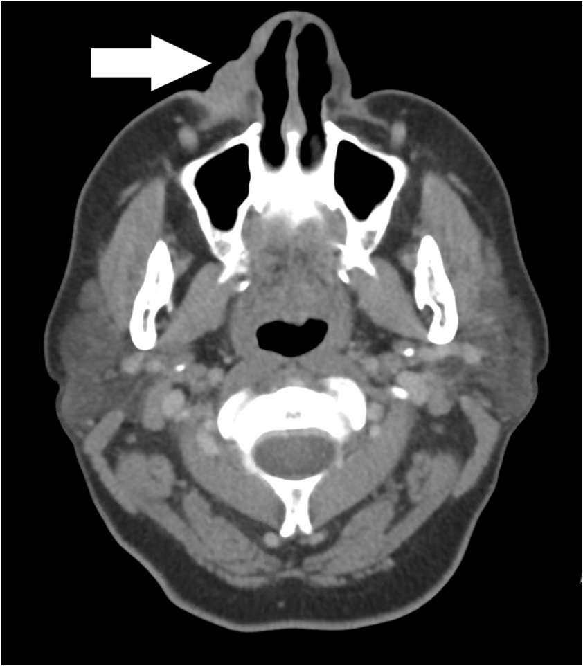
Contrast enhanced CT scan of the head & neck showed soft tissue attenuation mass within the nasolabial fold and lateral to right nasal vestibule (Figure 2).

Her presentation was concerning malignant pathology with basal cell, squamous cell, and adnexal tumor being in the differential diagnosis. Punch biopsy of the lesion was performed, and she was tentatively scheduled for partial rhinectomy and sentinel lymph node biopsy. However, punch biopsy revealed squamous epithelium with reactive changes and Gomori Methenamine-Silver (GMS) stain positive yeast organisms morphologically consistent with Blastomyces dermatitidis (Figure 3).

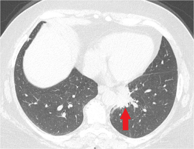
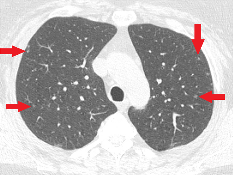
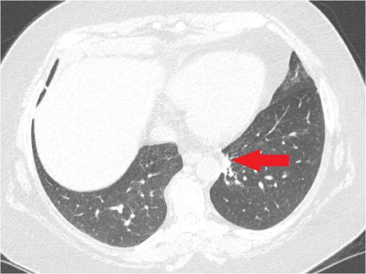
The patient was started on itraconazole. Her previous multiple abscesses were now thought to be secondary to Blastomyces as the bacterial cultures were negative and fungal cultures were not available. She was able to take itraconazole only for a day when she developed headache and right orbital pain. She was advised to go to the emergency department to get admitted and receive intravenous (IV) amphotericin B due to concern of CNS involvement and to get further work up for disseminated disease. CT chest without contrast showed 2.3 cm x 4 cm x 6.5 cm mass like consolidation with spiculated margins in left lower lobe and innumerable upper lobe predominant tiny nodules in both lungs consistent with pulmonary involvement (Figure 4 – Figure 5).


The Urine Blastomyces antigen was also positive. Further negative work up included MRI head, serum Blastomyces antigen, hepatitis B, C and HIV screen. She received IV amphotericin B for 7 days and was transitioned to oral itraconazole 200 mg twice daily on discharge. Follow up in the clinic after 6 weeks of antifungal therapy showed improvement in facial lesion and a repeat non-contrast CT scan of the chest at 14 weeks showed improvement of mass-like consolidation of the left lower lobe (Figure 6 - Figure 7). She will be completing 6 months of itraconazole.


Blastomycosis is a dimorphic fungus endemic in the Ohio and Mississippi river valley, Midwestern states and Saint Lawrence Riverway. 2 It is transmitted by inhalation of conidia usually from soil and decaying matter. Once inside the human body conidia changes to yeast which is resistant to phagocytosis. The incubation period varies depending on the primary site of inoculation ranging from days in case of primary skin infection to weeks in case of pulmonary infection. Pulmonary infection is most common and is seen in > 79% of cases. 3 Extrapulmonary spread occurs via hematogenous or lymphatic route in 20-50% of cases. Skin is the second most common organ involved usually by the hematogenous spread from the lungs. Rarely, direct skin inoculation can result in cutaneous inoculation blastomycosis. 4
Cutaneous involvement can take the form of single or multiple nodules, papules, or pustules. Lesions are usually on the exposed areas of the skin i.e., face, neck, and extremities. They may appear as verrucous or ulcerated having elevated borders and crusting with central scarring. Conditions mimicking cutaneous blastomycosis include other dimorphic fungi, cutaneous malignancies i.e., squamous, and basal cell cancers and pyoderma gangrenosum.
Diagnosis can be made by different techniques, quickest being the identification of dimorphic yeast under microscopy using special stains (Periodic acid-Schiff, 10% KOH and GMS). 5 Culturing the fungus from tissue or fluid samples remains the gold standard. Histopathology is needed to rule out malignancy. Typical histological findings include epidermal papillomatosis and intraepidermal micro abscesses with granulomatous or inflammatory reactions in the dermis. 6 These findings can be misleading unless special stains are used to demonstrate the broad-based budding yeast representative of Blastomyces dermatitidis. Serological testing is not useful due to poor sensitivity and cross-reactivity with other fungi. Antigen testing has better sensitivity but not great specificity.
Treatment depends upon the extent of the disease and immunological status. Isolated skin lesions can resolve spontaneously and may not warrant treatment. In the case of disseminated disease with cutaneous manifestation, initial treatment with amphotericin B for 1-2 weeks followed by maintenance with itraconazole for 6-12 months is warranted. Our patient continued to develop multiple and recurrent primary skin lesions without pulmonary symptoms. In such cases, secondary pulmonary involvement can be identified on surveillance to rule out disseminated disease after diagnosing cutaneous disease.
This case highlights the importance of considering non healing recurrent skin lesions as a manifestation of fungal infection especially in endemic areas even in the absence of pulmonary symptoms. Another important consideration is the possibility of negative bacterial cultures from skin lesions, making fungal cultures and biopsy necessary for definitive diagnosis as was seen in our patient. Where in such cases, very appropriately suspicion shifts towards malignancy as it mimics these nonhealing nodular exophytic skin lesions. Therefore, it is prudent to maintain a high suspicion for cutaneous blastomycosis to prevent major comorbidity and mortality.
Diagnosis of cutaneous blastomycosis remains challenging due to nonspecific manifestations and patients usually end up receiving multiple rounds of antibiotics before the diagnosis is made as was seen in our patient. A high degree of suspicion is required for early diagnosis and prompt treatment.
| [1] | Benedict, K., Roy, M., Chiller, T. and Davis, J.P.: Epidemiologic and Ecologic Features of Blastomycosis: A Review. Current Fungal Infection Reports. 2012, 6: 327-335. | ||
| In article | View Article | ||
| [2] | Klein BS, Vergeront JM, Davis JP: Epidemiologic aspects of blastomycosis, the enigmatic systemic mycosis. Semin Respir Infect. 1986, 1: 29-39. | ||
| In article | |||
| [3] | Chapman SW, Lin AC, Hendricks KA, et al.: Endemic blastomycosis in Mississippi: epidemiological and clinical studies. Semin Respir Infect. 1997, 12: 219-228. | ||
| In article | |||
| [4] | Steck WD. Blastomycosis. Dermatol Clin. 1989; 7(2): 241-250. | ||
| In article | View Article | ||
| [5] | Chapman SW. Blastomycosis dermatitidis. In: Mandell GL, Bennett JE, Dolin R, eds. Principles and Practice of Infectious Diseases. New York: Churchill Livingstone; 2000. pp. 2733-2746. | ||
| In article | |||
| [6] | Martin, D. S., & Smith, D. T.: Blastomycosis, American Blastomycosis, Gilchrist’s Disease: I. A Review of the Literature. American Review of the tuberculosis. 1939, 39: 275-304. | ||
| In article | |||
Published with license by Science and Education Publishing, Copyright © 2022 Shujaa Faryad, Habiba Hussain, Mawwra Faryad and Subramanyam Chittivelu
 This work is licensed under a Creative Commons Attribution 4.0 International License. To view a copy of this license, visit
http://creativecommons.org/licenses/by/4.0/
This work is licensed under a Creative Commons Attribution 4.0 International License. To view a copy of this license, visit
http://creativecommons.org/licenses/by/4.0/
| [1] | Benedict, K., Roy, M., Chiller, T. and Davis, J.P.: Epidemiologic and Ecologic Features of Blastomycosis: A Review. Current Fungal Infection Reports. 2012, 6: 327-335. | ||
| In article | View Article | ||
| [2] | Klein BS, Vergeront JM, Davis JP: Epidemiologic aspects of blastomycosis, the enigmatic systemic mycosis. Semin Respir Infect. 1986, 1: 29-39. | ||
| In article | |||
| [3] | Chapman SW, Lin AC, Hendricks KA, et al.: Endemic blastomycosis in Mississippi: epidemiological and clinical studies. Semin Respir Infect. 1997, 12: 219-228. | ||
| In article | |||
| [4] | Steck WD. Blastomycosis. Dermatol Clin. 1989; 7(2): 241-250. | ||
| In article | View Article | ||
| [5] | Chapman SW. Blastomycosis dermatitidis. In: Mandell GL, Bennett JE, Dolin R, eds. Principles and Practice of Infectious Diseases. New York: Churchill Livingstone; 2000. pp. 2733-2746. | ||
| In article | |||
| [6] | Martin, D. S., & Smith, D. T.: Blastomycosis, American Blastomycosis, Gilchrist’s Disease: I. A Review of the Literature. American Review of the tuberculosis. 1939, 39: 275-304. | ||
| In article | |||