This study evaluates the effect of fermentation duration on the phytochemical profile and bioactivities of the pericarp and rachis of cocoa beans. The residues were fermented for periods ranging from 0 to 120 hours, prior to the analysis of bioactive compounds and antioxidant and anti-inflammatory activities. The results show a significant increase in polyphenols between 0 and 48 hours, reaching, for example, 23.39% and 23.41 % in the pericarp and rachis respectively, followed by stabilisation. As for antioxidant activity, it reached high levels of around 95 %. At the same time, antinutritional compounds decreased significantly, with phytates falling from around 29 % to 11 %. These results indicate that fermentation promotes the release of bioactive compounds. Finally, the fermentation time that maximises the functional value of cocoa residues is between 48 and 72 hours.
Côte d’Ivoire is the world’s leading producer of cocoa (Theobroma cacao L.), accounting for nearly 40 per cent of global production. This sector is a major pillar of the national economy and generates significant quantities of agro-industrial by-products, notably the pericarp (cocoa pod), the shells and the midrib, which account for approximately 70–80 per cent of the fruit’s total biomass 1, 2, 3. These residues are still largely underutilised or left to waste on plantations despite their high potential for value-added use, which constitutes both an economic loss and a source of environmental pollution 2, 3.
These by-products are rich in dietary fibre and phytochemicals, notably polyphenols, flavonoids, tannins and anthocyanins, which are recognised for their antioxidant, anti-inflammatory and antimicrobial properties 4. This biochemical richness gives them great potential for use in the agri-food, animal feed, nutraceuticals, pharmaceutical and cosmetics sectors, as well as in the production of biomaterials and biofuels, in line with the principles of the circular economy 2, 3.
However, the use of fresh or unfermented pods remains limited by their high content of antinutritional factors, particularly phytates and oxalates, which reduce the bioavailability of nutrients 5. Their high moisture content also promotes microbial growth and reduces their storage stability, whilst their lignocellulosic structure limits the extraction and bioavailability of bioactive compounds 6. These constraints make it necessary to use processing methods capable of improving their nutritional and functional properties.
Among these processes, fermentation appears to be a particularly promising strategy. This biological process, driven by the successive action of yeasts, lactic acid bacteria, acetic acid bacteria and moulds, promotes the breakdown of cell walls, reduces antinutritional factors and enhances the release and bioavailability of phenolic compounds 7. These transformations enhance the antioxidant, anti-inflammatory and antimicrobial properties of the fermented by-products, thereby paving the way for their use in the formulation of functional foods, therapeutic herbal teas, food supplements, pharmaceutical and cosmetic ingredients, and animal feed with improved nutritional value 6.
Despite the extensive research devoted to the fermentation of cocoa beans 7, 8, the effects of this process on by-products, in particular the pericarp and the midrib, remain poorly documented. 2, 3. Against this background, the present study aims to assess the influence of fermentation duration on the phytochemical composition and on the antioxidant and anti-inflammatory activities of the cocoa pericarp and rachis, with a view to identifying the optimal conditions for their sustainable utilisation.
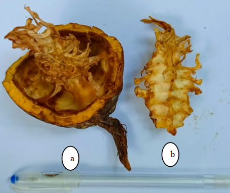
The plant material used in this study consists of cocoa residues (Figure 1), specifically pericarp (cocoa pod shells) and rachis, derived from cocoa pods (Theobroma cacao L.). These residues were collected following the shelling of pods harvested at physiological maturity in the village of Azaguié, located in the department of Agboville, in southern Côte d’Ivoire. The cocoa pods originated from cocoa production materials commonly cultivated in the region and represented a heterogeneous mixture of cocoa plant materials.
The pods were harvested manually using machetes, taking care to select only healthy fruits, free from visible signs of disease or deterioration. After harvesting, they were transported to Abidjan, to National Centre for Floristics at Félix Houphouët-Boigny University, where the shelling operations were carried out.
Following de-podding, the various residues were manually separated into distinct fractions, namely the pericarp and the pods.
2.2. MethodsThe fermentation of cocoa residues (pericarp and rachis) was carried out separately. After collecting the cocoa pods harvested at physiological maturity, the pericarp and rachis were separated. Each type of residue was then finely ground using a mechanical grinder to produce a homogeneous material.
After grinding, each type of residue was divided into homogeneous batches of sufficient mass to allow samples to be taken at each stage of fermentation without disturbing the other batches. The batches were placed separately in clean, hermetically sealed plastic containers. Fermentation was carried out at room temperature for a total duration of 120 hours, without the addition of external microorganisms, to allow the development of the indigenous microflora naturally present in the residues.
Six fermentation time points were studied: 0, 24, 48, 72, 96 and 120 hours. At each of these time points, the corresponding batch was sampled and 1 kg of fermented residue was collected for subsequent analysis. The use of separate batches for each fermentation time point allowed sampling to be carried out without disrupting the fermentation conditions of the other batches.
From 48 hours into fermentation, the substrates were mixed manually once every 24 hours to ensure the material was homogenised and to promote uniform fermentation. After each mixing, the containers were sealed tightly and kept at room temperature until the next sampling time.
The samples taken at each fermentation stage were then spread out and dried in the shade for 7 days at room temperature. After drying, the samples were analysed to determine their phytochemical parameters, antinutritional factors and biological activities.
Temperature and pH were not measured or recorded during the fermentation process.
Extraction of phenolic compounds was carried out according to the method of 9. One gram (1 g) of sample was homogenised in 10 mL of 70% (v/v) methanol, then centrifuged at 1000 rpm for 10 minutes. The resulting pellet was resuspended in 10 mL of the same solvent and centrifuged again under the same conditions. The supernatants obtained were combined in a Falcon tube to form the methanolic extract.
The total polyphenol content was determined according to method of 9. One millilitre (1 mL) of methanolic extract was mixed with 1 mL of Folin-Ciocalteu reagent, followed by a 3-minute stand. Next, 1 mL of a 20% (w/v) sodium carbonate solution was added, and the total volume adjusted to 10 mL with distilled water. After incubation in the dark for 30 minutes, the absorbance was measured at 725 nm. Quantification was performed using a calibration curve constructed with gallic acid (1 mg/mL).
The quantification of flavonoids was carried out according to the method described by 9. A volume of 0.5 mL of extract was successively mixed with 0.5 mL of distilled water, 0.5 mL of 10% (w/v) aluminium chloride, 0.5 mL of sodium acetate (1 M) and 2 mL of distilled water. After 30 minutes at a temperature of 37 ± 2 °C and in the dark, the absorbance was measured at 415 nm. The flavonoid concentration was determined using a standard curve established from quercetin (1 mg/mL).
Tannins were assayed using the method of 10. One millilitre (1 mL) of methanolic extract was added to 5 mL of a vanillin reagent (0.1 mg/mL in 70% sulphuric acid, v/v). After 20 minutes of incubation in the dark, the absorbance was measured at 500 nm. Quantification was performed using a calibration curve obtained with tannic acid (2 mg/mL).
The carotenoid content was determined using the method described by 11. One gram of sample was treated with 5 mL of ethanol containing 0.1% (w/v) BHT, then heated to 85 °C for 10 minutes. Next, 400 µL of 80% KOH was added for saponification, followed by a second identical heating. The mixture was rapidly cooled, then extracted with 3 mL of distilled water and 4 mL of hexane. After centrifugation at 4000 rpm for 10 minutes, the supernatant was collected and its absorbance measured at 450 nm, using hexane as the blank. The total carotenoid concentration (expressed in µg/g of material) was calculated using the following formula:
 | (1) |
where: V is the total volume of the extract (in mL); DO is the absorbance at 450 nm; m is the mass of the sample (in g); 2592 is the extinction coefficient of carotenoids in hexane.
The vitamin C content was determined in accordance with the method of 12. To do this, 10 g of sample were dissolved in 40 mL of a solution consisting of metaphosphoric acid and 2% (w/v) acetic acid. The resulting mixture was centrifuged at 3000 rpm for 20 minutes. The supernatant was then collected in a 50 mL volumetric flask and made up to volume with distilled water that had been previously boiled and then cooled in the dark. A 10 mL volume of this solution was taken to form the analytical sample. The vitamin C content was determined by titration using a 2,6-dichlorophenol-indophenol (2,6 DCPIP) solution, until the colour changed from blue to pale pink. The concentration was calculated using the following formula:
 | (2) |
Where V: the volume of 2,6 DCPIP (mL) and m: the mass of the sample
The anthocyanin content was determined using the differential method based on pH variation 13. Two buffers were prepared: a pH 1.0 buffer (0.025 M potassium chloride obtained by dissolving 1.86 g of KCl in 980 mL of distilled water, then adjusted to pH 1.0 (± 0.05) with 6.3 mL of hydrochloric acid), and a pH 4.5 buffer (0.4 M sodium acetate) prepared by dissolving 54.43 g of sodium acetate trihydrate in 960 mL of distilled water, then adjusted to pH 4.5 with 20 mL of HCl. For each analysis, one gram (1 g) of sample was mixed with 9 mL of each buffer. After centrifugation at 2000 rpm for 5 minutes, the absorbances of the supernatants were measured at wavelengths of 520 nm and 700 nm. This protocol was applied to the extracts prepared in both buffers in order to calculate the anthocyanin content by difference in absorbance.
The assessment of radical scavenging activity was carried out according to the method described by 13, using the stable free radical DPPH (2,2-diphenyl-1-picrylhydrazyl). For this purpose, 1.5 mL of methanolic extract was mixed with 1 mL of DPPH solution (3 mM in methanol). The mixture was incubated in the dark for 30 minutes, after which the absorbance was measured at 415 nm using a spectrophotometer. Antioxidant activity was expressed as the percentage inhibition of the DPPH radical, calculated using the following formula:
 | (3) |
where: DOc: absorbance of the control (1 mL DPPH + 2.5 mL methanol); DOe: absorbance of the test (1 mL DPPH + 2.5 mL extract); DOt: absorbance of the blank (1 mL methanol + 2.5 mL extract)
The anti-inflammatory activity of the cocoa by-product extracts was determined by the protein denaturation inhibition assay, following the method described by 14. The samples (cocoa bean shells and midribs) were dried at 50°C, ground into a fine powder, then extracted by maceration in 70% ethanol for 24 hours. The extracts were filtered, concentrated under vacuum using a rotary evaporator, and then diluted to various concentrations (63 to 1000 µg/mL). For the analysis, a reaction mixture was prepared using 0.45 mL of bovine serum albumin solution (5%) and 0.05 mL of extract. The pH was adjusted to 6.3 with hydrochloric acid (1N). The tubes were incubated at 37°C for 20 minutes, then heated to 57°C for 3 minutes to induce protein denaturation. After cooling, 2.5 mL of phosphate buffer (pH 6.3) was added, and the absorbance was measured at 416 nm. Diclofenac sodium served as the positive control and distilled water as the negative control. Protein denaturation inhibition was calculated using the following formula.
 | (4) |
The oxalate content was determined using potassium permanganate (KMnO₄ 0.05 M) until a solution was obtained, in accordance with the method described by 15. To do this, one gram (1 g) of sample powder was homogenised in 75 mL of sulphuric acid (3 M H₂SO₄) and stirred for one hour at a temperature of 37 ± 2 °C. After filtration through Whatman paper, 25 mL of the filtrate was taken for hot titration using a persistent pink colouring solution, indicating the end of the reaction. The oxalate content was determined using the following formula:
 | (5) |
The phytate concentration was determined using the method of 5. One gram (1 g) of sample was mixed with 20 mL of hydrochloric acid (0.65 N HCl) and stirred for 12 hours at room temperature (37 ± 2 °C). After centrifugation at 12,000 rpm for 40 minutes, 0.5 mL of the supernatant was removed and mixed with 3 mL of Wade's reagent. The tubes were left to stand for 15 minutes, then the optical density was measured at 490 nm. The phytate content was determined using a standard series prepared from a stock solution of sodium phytate (10 mg/mL), subjected to the same experimental conditions.
The qualitative analysis of triterpenes was carried out using the Slakowski test, as described by 18. To 5 mL of methanolic extract, 2 mL of chloroform and 3 mL of concentrated sulphuric acid were added. The appearance of a brown-red ring at the phase interface was interpreted as a positive indicator of the presence of triterpenes in the analysed extracts.
The determination of sterols and triterpene alcohols was carried out in accordance with the method described by 18. For each sample, 0.5 g of methanolic extract was mixed with 2 mL of acetic anhydride, followed by 2 mL of concentrated sulphuric acid (H₂SO₄). The occurrence of a colour change, from purple to blue or green, was interpreted as a qualitative indication of the presence of sterols and triterpene alcohols in the extracts analysed.
All experiments were carried out with at least three independent replicates. Data are presented as mean ± standard deviation (SD). Statistical analyses were performed using one-way analysis of variance (ANOVA). Differences were considered statistically significant at the 5 per cent level (p < 0.05). Statistical analyses were carried out using XLSTAT 2021 software.
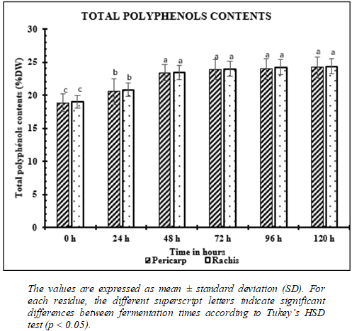
Figure 2 shows the change in total polyphenol content in the pericarp and rachis as a function of fermentation time. A significant increase in total polyphenol content is observed during the first 48 hours of fermentation. In the pericarp, the content rose from 18.87 ± 1.28 at 0 h to 20.56 ± 1.96 after 24 h, then reached 23.39 ± 1.22 at 48 h. A similar trend is observed for the rachis, where the content increases from 18.99 ± 0.90 to 20.80 ± 0.98, then to 23.41 ± 1.11 over the same period. Beyond 48 hours, total polyphenol content remains high and no longer shows any significant differences between 48 hours, 72 hours, 96 hours and 120 hours, for both the pericarp and the rachis, indicating that the levels have stabilised.
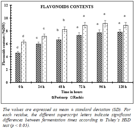
As illustrated in Figure 3, flavonoid content increased progressively and significantly in both cocoa pericarp and rachis throughout the fermentation process. In the pericarp, the flavonoid content rose from 4.60 ± 0.25 at 0 h to 5.97 ± 0.32 after 24 h, then to 6.65 ± 0.36 after 48 h. The levels continue to increase up to 72 hours (7.36 ± 0.39), after which there is no longer any significant difference between 72 hours, 96 hours (7.75 ± 0.42) and 120 hours (7.83 ± 0.42). A similar trend is observed for the rachis. Concentrations increased significantly from 6.28 ± 0.29 at 0 hours to 7.20 ± 0.34 after 24 hours, and then to 8.26 ± 0.39 after 48 hours. The highest values were recorded between 72 hours (8.91 ± 0.42) and 96 hours (9.19 ± 0.43), with no significant difference from the value observed at 120 hours (8.91 ± 0.42).
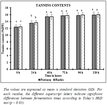
As shown in Figure 4, the tannin content increased markedly in both the pericarp and the rachis of the cocoa beans during the first few hours of fermentation. In the pericarp, the tannin content increased significantly, rising from 12.39 ± 0.66 at 0 h to 13.49 ± 0.72 after 24 h, reaching 15.38 ± 0.82 at 48 h. Similarly, in the rachis, the tannin concentration rose from 12.48 ± 0.58 at 0 h to 13.67 ± 0.64 after 24 h, reaching 15.38 ± 0.72 at 48 h. After 48 hours of fermentation, the tannin content remained relatively stable, with no significant differences observed between the values measured at 48, 72, 96 and 120 hours for either by-product.
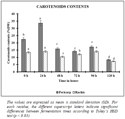
Figure 5 shows the change in carotenoid content in the pericarp and rachis as a function of fermentation time. In the pericarp, the carotenoid content increases significantly between 0 h (22.37 ± 1.20) and 24 h (33.55 ± 1.80), when it reaches its maximum value. Thereafter, a significant decrease is observed, reaching its lowest value at 120 h (8.31 ± 0.45).
In the rachis, a decrease in content was observed throughout fermentation. Its levels fell from 13.46 ± 0.63 (0 h) to 7.28 ± 0.34 (120 h).
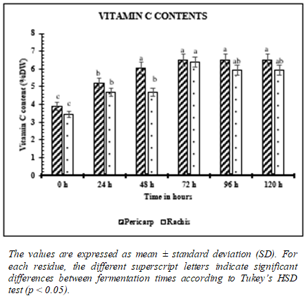
Figure 6 shows the change in vitamin C content of the pericarp and rachis as a function of fermentation time. A significant increase in vitamin C content is observed during the first few hours of fermentation. In the pericarp, the content rose from 3.90 ± 0.21 at 0 h to 6.04 ± 0.32 at 48 h. The levels then remained stable, with no significant difference between 48 h and 120 h (6.50 ± 0.35). In the rachis, the vitamin C content also increases significantly, rising from 3.44 ± 0.16 at 0 h to 6.38 ± 0.30 at 72 h. The levels recorded at 96 hours (5.92 ± 0.28) and 120 hours (5.92 ± 0.28) do not differ significantly from those observed at 72 hours or from those measured at 24–48 hours, indicating a slight decrease that remains statistically marginal.
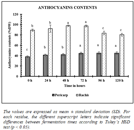
Figure 7 shows the change in anthocyanin content in the pericarp and rachis as a function of fermentation time. In the pericarp, the anthocyanin content increases gradually during fermentation. It rises from 37.83 ± 2.03 at 0 h to 44.37 ± 2.38 after 72 h. The levels then remain stable, with no significant difference between 72 h and 120 h. In the rachis, the levels increase significantly between 0 h (89.42 ± 2.16) and 72 h (97.39 ± 1.87). However, a decrease is observed from 96 h onwards.
Table 1 shows the changes in the antioxidant activity of the pericarp and rachis as a function of fermentation time. In the pericarp, antioxidant activity remains high at the start of fermentation, with no significant difference between 0 h (95.46 ± 2.80) and 48 h (94.88 ± 1.40). A significant decrease is then observed from 72 h (88.61 ± 1.20) to 120 h (79.19 ± 0.90), indicating a gradual reduction in antioxidant activity during fermentation. In the rachis, antioxidant activity also remained high at 0 h (98.54 ± 2.70) and 24 h (97.85 ± 2.10), with no significant difference between these two time points. A significant decrease was observed from 48 h (93.08 ± 2.10) onwards. Antioxidant activity decreased further at 96 hours (90.89 ± 2.10) and remained stable up to 120 hours (89.60 ± 2.10), with no significant difference between these last two time points.
The changes in the anti-inflammatory activity of the pericarp and rachis as a function of fermentation time are described in Table 2. In the pericarp, anti-inflammatory activity is highest at 0 h (96.42 ± 2.60). It decreases significantly up to 96 h (91.34 ± 2.20), whilst the lowest value is recorded at 120 h (88.81 ± 2.10). In the rachis, anti-inflammatory activity decreases gradually during fermentation. The highest values are observed at 0 h (85.74 ± 2.30), followed by a significant decrease between 24 h (77.46 ± 1.90) and 72 h (75.40 ± 1.90), followed by stabilisation up to 120 h.
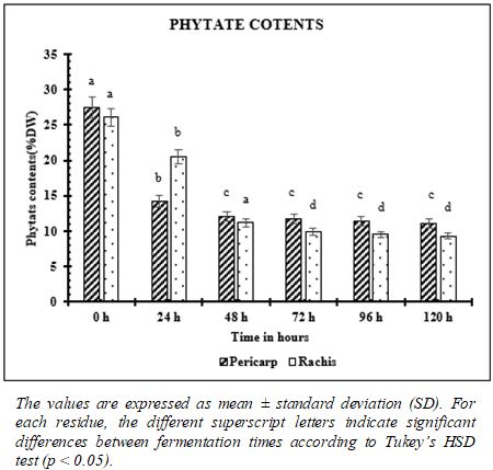
Figure 8 shows the changes in phytate content in the pericarp and rachis as a function of fermentation time. In the pericarp, the phytate content is highest at 0 h (27.51 ± 1.47), then decreases significantly until 48 h (12.03 ± 0.64), after which the levels remain stable until the end of fermentation, with no significant difference between 48 h and 120 h. In the rachis, a gradual decrease in phytate content was also observed. The values fell from 26.12 ± 1.22 at 0 h to 9.95 ± 0.46 at 72 h and then remained stable up to 120 h (9.33 ± 0.44).
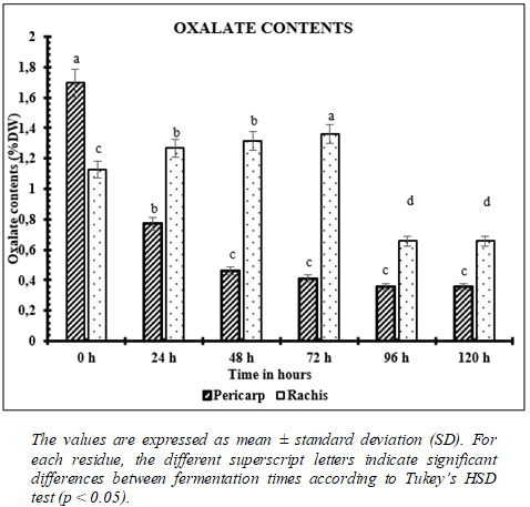
Figure 9 shows the changes in oxalate content in the pericarp and rachis as a function of fermentation time. In the pericarp, the oxalate content peaks at 0 h (1.70 ± 0.09), then decreases significantly to 0.46 ± 0.02 at 48 h, after which the levels remain stable until the end of fermentation, with no significant difference between 48 h and 72 h. In the rachis, the oxalate content increased slightly at the start of fermentation, rising from 1.13 ± 0.05 at 0 h to 1.31 ± 0.06 at 48 h. The maximum value is reached at 72 hours (1.36 ± 0.06). Subsequently, a significant decrease is observed at 96 hours (0.66 ± 0.03), a value which remains stable until 120 hours (0.66 ± 0.03).
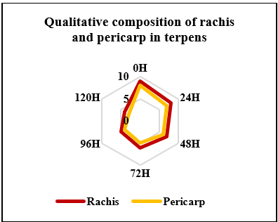
Figure 100 shows the changes in the qualitative composition of terpenes in the pericarp and rachis as a function of fermentation time. In both matrices, the profiles show a gradual decrease in the qualitative diversity of terpenes during the first few hours of fermentation. The highest levels are observed at 0 h, followed by a reduction at 24 h and 48 h, reaching a minimum at around 72 h. From 96 h onwards, a slight increase in the qualitative composition of terpenes is observed in both the pericarp and the rachis, followed by stabilisation at 120 h.
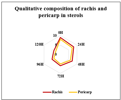
Figure 111 shows the changes in the qualitative composition of sterols in the pericarp and rachis as a function of fermentation time. In both matrices, the profiles reveal a gradual decrease in the qualitative composition of sterols during fermentation. The highest levels are observed at 0 h, then decrease gradually at 24 h and 48 h, reaching a minimum around 72 h.
From 96 h onwards, a slight increase in sterol levels is observed, followed by stabilisation at 120h.


The results obtained show that fermentation time significantly influences the phytochemical composition, biological activities and antinutritional factors of the cocoa pericarp and rachis.
The significant increase in the levels of total polyphenols, flavonoids, tannins and anthocyanins observed between 48 and 72 hours of fermentation reflects an improvement in the bioavailability of secondary metabolites. This change is mainly attributed to the synergistic action of yeasts, lactic acid bacteria and acetic acid bacteria, which secrete hydrolytic enzymes such as pectinases, cellulases, β-glucosidases and esterases, promoting the breakdown of cell walls and the release of phenolic compounds bound to the plant matrix 5, 6, 8. Similar observations have been reported in fermented cocoa as well as in other fermented plant matrices, where fermentation enhances the extractability of bioactive compounds 3.
The stabilisation of polyphenol, flavonoid and tannin levels after 72 hours suggest a balance between their release and their oxidative degradation. Indeed, the rise in temperature and the diffusion of oxygen into the fermented mass promote the activity of polyphenol oxidase, as well as the polymerisation and condensation reactions of flavanols, gradually reducing the quantity of extractable phenolic compounds 3, 20, 21. This phenomenon constitutes a key stage in cocoa fermentation and contributes to the modification of the biochemical profile of plant tissues.
Unlike polyphenols, carotenoids and vitamin C gradually decrease during fermentation, particularly after 48 to 72 hours. This trend is mainly due to their high sensitivity to oxidation reactions, rising temperatures and the action of oxidative enzymes. Carotenoids undergo isomerisation and oxidative degradation, whilst vitamin C is rapidly oxidised in the presence of oxygen 22. These observations are consistent with those reported for various plant matrices subjected to fermentation processes.
Antioxidant and anti-inflammatory activity varies in line with phenolic compound levels. The high values observed during the first few days of fermentation can be explained by the high concentrations of polyphenols, flavonoids and tannins, which are known for their ability to neutralise reactive oxygen species, inhibit lipid peroxidation and modulate inflammatory signalling pathways 23, 24. The gradual decrease in these activities after 72 hours is therefore directly linked to the reduction in the levels of bioactive phenolic compounds.
Fermentation also led to a significant reduction in phytate and oxalate levels, mainly during the first 48 hours. This reduction results from the activity of phytases and other microbial enzymes capable of gradually hydrolysing phytates into less complexing phosphorylated derivatives, thereby improving the bioavailability of essential minerals such as calcium, zinc and iron 19, 26, 25. The reduction in oxalates may also be linked to their enzymatic degradation or their utilisation as a carbon source by certain fermentative microorganisms 27. These results confirm that fermentation is an effective process for reducing antinutritional factors and improving the nutritional quality of cocoa by-products.
Finally, the qualitative profiles of terpenes and sterols show a gradual decrease up to 72 hours, followed by a slight stabilisation between 96 and 120 hours. This trend can be attributed to the volatilisation, oxidation and enzymatic biotransformation of lipophilic compounds during fermentation 28. The higher concentrations observed in the rachis compared with the pericarp indicate a differential distribution of secondary metabolites amongst the different tissues of the cocoa fruit, as also reported 3, 20.
All these results confirm the significant potential of these by-products as sources of functional ingredients.
The authors would like to thank CEMOI-CI for its financial support, the Université Félix Houphouet Boigny for hosting this work, and the agrifood biotechnology laboratory where all the manipulations were carried out.
Marlène OUALI wrote the manuscript, Sébastien NAIMKÉ and Hadja Djeneba OUATTARA critically reviewed the manuscript and have given their approval for the publication of the article.
All data generated or analyzed during this study are included in this published article. Declaration of Conflicting Interests The authors had no potential conflicts of interest in the research, writing and/or publication of this article.
The Laboratory of Biotechnology, Agriculture and Valorization of Biological Resources of the University Félix Houphouet Boigny of Cocody approved the study protocol. The work had the written or oral consent of all participants.
| [1] | International Cocoa Organization (ICCO), Annual Report 2022, Abidjan, Côte d'Ivoire, 2022. | ||
| In article | |||
| [2] | E. O. Afoakwa, A. Paterson, M. Fowler, and A. Ryan, “Flavor formation and character in cocoa and chocolate: A critical review,” Critical Reviews in Food Science and Nutrition, vol. 53, no. 7, pp. 739–753, 2013. | ||
| In article | |||
| [3] | J. E. Kongor, M. Hinneh, D. Van de Walle, E. O. Afoakwa, P. Boeckx, and K. Dewettinck, “Factors influencing quality variation in cocoa (Theobroma cacao) bean flavour profile: A review,” Food Research International, vol. 82, pp. 44–52, 2016. | ||
| In article | View Article | ||
| [4] | M. Naczk and F. Shahidi, “Extraction and analysis of phenolics in food,” Journal of Chromatography A, vol. 1054, no. 1–2, pp. 95–111, 2004. | ||
| In article | View Article PubMed | ||
| [5] | M. Latta and M. Eskin, “A simple and rapid colorimetric method for phytate determination,” Journal of Agricultural and Food Chemistry, vol. 28, no. 6, pp. 1313–1315, 1980. | ||
| In article | View Article | ||
| [6] | L. De Vuyst and F. Leroy, “Functional role of yeasts, lactic acid bacteria and acetic acid bacteria in cocoa fermentation processes,” FEMS Microbiology Reviews, vol. 44, no. 4, pp. 432–453, 2020. | ||
| In article | View Article PubMed | ||
| [7] | L. De Vuyst and S. Weckx, “The cocoa bean fermentation process: From ecosystem analysis to starter culture development,” Journal of Applied Microbiology, vol. 121, no. 1, pp. 5–17, 2016. | ||
| In article | View Article PubMed | ||
| [8] | D. S. Nielsen, O. D. Teniola, L. Ban-Koffi, M. Owusu, T. S. Andersson, and W. H. Holzapfel, “The microbiology of Ghanaian cocoa fermentations analysed using culture-dependent and culture-independent methods,” International Journal of Food Microbiology, vol. 114, no. 2, pp. 168–186, 2007. | ||
| In article | View Article PubMed | ||
| [9] | V. L. Singleton, R. Orthofer, and R. M. Lamuela-Raventós, “Analysis of total phenols and other oxidation substrates and antioxidants by means of Folin–Ciocalteu reagent,” Methods in Enzymology, vol. 299, pp. 152–178, 1999. | ||
| In article | View Article | ||
| [10] | A. Meda, C. E. Lamien, M. Romito, J. Millogo, and O. G. Nacoulma, “Determination of the total phenolic, flavonoid and proline contents in Burkina Faso honey, as well as their radical scavenging activity,” Food Chemistry, vol. 91, no. 3, pp. 571–577, 2005. | ||
| In article | View Article | ||
| [11] | Z. Bainbridge, K. Tomlins, and A. Westby, “Analysis of condensed tannins using acidified vanillin,” Journal of the Science of Food and Agriculture, vol. 29, pp. 77–79, 1996. | ||
| In article | |||
| [12] | J. A. Howe and S. A. Tanumihardjo, “Evaluation of analytical methods for carotenoid extraction from biofortified maize,” Journal of Agricultural and Food Chemistry, vol. 54, no. 21, pp. 7992–7997, 2006. | ||
| In article | View Article PubMed | ||
| [13] | G. Pongracz, H. Weiser, and D. Matzinger, “Tocopherols—Antioxidant,” Fat Science and Technology, vol. 97, pp. 90–104, 1995. | ||
| In article | View Article | ||
| [14] | J. Lee, R. W. Durst, and R. E. Wrolstad, “Determination of total monomeric anthocyanin pigment content of fruit juices, beverages, natural colorants, and wines by the pH differential method: Collaborative study,” Journal of AOAC International, vol. 88, no. 5, pp. 1269–1278, 2005. | ||
| In article | View Article PubMed | ||
| [15] | C. W. Choi, S. C. Kim, S. S. Hwang, B. K. Choi, H. J. Ahn, M. Y. Lee, S. H. Park, and S. K. Kim, “Antioxidant activity and free radical scavenging capacity between Korean medicinal plants and flavonoids by assay-guided comparison,” Plant Science, vol. 163, no. 6, pp. 1161–1168, 2002. | ||
| In article | View Article | ||
| [16] | K. D. Gunathilake, K. K. Ranaweera, and H. P. Rupasinghe, “In vitro anti-inflammatory properties of selected green leafy vegetables,” Biomedicines, vol. 6, no. 4, Art. 107, 2018. | ||
| In article | View Article PubMed | ||
| [17] | R. A. Day and A. L. Underwood, Quantitative Analysis, 5th ed., Prentice Hall, Englewood Cliffs, NJ, USA, 1986. | ||
| In article | |||
| [18] | H. O. Edeoga, D. E. Okwu, and B. O. Mbaebie, “Phytochemical constituents of some Nigerian medicinal plants,” African Journal of Biotechnology, vol. 4, no. 7, pp. 685–688, 2005. | ||
| In article | View Article | ||
| [19] | O. A. Adebo and I. G. Medina-Meza, “Impact of fermentation on the phenolic compounds and antioxidant activity of whole cereal grains: A mini review,” Molecules, vol. 25, no. 4, Art. 927, 2020. | ||
| In article | View Article PubMed | ||
| [20] | A. C. Aprotosoaie, S. V. Luca, and A. Miron, “Flavor chemistry of cocoa and cocoa products—An overview,” Comprehensive Reviews in Food Science and Food Safety, vol. 15, pp. 73–91, 2016. | ||
| In article | View Article PubMed | ||
| [21] | D. Kadow, N. Niemenak, S. Rohn, and R. Lieberei, “Fermentation-like incubation of cocoa seeds (Theobroma cacao L.): Reconstruction and guidance of the fermentation process,” LWT – Food Science and Technology, vol. 62, pp. 357–361, 2015. | ||
| In article | View Article | ||
| [22] | M. Nowacka, U. Tylewicz, S. Romani, M. Dalla Rosa, and D. Witrowa-Rajchert, “Influence of drying techniques on bioactive compounds in fruits and vegetables,” Trends in Food Science & Technology, vol. 78, pp. 77–90, 2018. | ||
| In article | |||
| [23] | F. Shahidi and P. Ambigaipalan, “Phenolics and polyphenolics in foods, beverages and spices: Antioxidant activity and health effects—A review,” Journal of Functional Foods, vol. 18, pp. 820–897, 2015. | ||
| In article | View Article PubMed | ||
| [24] | A. N. Panche, A. D. Diwan, and S. R. Chandra, “Flavonoids: An overview,” Journal of Nutritional Science, vol. 5, Art. e47, 2016. | ||
| In article | View Article PubMed | ||
| [25] | X. G. Lei, J. D. Weaver, E. Mullaney, A. H. J. Ullah, and M. J. Azain, “Phytase, a new life for an ‘old’ enzyme,” Annual Review of Animal Biosciences, vol. 1, pp. 283–309, 2013. | ||
| In article | View Article PubMed | ||
| [26] | M. Samtiya, R. E. Aluko, T. Dhewa, and J. M. Moreno-Rojas, “Potential health benefits of plant food-derived bioactive components: An overview,” Foods, vol. 9, Art. 1302, 2020. | ||
| In article | View Article PubMed | ||
| [27] | H. V. H. Nguyen, G. P. Savage, et al., “Effects of maturity at harvesting and primary processing of cocoa beans on oxalate contents of cocoa powder,” Journal of Food Composition and Analysis, vol. 67, pp. 86–90, 2018. | ||
| In article | View Article | ||
| [28] | M. A. M. El Hadi, F. J. Zhang, F. F. Wu, C. H. Zhou, and J. Tao, “Advances in fruit aroma volatile research,” Molecules, vol. 18, pp. 8200–8229, 2013. | ||
| In article | View Article PubMed | ||
Published with license by Science and Education Publishing, Copyright © 2026 Marlène OUALI, Hadja Djeneba OUATTARA and Sébastien NIAMKÉ
 This work is licensed under a Creative Commons Attribution 4.0 International License. To view a copy of this license, visit
http://creativecommons.org/licenses/by/4.0/
This work is licensed under a Creative Commons Attribution 4.0 International License. To view a copy of this license, visit
http://creativecommons.org/licenses/by/4.0/
| [1] | International Cocoa Organization (ICCO), Annual Report 2022, Abidjan, Côte d'Ivoire, 2022. | ||
| In article | |||
| [2] | E. O. Afoakwa, A. Paterson, M. Fowler, and A. Ryan, “Flavor formation and character in cocoa and chocolate: A critical review,” Critical Reviews in Food Science and Nutrition, vol. 53, no. 7, pp. 739–753, 2013. | ||
| In article | |||
| [3] | J. E. Kongor, M. Hinneh, D. Van de Walle, E. O. Afoakwa, P. Boeckx, and K. Dewettinck, “Factors influencing quality variation in cocoa (Theobroma cacao) bean flavour profile: A review,” Food Research International, vol. 82, pp. 44–52, 2016. | ||
| In article | View Article | ||
| [4] | M. Naczk and F. Shahidi, “Extraction and analysis of phenolics in food,” Journal of Chromatography A, vol. 1054, no. 1–2, pp. 95–111, 2004. | ||
| In article | View Article PubMed | ||
| [5] | M. Latta and M. Eskin, “A simple and rapid colorimetric method for phytate determination,” Journal of Agricultural and Food Chemistry, vol. 28, no. 6, pp. 1313–1315, 1980. | ||
| In article | View Article | ||
| [6] | L. De Vuyst and F. Leroy, “Functional role of yeasts, lactic acid bacteria and acetic acid bacteria in cocoa fermentation processes,” FEMS Microbiology Reviews, vol. 44, no. 4, pp. 432–453, 2020. | ||
| In article | View Article PubMed | ||
| [7] | L. De Vuyst and S. Weckx, “The cocoa bean fermentation process: From ecosystem analysis to starter culture development,” Journal of Applied Microbiology, vol. 121, no. 1, pp. 5–17, 2016. | ||
| In article | View Article PubMed | ||
| [8] | D. S. Nielsen, O. D. Teniola, L. Ban-Koffi, M. Owusu, T. S. Andersson, and W. H. Holzapfel, “The microbiology of Ghanaian cocoa fermentations analysed using culture-dependent and culture-independent methods,” International Journal of Food Microbiology, vol. 114, no. 2, pp. 168–186, 2007. | ||
| In article | View Article PubMed | ||
| [9] | V. L. Singleton, R. Orthofer, and R. M. Lamuela-Raventós, “Analysis of total phenols and other oxidation substrates and antioxidants by means of Folin–Ciocalteu reagent,” Methods in Enzymology, vol. 299, pp. 152–178, 1999. | ||
| In article | View Article | ||
| [10] | A. Meda, C. E. Lamien, M. Romito, J. Millogo, and O. G. Nacoulma, “Determination of the total phenolic, flavonoid and proline contents in Burkina Faso honey, as well as their radical scavenging activity,” Food Chemistry, vol. 91, no. 3, pp. 571–577, 2005. | ||
| In article | View Article | ||
| [11] | Z. Bainbridge, K. Tomlins, and A. Westby, “Analysis of condensed tannins using acidified vanillin,” Journal of the Science of Food and Agriculture, vol. 29, pp. 77–79, 1996. | ||
| In article | |||
| [12] | J. A. Howe and S. A. Tanumihardjo, “Evaluation of analytical methods for carotenoid extraction from biofortified maize,” Journal of Agricultural and Food Chemistry, vol. 54, no. 21, pp. 7992–7997, 2006. | ||
| In article | View Article PubMed | ||
| [13] | G. Pongracz, H. Weiser, and D. Matzinger, “Tocopherols—Antioxidant,” Fat Science and Technology, vol. 97, pp. 90–104, 1995. | ||
| In article | View Article | ||
| [14] | J. Lee, R. W. Durst, and R. E. Wrolstad, “Determination of total monomeric anthocyanin pigment content of fruit juices, beverages, natural colorants, and wines by the pH differential method: Collaborative study,” Journal of AOAC International, vol. 88, no. 5, pp. 1269–1278, 2005. | ||
| In article | View Article PubMed | ||
| [15] | C. W. Choi, S. C. Kim, S. S. Hwang, B. K. Choi, H. J. Ahn, M. Y. Lee, S. H. Park, and S. K. Kim, “Antioxidant activity and free radical scavenging capacity between Korean medicinal plants and flavonoids by assay-guided comparison,” Plant Science, vol. 163, no. 6, pp. 1161–1168, 2002. | ||
| In article | View Article | ||
| [16] | K. D. Gunathilake, K. K. Ranaweera, and H. P. Rupasinghe, “In vitro anti-inflammatory properties of selected green leafy vegetables,” Biomedicines, vol. 6, no. 4, Art. 107, 2018. | ||
| In article | View Article PubMed | ||
| [17] | R. A. Day and A. L. Underwood, Quantitative Analysis, 5th ed., Prentice Hall, Englewood Cliffs, NJ, USA, 1986. | ||
| In article | |||
| [18] | H. O. Edeoga, D. E. Okwu, and B. O. Mbaebie, “Phytochemical constituents of some Nigerian medicinal plants,” African Journal of Biotechnology, vol. 4, no. 7, pp. 685–688, 2005. | ||
| In article | View Article | ||
| [19] | O. A. Adebo and I. G. Medina-Meza, “Impact of fermentation on the phenolic compounds and antioxidant activity of whole cereal grains: A mini review,” Molecules, vol. 25, no. 4, Art. 927, 2020. | ||
| In article | View Article PubMed | ||
| [20] | A. C. Aprotosoaie, S. V. Luca, and A. Miron, “Flavor chemistry of cocoa and cocoa products—An overview,” Comprehensive Reviews in Food Science and Food Safety, vol. 15, pp. 73–91, 2016. | ||
| In article | View Article PubMed | ||
| [21] | D. Kadow, N. Niemenak, S. Rohn, and R. Lieberei, “Fermentation-like incubation of cocoa seeds (Theobroma cacao L.): Reconstruction and guidance of the fermentation process,” LWT – Food Science and Technology, vol. 62, pp. 357–361, 2015. | ||
| In article | View Article | ||
| [22] | M. Nowacka, U. Tylewicz, S. Romani, M. Dalla Rosa, and D. Witrowa-Rajchert, “Influence of drying techniques on bioactive compounds in fruits and vegetables,” Trends in Food Science & Technology, vol. 78, pp. 77–90, 2018. | ||
| In article | |||
| [23] | F. Shahidi and P. Ambigaipalan, “Phenolics and polyphenolics in foods, beverages and spices: Antioxidant activity and health effects—A review,” Journal of Functional Foods, vol. 18, pp. 820–897, 2015. | ||
| In article | View Article PubMed | ||
| [24] | A. N. Panche, A. D. Diwan, and S. R. Chandra, “Flavonoids: An overview,” Journal of Nutritional Science, vol. 5, Art. e47, 2016. | ||
| In article | View Article PubMed | ||
| [25] | X. G. Lei, J. D. Weaver, E. Mullaney, A. H. J. Ullah, and M. J. Azain, “Phytase, a new life for an ‘old’ enzyme,” Annual Review of Animal Biosciences, vol. 1, pp. 283–309, 2013. | ||
| In article | View Article PubMed | ||
| [26] | M. Samtiya, R. E. Aluko, T. Dhewa, and J. M. Moreno-Rojas, “Potential health benefits of plant food-derived bioactive components: An overview,” Foods, vol. 9, Art. 1302, 2020. | ||
| In article | View Article PubMed | ||
| [27] | H. V. H. Nguyen, G. P. Savage, et al., “Effects of maturity at harvesting and primary processing of cocoa beans on oxalate contents of cocoa powder,” Journal of Food Composition and Analysis, vol. 67, pp. 86–90, 2018. | ||
| In article | View Article | ||
| [28] | M. A. M. El Hadi, F. J. Zhang, F. F. Wu, C. H. Zhou, and J. Tao, “Advances in fruit aroma volatile research,” Molecules, vol. 18, pp. 8200–8229, 2013. | ||
| In article | View Article PubMed | ||