Voltage-gated sodium (Nav) channels are important pharmacological targets, with Nav1.7 playing a central role in pain signaling. Unlike conventional pore blockers, aryl sulfonamide gating modifiers target the voltage-sensing domain 4 (VSD4) through a state-dependent voltage-sensor trapping mechanism, preferentially interacting with the activated VSD4 and its R4 gating-charge residue. Targeting the relatively less-conserved VSD4 region may offer greater Nav1.7 isoform selectivity and reduced off-target effects. In this study, molecular docking was employed to investigate the interactions of two amide ligands and their corresponding copper(II) and cobalt(II) complexes with human Nav1.7 VSD4, using the selective VSD4 inhibitor GX-936 as a reference. Docking was performed using AutoDock following standard receptor and ligand preparation procedures. Among the investigated compounds, Cobalt Amide ligand 1 yielded the most negative docking score among the tested compounds. Cobalt Amide ligand 1 formed multiple predicted hydrogen-bonding electrostatic, π–π and hydrophobic interactions with key VSD4 residues, including Tyr1537, Trp1538, Arg1602, Arg1605, and particularly Arg1608, with a predicted electrostatic interaction distance of 4.48 Å. The better docking behavior of the cobalt complex may be associated with metal coordination-induced changes in molecular geometry, electronic distribution, and conformational properties. Overall, these findings identify Cobalt Amide ligand 1 as a promising candidate for further investigation as a Nav1.7 VSD4 modulator, with potential applications in the development of novel analgesic or anesthetic agents
Voltage-gated sodium (Nav) channels are integral membrane proteins responsible for the initiation and propagation of action potentials in electrically excitable cells. The rapid influx of sodium ions through these channels regulates neuronal signaling, sensory transmission, skeletal muscle excitation, and cardiac electrical activity 1, 2. Consequently, Nav channels are important therapeutic targets for several neurological and cardiovascular disorders and have attracted considerable interest in the development of analgesic and anesthetic agents 2, 3, 4.
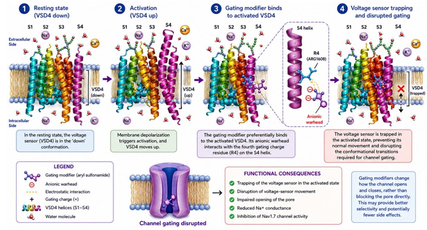
Structural and pharmacological studies of human Nav1.7 have provided important insights into the mechanism of VSD4-targeting inhibitors. The crystal structure of human Nav1.7 VSD4 (PDB ID: 5EK0) revealed a pharmacologically relevant binding site for aryl sulfonamide inhibitors, including GX-936 (3-cyano-4-[2-[2-(1-ethylazetidin-3-yl)pyrazol-3-yl]-4-(trifluoromethyl)phenoxy]-N-(1,2,4-thiadiazol-5-yl)benzenesulfonamide) 5. Unlike conventional pore blockers that directly inhibit Na⁺ conductance, aryl sulfonamides act as gating modifiers by preferentially binding to the activated conformation of VSD4. Their anionic sulfonamide group interacts with the fourth gating-charge residue (R4) of the S4 helix, thereby opposing voltage-sensor deactivation, trapping the voltage sensor, and disrupting the conformational transitions required for normal channel gating (Figure 1) 5. In contrast, outer-pore blockers such as tetrodotoxin prevent Na⁺ permeation, whereas local anesthetics primarily interact with the inner pore and stabilize non-conducting or inactivated channel states 7, 8. Importantly, the greater sequence variability of VSD4 relative to the highly conserved Nav pore, together with interactions involving residues in the S2 and S3 helices, may contribute to improved isoform selectivity and reduced off-target activity 5. The involvement of phospholipids in the VSD4 binding environment further emphasizes the importance of the membrane context in Nav1.7 pharmacology 5. Collectively, these structural features establish VSD4 as a promising target for the development of novel Nav1.7-selective molecular scaffolds and provide a rational framework for investigating alternative compounds capable of modulating voltage-sensor function. Nav channel inhibition is also an important mechanism underlying the action of local anesthetics. Local anesthetic compounds reduce nerve impulse propagation by modulating sodium-channel-mediated electrical signaling, resulting in reversible loss of sensation 7, 8. Based on their chemical structures, local anesthetics are commonly classified as ester- or amide-type compounds. Amide-type local anesthetics, including lidocaine, bupivacaine, mepivacaine, and ropivacaine, are widely used clinically 7, 8, 9. The amide functionality contributes to the molecular architecture, polarity, chemical stability, and hydrogen-bonding properties of these compounds. Their molecular framework generally contains both hydrophobic and polar components, allowing interactions with complementary regions of sodium-channel binding environments 7, 8, 9.

The identification of new amide-containing scaffolds is therefore of interest for the computational exploration of compounds that may interact with Nav channels. In addition to conventional structural modification, coordination with transition metals can substantially alter the physicochemical characteristics of an organic ligand. Metal coordination may modify molecular geometry, charge and electronic distribution, conformational flexibility, rigidity, and the spatial orientation of functional groups 10, 11. Consequently, copper and cobalt complexation provides an approach for generating molecular architectures distinct from the corresponding free ligands.
Copper and cobalt are particularly relevant transition metals because of their versatile coordination chemistry with nitrogen- and oxygen-containing ligands 10, 11, 12. Coordination with these metals can influence the electronic properties and three-dimensional arrangement of an amide-based ligand, potentially altering its molecular recognition by biological macromolecules. The resulting copper and cobalt complexes may therefore establish different interaction profiles from those of their parent ligands. However, the biological effect of metal coordination is dependent on the specific ligand, coordination environment, complex stability, and properties of the target protein. Importantly, neither the presence of copper or cobalt nor a favorable docking score alone establishes anesthetic activity or sodium-channel inhibition.
Molecular docking is a useful computational method for preliminary investigation of possible protein–ligand interactions. It predicts plausible ligand-binding orientations and provides estimates of relative binding favorability based on a defined scoring function 13, 14. Docking can additionally identify amino acid residues potentially involved in hydrogen bonding, hydrophobic contacts, electrostatic interactions, and aromatic interactions. Comparative docking of parent ligands and metal complexes can therefore provide preliminary information regarding the influence of metal coordination on predicted receptor recognition.
Accordingly, the present study investigated two amide ligands and their corresponding copper and cobalt complexes against the Nav1.7 VSD4 receptor structure represented by PDB ID 5EK0. Chain A was selected for receptor preparation and docking. The parent amide ligands, their Cu and Co complexes, and the reference inhibitor GX-936 were comparatively evaluated. The objective was to determine whether copper and cobalt coordination influenced the predicted binding behavior of the amide-based molecular compounds.scaffolds and to identify key non-covalent interactions associated with the best-ranked compounds.
The compounds used were prepared and characterized according to the literature 15. Two amide ligands were used. The compounds are illustrated as structures (1)-(6): Amide ligand 1 (1), Amide ligand 2 (2), Copper Amide ligand 1 (3), Cobalt Amide ligand 1 (4), Copper Amide ligand 2 (5), Cobalt Amide ligand 2 (6).
|
The crystal structure of the human Nav1.7 voltage-sensor domain IV (VSD4) was obtained from the Protein Data Bank under the accession number 5EK0 5. For the molecular docking study, chain A was selected as the receptor structure.
Protein preparation was performed using MGLTools and AutoDockTools 16. Crystallographic water molecules and non-essential heteroatoms were removed from the receptor structure before docking preparation. Polar hydrogen atoms were added, and Kollman charges were assigned to the protein. The prepared receptor was subsequently saved in PDBQT format for molecular docking.
2.3. Ligand Structure Generation and PreparationThe investigated compounds consisted of Amide ligand 1, Amide ligand 2, Cobalt Amide ligand 1, Cobalt Amide ligand 2, Copper Amide ligand 1, Copper Amide ligand 2, and the reference Nav1.7 inhibitor GX-936.
The chemical structures were generated and converted into three-dimensional molecular structures using ChemDraw. The generated ligand structures were subsequently prepared using MGLTools/AutoDockTools. Hydrogen atoms were added, and Gasteiger partial charges were assigned. Rotatable bonds were defined where applicable, and the prepared ligands were converted into PDBQT format.
For the copper and cobalt complexes, the coordinated structures were retained during ligand preparation to preserve the coordination architecture used in the computational docking calculations.
2.4. Molecular DockingMolecular docking was performed using the AutoDock program 13. The prepared chain A of PDB ID 5EK0 was used as the receptor. A docking grid was positioned to encompass the selected pharmacologically relevant receptor region associated with the VSD4 binding site.
Each ligand was independently docked against the prepared receptor structure. Multiple possible ligand conformations and orientations were generated and ranked according to the AutoDock scoring function. The docking conformation with the lowest predicted binding energy was selected as the best-ranked pose for further analysis.
Docking scores were expressed in kcal/mol, and more negative values were interpreted as more favorable predicted interactions according to the applied AutoDock scoring function. The docking scores were used for comparative ranking and were not interpreted as direct measurements of experimental binding affinity or pharmacological potency.
2.5. Protein–Ligand Interaction AnalysisThe best-ranked docking poses were analyzed to identify predicted non-covalent interactions between the ligands and residues of the Nav1.7 VSD4 receptor. Hydrogen bonds, carbon–hydrogen bonds, electrostatic interactions, hydrophobic contacts, π–π interactions, π-alkyl interactions, amide–π interactions, π-sulfur interactions, and other relevant contacts were examined.
Particular attention was given to Cobalt Amide ligand 1, which produced the most favorable docking score, and to the reference compound GX-936. Interaction distances were recorded in angstroms (Å).
2.6. Data AnalysisThe docking scores of the parent amide ligands were compared with those of their respective cobalt and copper complexes to assess the influence of metal coordination on the predicted binding behavior. The docking scores and interaction profiles were additionally compared with those obtained for GX-936.
The results were interpreted as computational predictions of relative binding favorability under the applied docking protocol. Further experimental and computational validation would be required to establish actual binding affinity, channel inhibition, anesthetic activity, or therapeutic potential.
Molecular docking was performed to investigate the binding potential of two amide ligands and their corresponding copper and cobalt complexes against the voltage-sensor domain IV (VSD4) of the human voltage-gated sodium channel Nav1.7. The Nav1.7 VSD4 structure (PDB ID: 5EK0) was used as the receptor, and 3-cyano-4-[2-[2-(1-ethylazetidin-3-yl)pyrazol-3-yl]-4-(trifluoromethyl)phenoxy]-N-(1,2,4-thiadiazol-5-yl)benzenesulfonamide (GX-936) was included as the reference Nav1.7 inhibitor. The docking results were evaluated based on predicted docking scores and ligand–receptor interactions to identify compounds with favorable binding characteristics.
The predicted docking scores of the investigated compounds are presented in Table 1. Among the tested compounds, Cobalt Amide ligand 1 exhibited the most negative docking scores (-9.1 kcal/mol), followed by Cobalt Amide ligand 2 (-8.6 kcal/mol), Amide ligand 1 (-8.3 kcal/mol), Copper Amide ligand 1 (-7.8 kcal/mol), Copper Amide ligand 2 (-7.4 kcal/mol), and Amide ligand 2 (-7.2 kcal/mol), (Table 1). The reference inhibitor GX-936 showed a docking score of -5.9 kcal/mol under the applied computational protocol.
The more negative docking score observed for Cobalt Amide ligand 1 is interpreted as in a more favorable predicted binding pose according to the applied scoring function. Cobalt Amide ligand 1 yielded a docking score that was 3.2 kcal/mol more negative than that of GX- 936; however, this difference does not imply a corresponding difference in free energy of binding. However, these values represent computational scoring estimates and should not be interpreted as direct experimental binding affinities or potency measurements.
The relatively favorable docking scores observed (Table 1) for the metal complexes compared with their corresponding parent ligands are consistent with the hypothesis that metal coordination may influence the predicted receptor-binding behavior of the amide scaffold. In particular, cobalt coordination resulted in the most favorable docking scores among the investigated compounds. This may be associated with changes in molecular geometry, electronic distribution, conformational rigidity, charge distribution, and the spatial orientation of ligand functional groups produced by metal coordination. Details for the remaining compounds are provided in the Supplementary Material.
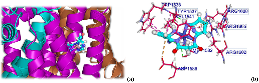
The favorable docking score of Cobalt Amide ligand 1 was supported by an extensive network of predicted hydrogen-bonding electrostatic, and hydrophobic interactions within the Nav1.7 VSD4 binding region (Table 2) (Figure 2). Importantly, Cobalt Amide ligand 1 interacted with the key VSD4 residues TYR1537, TRP1538, ARG1602, ARG1605, and ARG1608.
Two conventional hydrogen bonds were predicted between Cobalt Amide ligand 1 and TYR1537, with distances of 2.09 Å and 2.18 Å. TYR1537 also formed a π–π stacked hydrophobic interaction with the ligand at 4.51 Å. These interactions suggest that Tyr1537 may contribute to recognition and stabilization of the predicted ligand–VSD4 complex.
The ligand also showed multiple interactions with TRP1538, including an amide–π stacked interaction at 4.69 Å and π–π T-shaped interactions at 4.93 Å and 5.33 Å. These aromatic interactions may assist in positioning and stabilization of the ligand within the hydrophobic portion of the VSD4 binding environment.
Interactions with the positively charged ARG1602 included a π–cation electrostatic interaction at 4.72 Å and a π-alkyl hydrophobic interaction at 5.32 Å. Similarly, ARG1605 participated in a π-alkyl interaction at 4.77 Å.

Of particular significance, Cobalt Amide ligand 1 established an electrostatic π-cation interaction with ARG1608 at a distance of 4.48 Å. ARG1608 corresponds to the R4 gating-charge residue of the VSD4 S4 segment and is therefore an especially important residue when considering voltage-sensor modulation. The predicted interaction between Cobalt Amide ligand 1 and ARG1608 provides a potentially relevant structural basis for its recognition of the Nav1.7 VSD4 region.
Additional hydrogen-bonding interactions were predicted with MET1582, at distances of 2.36 Å and 2.68 Å, while a carbon-hydrogen interaction involving ASP1586 was observed at 2.83 Å. ASP1586 also participated in an electrostatic π–anion interaction at 4.07 Å. Additional hydrophobic contacts were observed with VAL1541 (4.92 Å) and other residues within the binding region.
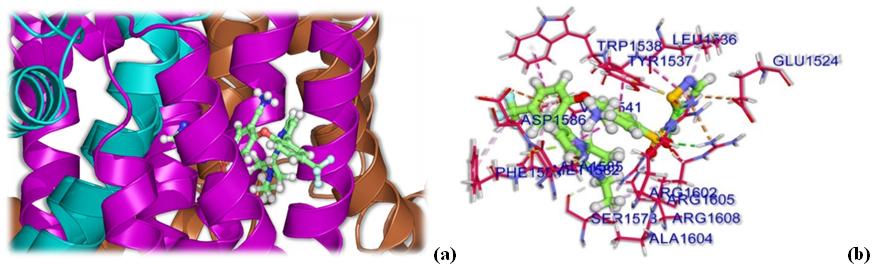
The interaction profile of Cobalt Amide ligand 1 was further compared with that of GX-936 (Table 3) (Figure 3). GX-936 showed predicted interactions with several residues also contacted by Cobalt Amide ligand 1, including TYR1537, TRP1538, ARG1602, ARG1605, and ARG1608. GX-936 formed hydrogen-bonding interactions involving ARG1602, ARG1605, and ARG1608, together with electrostatic interactions involving ARG1605, ASP1586, and GLU1524. It also showed aromatic and hydrophobic interactions with TYR1537, TRP1538, LEU1536, VAL1541, MET1582, PHE1583, and ALA1604.
The overlap in the interaction residues is particularly noteworthy. Both Cobalt Amide ligand 1 and GX-936 engage TYR1537, TRP1538, ARG1602, ARG1605, and ARG1608, suggesting that Cobalt Amide ligand 1 occupies a functionally relevant region of the VSD4 binding environment. Most importantly, the predicted interaction with ARG1608 at 4.48 Å provides a hypothesis for a mechanistic link to the VSD4 region targeted by GX-936. Nevertheless, the present docking analysis cannot establish that Cobalt Amide ligand 1 produces the same voltage-sensor trapping mechanism as GX-936. Electrophysiological experiments would be required to determine the functional consequences of the predicted interaction with ARG1608.

The incorporation of copper and cobalt into the amide scaffold represents an important structural modification that can substantially alter the physicochemical and conformational properties of the parent ligand. Transition-metal coordination may modify molecular geometry, electronic distribution, conformational rigidity, charge distribution, and the orientation of functional groups available for protein recognition. These changes can consequently influence hydrogen bonding, electrostatic interactions, π-interactions, and hydrophobic contacts within a receptor binding site.
The present docking results provide evidence that metal coordination may influence the predicted interaction of the amide derivatives with Nav1.7 VSD4. Cobalt Amide ligand 1 produced the most favorable docking score (-9.1 kcal/mol), whereas the corresponding Amide ligand 1 showed a score of -8.3 kcal/mol. Similarly, the copper complexes displayed favorable docking scores, although they were less favorable than those of the cobalt complexes in the present analysis. These differences suggest that the nature of the coordinated metal may influence the three-dimensional and electronic properties of the resulting complexes and consequently their predicted receptor interactions 11, 18, 19, 20.
The particularly favorable docking scores of Cobalt Amide ligand 1 may therefore arise from a combination of metal-induced conformational organization and favorable presentation of its interacting groups toward the VSD4 binding region. Its extensive interaction network, including contacts with the key residues TYR1537, TRP1538, ARG1602, ARG1605, and ARG1608, further supports this interpretation.
Taken together, the docking analysis identifies Cobalt Amide ligand 1 as the top-ranked compound, with a predicted docking score of -9.1 kcal/mol and an extensive interaction network within Nav1.7 VSD4. Particularly important are its predicted interactions with TYR1537, TRP1538, ARG1602, ARG1605, and especially ARG1608. The interaction with ARG1608 at 4.48 Å is noteworthy because this residue represents the R4 gating charge of the VSD4 S4 segment and is involved in recognition of the reference VSD4 inhibitor GX-936.
The results suggest that cobalt coordination may improve the docking score of the amide scaffold with the Nav1.7 VSD4 region, potentially through changes in molecular geometry, electronic properties, and conformational organization. The similarity between the interaction residues of Cobalt Amide ligand 1 and GX-936 further supports the possibility that the cobalt complex may engage a functionally relevant region of the Nav1.7 voltage sensor.
Accordingly, Cobalt Amide ligand 1 represents a promising candidate for further investigation as a potential Nav1.7 VSD4-modulating compound and as a candidate for anesthetic or analgesic applications. Nevertheless, molecular docking alone cannot establish Nav1.7 inhibition, voltage-sensor trapping, anesthetic activity, or clinical efficacy. Molecular dynamics simulations, quantum-mechanical/QM-MM studies, experimental binding assays, electrophysiological measurements, and toxicity/pharmacological evaluation are required to determine whether the predicted interactions translate into functional Nav1.7 modulation and potential anesthetic activity.
The present molecular docking study investigated two amide ligands and their corresponding copper(II) and cobalt(II) complexes as potential modulators of the voltage-sensing domain IV (VSD4) of human Nav1.7. Among the tested compounds, Cobalt Amide ligand 1 exhibited the best docking score (−9.1 kcal/mol), followed by Cobalt Amide ligand 2 (−8.6 kcal/mol) and Amide ligand 1 (−8.3 kcal/mol), compared with −5.9 kcal/mol for the reference inhibitor GX-936 under the applied conditions. Cobalt Amide ligand 1 formed multiple predicted interactions with key VSD4 residues, including TYR1537, TRP1538, ARG1602, ARG1605, and particularly ARG1608, the R4 gating-charge residue, with a predicted interaction distance of 4.48 Å. Its interaction with this functionally important residue suggests that the cobalt complex may engage a pharmacologically relevant region of the Nav1.7 voltage sensor. Comparison of the free ligands and their metal complexes further indicates that metal coordination may alter molecular geometry, electronic distribution, conformational rigidity, and functional-group orientation, thereby influencing receptor interactions. Unlike conventional pore blockers that directly occlude the sodium-conducting pore, gating modifiers regulate voltage-sensor movement and channel gating, which may provide greater isoform selectivity and potentially reduce unwanted effects on other Nav channels. Overall, Cobalt Amide ligand 1 emerges as a promising computational candidate for Nav1.7 VSD4 modulation, warranting further experimental validation through biochemical, electrophysiological, and molecular dynamics studies. When developing metal complexes as potential pharmaceuticals or anesthetics, their thermodynamic and kinetic stability under physiological conditions, represent critical parameters. In general, transition metal ions such as Co(II) and Cu(II) can be effectively stabilized by well-optimized chelating ligands, which significantly minimizes premature dissociation in biological media and suppresses the cytotoxicity associated with free metal ions. The amide ligands investigated in this study were strategically designed to form robust chelate rings with the metal centers via multiple nitrogen and oxygen donor atoms, which is expected to impart reasonable stability under physiological environments. Nevertheless, a comprehensive assessment of their real-time biostability, neurotoxicity, and general cytotoxicity remains beyond the scope of this preliminary docking study. Future works must incorporate solution stability assays in physiological buffers, along with in vitro cell viability studies, to rigorously balance the anesthetic efficacy and safety profiles of these coordination compounds.
| [1] | Catterall WA. Voltage-gated sodium channels at 60: structure, function and pathophysiology. The Journal of Physiology. 2012; 590: 2577–2589. | ||
| In article | View Article PubMed | ||
| [2] | Catterall WA. From ionic currents to molecular mechanisms: the structure and function of voltage-gated sodium channels. Neuron. 2000; 26: 13–25. | ||
| In article | View Article PubMed | ||
| [3] | Dib-Hajj SD, Yang Y, Black JA, Waxman SG. The NaV1.7 sodium channel: from molecule to man. Nature Reviews Neuroscience. 2013; 14: 49–62. | ||
| In article | View Article PubMed | ||
| [4] | Hille B. Ion Channels of Excitable Membranes. 3rd ed. Sunderland, MA: Sinauer Associates; 2001. | ||
| In article | |||
| [5] | Ahuja S, Mukund S, Deng L, et al. Structural basis of Nav1.7 inhibition by an isoform-selective small-molecule antagonist. Science. 2015; 350(6267): aac5464. | ||
| In article | |||
| [6] | Cox JJ, Reimann F, Nicholas AK, et al. An SCN9A channelopathy causes congenital inability to experience pain. Nature. 2006; 444: 894–898. | ||
| In article | View Article PubMed | ||
| [7] | Becker DE, Reed KL. Local anesthetics: review of pharmacological considerations. Anesthesia Progress. 2012; 59: 90–102. | ||
| In article | View Article PubMed | ||
| [8] | Scholz A. Mechanisms of (local) anaesthetics on voltage-gated sodium and other ion channels. British Journal of Anaesthesia. 2002; 89: 52–61. | ||
| In article | View Article PubMed | ||
| [9] | Butterworth JF. Models and mechanisms of local anesthetic action. Journal of Clinical Anesthesia. 1989; 1: 275–284. | ||
| In article | |||
| [10] | Gasser G, Metzler-Nolte N. The potential of organometallic complexes in medicinal chemistry. Current Opinion in Chemical Biology. 2012; 16: 84–91. | ||
| In article | View Article PubMed | ||
| [11] | Barry NPE, Sadler PJ. Exploration of the medical periodic table: towards new targets. Chemical Communications. 2013; 49: 5106–5131. | ||
| In article | View Article PubMed | ||
| [12] | Kostova I. Ruthenium complexes as anticancer agents. Current Medicinal Chemistry. 2006; 13: 1085–1107. | ||
| In article | View Article PubMed | ||
| [13] | Morris GM, Huey R, Lindstrom W, et al. AutoDock4 and AutoDockTools4: automated docking with selective receptor flexibility. Journal of Computational Chemistry. 2009; 30: 2785–2791. | ||
| In article | View Article PubMed | ||
| [14] | Goodsell DS, Olson AJ. Automated docking of substrates to proteins by simulated annealing. Proteins. 1990; 8: 195–202. | ||
| In article | View Article PubMed | ||
| [15] | Aiyelabola TO, Pradhan S, Akitsu T. Synthesis, characterization and anti-inflammatory activity of two novel amides and their Cu(II) and Co(II) complexes. ES Chemistry and Sustainability. 2026; 7: 2195. | ||
| In article | View Article | ||
| [16] | Sanner MF. Python: a programming language for software integration and development. Journal of Molecular Graphics and Modelling. 1999; 17: 57–61. | ||
| In article | |||
| [17] | Roopashree B. A review study on computational insights into transition metal complex cytotoxicity in neurobiology. Developmental Neurobiology. 2025. | ||
| In article | |||
| [18] | Margariti A, Papakonstantinou VD, Stamatakis G, et al. First-row transition metal complexes incorporating the 2-(2′-pyridyl)quinoxaline ligand (pqx), as potent inflammatory mediators: cytotoxic properties and biological activities against the platelet-activating factor (PAF) and thrombin. Molecules. 2023; 28: 6899. | ||
| In article | View Article PubMed | ||
| [19] | Pöthig A, Casini A. Recent developments of supramolecular metal-based structures for applications in cancer therapy and imaging. Theranostics. 2019; 9: 3150–3169. | ||
| In article | View Article PubMed | ||
| [20] | Zhang R, Pan Y, Ahmed L, et al. A multispecific investigation of the metal effect in mammalian odorant receptors for sulfur-containing compounds. Chemical Senses. 2018; 43: 357–366. | ||
| In article | View Article PubMed | ||
Published with license by Science and Education Publishing, Copyright © 2026 Temitayo O. Aiyelabola, Sayantan Pradhan and Takashiro Akitsu
 This work is licensed under a Creative Commons Attribution 4.0 International License. To view a copy of this license, visit
http://creativecommons.org/licenses/by/4.0/
This work is licensed under a Creative Commons Attribution 4.0 International License. To view a copy of this license, visit
http://creativecommons.org/licenses/by/4.0/
| [1] | Catterall WA. Voltage-gated sodium channels at 60: structure, function and pathophysiology. The Journal of Physiology. 2012; 590: 2577–2589. | ||
| In article | View Article PubMed | ||
| [2] | Catterall WA. From ionic currents to molecular mechanisms: the structure and function of voltage-gated sodium channels. Neuron. 2000; 26: 13–25. | ||
| In article | View Article PubMed | ||
| [3] | Dib-Hajj SD, Yang Y, Black JA, Waxman SG. The NaV1.7 sodium channel: from molecule to man. Nature Reviews Neuroscience. 2013; 14: 49–62. | ||
| In article | View Article PubMed | ||
| [4] | Hille B. Ion Channels of Excitable Membranes. 3rd ed. Sunderland, MA: Sinauer Associates; 2001. | ||
| In article | |||
| [5] | Ahuja S, Mukund S, Deng L, et al. Structural basis of Nav1.7 inhibition by an isoform-selective small-molecule antagonist. Science. 2015; 350(6267): aac5464. | ||
| In article | |||
| [6] | Cox JJ, Reimann F, Nicholas AK, et al. An SCN9A channelopathy causes congenital inability to experience pain. Nature. 2006; 444: 894–898. | ||
| In article | View Article PubMed | ||
| [7] | Becker DE, Reed KL. Local anesthetics: review of pharmacological considerations. Anesthesia Progress. 2012; 59: 90–102. | ||
| In article | View Article PubMed | ||
| [8] | Scholz A. Mechanisms of (local) anaesthetics on voltage-gated sodium and other ion channels. British Journal of Anaesthesia. 2002; 89: 52–61. | ||
| In article | View Article PubMed | ||
| [9] | Butterworth JF. Models and mechanisms of local anesthetic action. Journal of Clinical Anesthesia. 1989; 1: 275–284. | ||
| In article | |||
| [10] | Gasser G, Metzler-Nolte N. The potential of organometallic complexes in medicinal chemistry. Current Opinion in Chemical Biology. 2012; 16: 84–91. | ||
| In article | View Article PubMed | ||
| [11] | Barry NPE, Sadler PJ. Exploration of the medical periodic table: towards new targets. Chemical Communications. 2013; 49: 5106–5131. | ||
| In article | View Article PubMed | ||
| [12] | Kostova I. Ruthenium complexes as anticancer agents. Current Medicinal Chemistry. 2006; 13: 1085–1107. | ||
| In article | View Article PubMed | ||
| [13] | Morris GM, Huey R, Lindstrom W, et al. AutoDock4 and AutoDockTools4: automated docking with selective receptor flexibility. Journal of Computational Chemistry. 2009; 30: 2785–2791. | ||
| In article | View Article PubMed | ||
| [14] | Goodsell DS, Olson AJ. Automated docking of substrates to proteins by simulated annealing. Proteins. 1990; 8: 195–202. | ||
| In article | View Article PubMed | ||
| [15] | Aiyelabola TO, Pradhan S, Akitsu T. Synthesis, characterization and anti-inflammatory activity of two novel amides and their Cu(II) and Co(II) complexes. ES Chemistry and Sustainability. 2026; 7: 2195. | ||
| In article | View Article | ||
| [16] | Sanner MF. Python: a programming language for software integration and development. Journal of Molecular Graphics and Modelling. 1999; 17: 57–61. | ||
| In article | |||
| [17] | Roopashree B. A review study on computational insights into transition metal complex cytotoxicity in neurobiology. Developmental Neurobiology. 2025. | ||
| In article | |||
| [18] | Margariti A, Papakonstantinou VD, Stamatakis G, et al. First-row transition metal complexes incorporating the 2-(2′-pyridyl)quinoxaline ligand (pqx), as potent inflammatory mediators: cytotoxic properties and biological activities against the platelet-activating factor (PAF) and thrombin. Molecules. 2023; 28: 6899. | ||
| In article | View Article PubMed | ||
| [19] | Pöthig A, Casini A. Recent developments of supramolecular metal-based structures for applications in cancer therapy and imaging. Theranostics. 2019; 9: 3150–3169. | ||
| In article | View Article PubMed | ||
| [20] | Zhang R, Pan Y, Ahmed L, et al. A multispecific investigation of the metal effect in mammalian odorant receptors for sulfur-containing compounds. Chemical Senses. 2018; 43: 357–366. | ||
| In article | View Article PubMed | ||