 OPEN ACCESS
 PEER-REVIEWED
Reproductive Effects of 3-Monochloropropane-1, 2-diol on Mice Sperm Function and Early Embryonic Development In Vitro
Luona Wen1, Jianxia Sun2, Shun Bai1, Yunfeng Hu1, Shi Wu1, Rui Jiao1, Shiyi Ou1, Weibin Bai1
1Department of Food Science and Engineering, Department of Developmental and Regenerative Biology, Biopharmaceutical R&D Center, Jinan University, Guangzhou, China
2Faculty of Chemical Engineering and Light Industry, Guangdong University of Technology, Guangzhou, China
Abstract
3-Monochloropropane-1, 2-diol (3-MCPD) is a well-known chloropropanol that is synthesized during food processing contamination. While evaluating sperm quality, sperm penetration into oocytes and early embryonic development, the present study investigated the effects of 3-MCPD onmicereproduction in vitro. During thein vitro fertilization (IVF) process, zygotes and 2-cell embryos were incubated with 3-MCPD-supplemented medium until 8-cell embryo formation to evaluate the reproductive toxicity of 3-MCPD. Our data showed that compared with the control group, the fertilization rate and cleavage rate remarkably decreased in groups treated with 1.802 and 3.160 mM 3-MCPD. The percentage of 2-cell embryo and 4-cell embryo formation also significantly decreased (P < 0.05). Moreover, 8-cell embryos were not observed in any of the treated groups. Taken together, these results suggest that 3-MCPD exposure has hazardous effects on the micesperm fertilization ability as well as embryonic development.
Keywords: 3-Monochloropropane-1, 2-diol; mouse sperm function; early embryonic development
Received June 04, 2015; Revised June 22, 2015; Accepted July 09, 2015
Copyright © 2015 Science and Education Publishing. All Rights Reserved.Cite this article:
- Luona Wen, Jianxia Sun, Shun Bai, Yunfeng Hu, Shi Wu, Rui Jiao, Shiyi Ou, Weibin Bai. Reproductive Effects of 3-Monochloropropane-1, 2-diol on Mice Sperm Function and Early Embryonic Development In Vitro. Journal of Food and Nutrition Research. Vol. 3, No. 6, 2015, pp 405-409. http://pubs.sciepub.com/jfnr/3/6/8
- Wen, Luona, et al. "Reproductive Effects of 3-Monochloropropane-1, 2-diol on Mice Sperm Function and Early Embryonic Development In Vitro." Journal of Food and Nutrition Research 3.6 (2015): 405-409.
- Wen, L. , Sun, J. , Bai, S. , Hu, Y. , Wu, S. , Jiao, R. , Ou, S. , & Bai, W. (2015). Reproductive Effects of 3-Monochloropropane-1, 2-diol on Mice Sperm Function and Early Embryonic Development In Vitro. Journal of Food and Nutrition Research, 3(6), 405-409.
- Wen, Luona, Jianxia Sun, Shun Bai, Yunfeng Hu, Shi Wu, Rui Jiao, Shiyi Ou, and Weibin Bai. "Reproductive Effects of 3-Monochloropropane-1, 2-diol on Mice Sperm Function and Early Embryonic Development In Vitro." Journal of Food and Nutrition Research 3, no. 6 (2015): 405-409.
| Import into BibTeX | Import into EndNote | Import into RefMan | Import into RefWorks |
At a glance: Figures
1. Introduction
3-MCPD, which belongs to a group of compounds known as chloropropanol, is a well-known food-processing contaminant. It was first identified in soy sauce prepared using acid-hydrolyzed vegetable proteins (acid-HVP) [1, 2] and has also been detected in various heat-processed foods [3, 4]. Over the past few years, free 3-MCPD has been found to be released from 3-MCPD esters in the gastrointestinal tract [5]. The 3-MCPD esters presented in refined oil at a concentration of 0.3-10 mg/kg was significantly higher compared to those present in soy sauce [6, 7, 8, 9].
In addition, 3-MCPD can cross the blood-brain barrier and blood-testis barrier, which results in a wide distribution of 3-MCPD in body fluids[10]. A renal tubule hyperplasia study determined the contaminant’s lowest observed adverse effect level at 1.1 mg/kg body weight per day [11]. This finding was based on a maximum tolerable daily intake of 3-MCPD, which was recommended at 2 μg/kg body weight by the Scientific Committee on Food of the European Union.
3-MCPD has also been reported to exhibit reproductive toxicity in rats [12], monkeys [13] cats and cheetahs [14]. Low dose exposure to 3-MCPD has been reported to cause temporary sterility, while high dose exposure to 3-MCPD induced spermatocele, which resulted in blockade of the sperm channel in the testis and further caused permanent infertility over time [15]. A 7-day repeated oral dose of 3-MCPD in rats showed that 3-MCPD elicited spermatotoxicity in the epididymis at≥10 mg/kg/day[16]. Moreover, several studies have shown that 3-MCPD affected the energy metabolic pathways of epididymal sperm, resulting in a decrease in mating frequency, birth index and number of alive newborns [17, 18, 19, 20].
Various toxicity studies have been performed to investigate the toxicity of 3-MCPD during the development of the embryo and reproductive organs. 3-MCPD acts both as an epididymal toxicant and as a toxicant that is capable of directly affecting sperm metabolism and sperm motility [21, 22]. However, the effects of 3-MCPD on fertilization and early embryonic development have not yet been determined. The aim of this study was to evaluate the effects of 3-MCPD on fertilization and early embryonic development, which was examined by comparing the number of fertilized oocytes and the cleavage of fertilized oocytes in control and 3-MCPD-treated groups. Validating these effects would be of great importance in the evaluation of reproductive and developmental toxicology of 3-MCPD.
2. Materials and Methods
2.1. Chemicals3-MCPD was obtained from Aladdin (Shanghai, China). Based on a dose−response curve study performed in our previous study, the IC25, IC50, and IC75 of 3-MCPD were 1.027, 1.802, and 3.160 mM, respectively [23]. Pregnant mare serum gonadotropin (PMSG) and human chorionic gonadotropin (HCG) were purchased from Ningbo second hormone factory (Ningbo, Zhejiang, China). Human tubal fluid (HTF) medium and cleavage medium were purchased from SAGE (Louisiana, America) and supplemented with 10 % fetal bovine serum (FBS). FBS was acquired from Gibco (California, America), and stored at -20°C after immersion in a water bath at 56°C for 30 min. Paraffin oil was acquired from Sigma (Santa Clara, California, America).
2.2. AnimalsIn this study, 6-8 weeks old female and male Kunming mice were obtained from Guangdong Medical Laboratory Animal Center and used as oocyte and semen donors, respectively. All mice were housed under controlled light conditions of a 12-h light-dark cycle in the experimental animal room at room temperature with free access to food and water.
2.3. Collection of OocytesSuperovulation and oocyte collection were performed as previously described, with slight modifications [24]. Eight-10 week old female mice were injected with 10 IU PMSG in 100 μL of saline at 21:00 (GMT+8), and 10 IU HCG in 100 μL of saline 48 h later. Both fallopian tubes were isolated 14-16 h after HCG administration and maintained in 1 mL of HTF medium, which was previously equilibrated in a humidified atmosphere of 5% CO2 in air at 37°C. Next, cumulus oocyte complexes (COCs) were collected from the removed oviducts and washed in 10% FBS supplemented with HTF medium droplet 6 times.
2.4. Collection of Spermatozoa.The collection of spermatozoa and the IVF procedure were performed according to a previously described method, with minor modifications [25]. Eight -10 weeks old male mice were sacrificed 14 h after HCG administration of female mice. Each caudaepididymidis was isolated aseptically from the mice and placed in petri dish with 1 mL of HTF supplemented with 10 % FBS. The caudaepididymidis was excised using ophthalmic scissors, and a dense mass of sperm was squeezed out and subsequently incubated in 1 mL of HTF supplemented with 10 % FBS. Next, the caudaepididymidis and medium in the petri dish were placed into a 1.5-mL centrifuge tube and placed in an incubator at 37°C for 60-90 min to obtain the capacitated spermatozoa.
2.5. In Vitro FertilizationOne day prior to the experiment, the HTF medium and cleavage medium were equilibrated in a 37°C in a 5 % CO2 incubator. On the next day, mature oocytes were added to a drop of 100 μL of HTF (with 10% FBS) medium containing 3-MCPD at cdifferent concentrations, and the capacitated sperm suspension was added to the droplet to achieve a final sperm concentration of 1×10⁶ /ml. Fertilized oocytes were examined 6 h after fertilization by extrusion of the second polar body and the presence of two pronuclei. The number of fertilized oocytes was recorded.
2.6. In vitro Early Embryonic Development ProcedureWhen fertilization was completed, the fertilized oocytes were washed in HTF medium droplet supplemented with 10 % FBS 6 times to remove the unfertilized oocytes and spermatozoa. Next, the fertilized oocytes were moved to cleavage-FBS medium containing 3-MCPD at different concentrations and observed using an inverted microscope. The number of 2-cell embryos, 4-cell embryos and 8-cell embryos were recorded.
2.7. Statistical AnalysisThe values were expressed as the mean of triplicate measurements for all experiments. SPSS 19.0 and GraphPad Prism 5 were used for statistical analysis. Results were considered statistically significant at P < 0.05.
3. Results
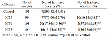
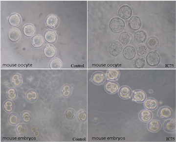
3.1. Effects of Chemical Exposure on the Fertilization RateOocytes were observed for fertilization 6 h after IVF using an inverted microscope. The fertilized oocytes were evaluated to be normal by the appearance of two pronuclei or two polar bodies. However, some of the fertilized oocytes appeared with only one polar body, which may be potentially due to it located in the back of the oocyte and being hidden by another polar body (Figure 1). When fertilization was performed with spermatozoa pretreated with 3-MCPD at the concentration of IC25, IC50 or IC75, the fertilization rate and fracture rate significantly decreased with an increase in the 3-MCPD concentration (P<0.01 and P<0.05, respectively) (Table 1). During in vitro fertilization, 3-MCPD induced a decrease in the fertilization rate, particularly at high concentrations of IC50 and IC75, which resulted in a greater than 73% decrease compared with the control group. Moreover, the fracture rate of oocytes in the IC50 and IC75 group was higher compared to the control group, in which the fracture rate of oocytes in the IC75 group was 46.15 %. In addition, when 3-MCPD-treated fertilized oocytes were moved to normal cleavage medium after fertilization, no 2-cell embryos were observed (data not shown). Thus, 3-MCPD could affect fertilized oocyte development and subsequent 2-cell formation.
 Download as
 Download as
Fertilized oocytes were moved into cleavage medium (with 10 % FBS) after normal fertilization and observed using an inverted microscope on the third day (Figure 2). As shown in Table 2, the number of 4-cell embryos and 8-cell embryos was remarkably less compared to the number of 2-cell embryos in both the control and 3-MCPD-treated groups, indicating a developmental block due to the mouse embryo itself. 3-MCPD induced a significant decrease in the percentage of embryos at 2- and 4-cell stage formation (P< 0.05). The 4-cell formation rate of the 3-MCPD-treated group significantly decreased, indicating that 3-MCPD induced a developmental block in 2-cell embryos. In addition, 8-cell embryos were not found in any of the treated groups indicating a remarkable inhibition of 3-MCPD on the formation of 8-cell embryos.
 Download as
 Download as
4. Discussion
The content of 3-MCPD in food is critical for health [26]. In this context, the reproductive toxicity of 3-MCPD was evaluated regarding its effect on fertilization and preimplantation embryonic development.
Kunming mice were selected due to their short reproductive cycle, rapid cleavage, and easily manipulated growing environment. PMSG and HCG were injected to induce ovulation prior to COC collection. Importantly, the time of exposure of oocytes and spermatozoa in air during their collection should be as short as possible, and the temperature, light and pH of medium should be appropriately maintained.
Sperm motility is important in male fertility and early embryonic development [27], and it is also a quick and sensitive index for reproductive toxicity evaluation [28]. However, there is still no consensus on whether the exposure of sperm to 3-MCPD induces sperm motility. Jelks found that the parameters of sperm motility were not affected at 10 mg/kg 3-MCPD in Sprague Dawley rats but that the ATP levels were significantly decreased [21]. In contrast, Xie et al. detected a significant increase in the percentage of abnormal sperm at a 10 mg/kg 3-MCPD in Sprague Dawley rats, which may be attributed to a deficiency in ATP [29]. However, other reports have also shown a decrease in sperm motility upon exposure to 3-MCPD [14, 30]. Our results showed that sperm motility was not statistically significant between the 3-MCPD-treated and control groups. Thus, 3-MCD did not induce any toxicity in sperm motility within a short time period. Because 3-MCPD could trigger a significant increase in the fracture rate, we proposed that the effect of fertility by 3-MCPD could be revealed using oocytes instead of sperm pathway in vitro. 3-MCPD combined with glutathione in vivo forms oxalate [31], which results in a decrease in GSH levels, which serves as an indicator of cytoplasmic maturation in oocytes [32, 33].
During in vitro fertilization, 3-MCPD induced a decrease in the fertilization rate and an increase in the oocyte fracture rate, particularly at high concentrations of IC50 and IC75. 3-MCPD has also been reported to inhibit the expression of genes affecting glycogen synthetase activity and to generate a decrease in the sperm's storage capacity, resulting in dysfunctional sperm-oocyte binding and a subsequent decrease in fertility [21].
Early embryonic developmental procedures, including the formation of 2-cell embryos, 4-cell embryos and 8-cell embryos, were adversely affected by 3-MCPD treatment. In this study, 3-MCPD significantly decreased the formation rates of 4-cell embryos. Cleavage terminated after the formation of 4-cell embryos; that is, no 8-cell embryos formed in the IC75 group. Taken together, these results suggested that early embryonic development was remarkably inhibited by 3-MCPD.
Developmental blockade, which normally occurs during embryonic development in vitro, was observed at the 2-cell stage in the present study. A lack of some components in the culture medium and/or inappropriate environmental conditions for embryonic development could result in a developmental block of early embryos [34], which often occurs in 2-cell embryos in mice [35]. A family of genes known as Bcl-2 or nuclear fragmentation may be the cause of this blockade phenomenon [36], and further studies are warranted.
5. Conclusions
Our results indicated that 3-MCPD resulted in a decrease in the fertilization rate and an increase in oocyte fracture rate.3-MCPD exposure has hazardous effects on the mice embryonic development by reducing the percentage of 2-cell embryo and 4-cell embryo formation. Moreover, a developmental block at the two-cell stage exists in mouse embryos.
Acknowledgements
This work was supported by the National Science Foundation of China (NSFC) under Grant 31471588; and the Program for New Century Excellent Talents in University (NCET) under Grant 31201340.
Statement of Competing Interests
The authors have no competing interests.
List of Abbreviations
3-Monochloropropane-1, 2-diol, 3-MCPD; In vitro fertilization, IVF; Acid-hydrolyzed vegetable proteins, acid-HV; Pregnant mare serum gonadotropin, PMSG; Human chorionic gonadotropin, HCG; Human tubal fluid, HTF; Fetal bovine serum, FBS; Cumulus oocyte complexes, COCs; Inhibition Concentration, IC.
References
| [1] | Velíšek J, Davidek J, Hajšlová J, Kubelka V, Janíček G, Mánková B. Chlorohydrins in protein hydrolysates. Zeitschrift für Lebensmittel-Untersuchung und Forschung 1978;167:241-4. | ||
 In article In article | View Article PubMed | ||
| [2] | Davidek J, Velíšek J, Kubelka V, Janíček G, Šimicová Z. Glycerol chlorohydrins and their esters as products of the hydrolysis of tripalmitin, tristearin and triolein with hydrochloric acid. Zeitschrift für Lebensmittel-Untersuchung und Forschung 1980;171:14-7. | ||
| In article | View Article | ||
| [3] | Baer I, de la Calle B, Taylor P. 3-MCPD in food other than soy sauce or hydrolysed vegetable protein (HVP). Anal Bioanal Chem 2010;396:443-56. | ||
| In article | View Article PubMed | ||
| [4] | Wenzl T, Lachenmeier DW, Gokmen V. Analysis of heat-induced contaminants (acrylamide, chloropropanols and furan) in carbohydrate-rich food. Anal Bioanal Chem 2007;389:119-37. | ||
| In article | View Article PubMed | ||
| [5] | Hamlet CG, Asuncion L, Velíšek J, Doležal M, Zelinková Z, Crews C. Formation and occurrence of esters of 3-chloropropane-1, 2-diol (3-CPD) in foods: What we know and what we assume. European journal of lipid science and technology 2011;113:279-303. | ||
| In article | View Article | ||
| [6] | Liu Q, Han F, Xie K, Miao H, Wu Y. Simultaneous determination of total fatty acid esters of chloropropanols in edible oils by gas chromatography–mass spectrometry with solid-supported liquid–liquid extraction. Journal of Chromatography A 2013;1314:208-15. | ||
| In article | View Article PubMed | ||
| [7] | Yamazaki K, Ogiso M, Isagawa S, Urushiyama T, Ukena T, Kibune N. A new, direct analytical method using LC-MS/MS for fatty acid esters of 3-chloro-1, 2-propanediol (3-MCPD esters) in edible oils. Food Additives & Contaminants: Part A 2013;30:52-68. | ||
| In article | View Article PubMed | ||
| [8] | Schilter B, Scholz G, Seefelder W. Fatty acid esters of chloropropanols and related compounds in food: toxicological aspects. European journal of lipid science and technology 2011;113:309-13. | ||
| In article | View Article | ||
| [9] | Küsters M, Bimber U, Reeser S, Gallitzendörfer R, Gerhartz M. Simultaneous determination and differentiation of glycidyl esters and 3-monochloropropane-1, 2-diol (MCPD) esters in different foodstuffs by GC-MS. Journal of agricultural and food chemistry 2011;59:6263-70. | ||
| In article | View Article PubMed | ||
| [10] | Li Y, Liu S, Wang C, Li K, Shan Y-J, Wang X-J, et al. Novel biomarkers of 3-chloro-1, 2-propanediol exposure by ultra performance liquid chromatography/mass spectrometry based metabonomic analysis of rat urine. Chemical research in toxicology 2010;23:1012-7. | ||
| In article | View Article PubMed | ||
| [11] | Commission E, No R. 2006 of 19 December 2006, Setting Maximum Levels for certain Contaminants in Foodstuffs. Official Journal of the European Union 2006. | ||
| In article | |||
| [12] | Madhu NR, Sarkar B, Biswas SJ, Behera BK, Patra A. Evaluating the anti-fertility potential of alpha-chlorohydrin on testis and spermatozoa in the adult male wild Indian house rat (Rattus rattus). Journal of environmental pathology, toxicology and oncology : official organ of the International Society for Environmental Toxicology and Cancer 2011;30:93-102. | ||
| In article | View Article | ||
| [13] | Hung PH, Miller MG, Meyers SA, VandeVoort CA. Sperm mitochondrial integrity is not required for hyperactivated motility, zona binding, or acrosome reaction in the rhesus macaque. Biol Reprod 2008;79:367-75. | ||
| In article | View Article PubMed | ||
| [14] | Terrell KA, Wildt DE, Anthony NM, Bavister BD, Leibo SP, Penfold LM, et al. Glycolytic enzyme activity is essential for domestic cat (Felis catus) and cheetah (Acinonyx jubatus) sperm motility and viability in a sugar-free medium. Biol Reprod 2011;84:1198-206. | ||
| In article | View Article PubMed | ||
| [15] | Jones A, Milton D, Murcott C. The oxidative metabolism of α-chlorohydrin in the male rat and the formation of spermatocoeles. Xenobiotica 1978;8:573-82. | ||
| In article | View Article PubMed | ||
| [16] | Kim S-H, Lee I-C, Lim J-H, Moon C, Bae C-S, Kim S-H, et al. Spermatotoxic effects of α-chlorohydrin in rats. Laboratory animal research 2012;28:11-6. | ||
| In article | View Article PubMed | ||
| [17] | Kawaguchi T, Kawachi M, Morikawa M, Kazuta H, Shibata K, Ishida M, et al. Key parameters of sperm motion in relation to male fertility in rats given alpha-chlorohydrin or nitrobenzene. The Journal of toxicological sciences 2004;29:217-31. | ||
| In article | View Article PubMed | ||
| [18] | Kwack SJ, Kim SS, Choi YW, Rhee GS, Lee RD, Seok JH, et al. Mechanism of antifertility in male rats treated with 3-monochloro-1, 2-propanediol (3-MCPD). Journal of Toxicology and Environmental Health, Part A 2004;67:2001-4. | ||
| In article | View Article PubMed | ||
| [19] | Kim SH, Lee IC, Lim JH, Moon C, Bae CS, Kim SH, et al. Spermatotoxic effects of alpha-chlorohydrin in rats. Lab Anim Res 2012;28:11-6. | ||
| In article | View Article PubMed | ||
| [20] | Zhang H, Yu H, Wang X, Zheng W, Yang B, Pi J, et al. (S)-alpha-chlorohydrin inhibits protein tyrosine phosphorylation through blocking cyclic AMP - protein kinase A pathway in spermatozoa. PLoS One 2012;7:e43004. | ||
| In article | View Article PubMed | ||
| [21] | Jelks K, Berger T, Horner C, Miller MG. α-Chlorohydrin induced changes in sperm fertilizing ability in the rat: association with diminished sperm ATP levels and motility. Reproductive toxicology 2000;15:11-20. | ||
| In article | View Article | ||
| [22] | Kim SH, Lee IC, Baek HS, Moon C, Bae CS, Kim SH, et al. Ameliorative effects of pine bark extract on spermatotoxicity by alpha-chlorohydrin in rats. Phytotherapy research : PTR 2014;28:451-7. | ||
| In article | View Article PubMed | ||
| [23] | Sun J, Bai S, Bai W, Zou F, Zhang L, Su Z, et al. Toxic Mechanisms of 3-Monochloropropane-1, 2-Diol on Progesterone Production in R2C Rat Leydig Cells. Journal of agricultural and food chemistry 2013;61:9955-60. | ||
| In article | View Article PubMed | ||
| [24] | Gu Y-h, Li Y, Huang X-f, Zheng J-f, Yang J, Diao H, et al. Reproductive Effects of Two Neonicotinoid Insecticides on Mouse Sperm Function and Early Embryonic Development In Vitro. PloS one 2013;8:e70112. | ||
| In article | View Article PubMed | ||
| [25] | Wakayama S, Kawahara Y, Li C, Yamagata K, Yuge L, Wakayama T. Detrimental effects of microgravity on mouse preimplantation development in vitro. PloS one 2009;4:e6753. | ||
| In article | View Article PubMed | ||
| [26] | Crews C, Brereton P, Davies A. The effects of domestic cooking on the levels of 3-monochloropropanediol in foods. Food Additives & Contaminants 2001;18:271-80. | ||
| In article | View Article PubMed | ||
| [27] | D’Occhio M, Hengstberger K, Johnston S. Biology of sperm chromatin structure and relationship to male fertility and embryonic survival. Animal reproduction science 2007;101:1-17. | ||
| In article | View Article PubMed | ||
| [28] | Claassens OE, Wehr JB, Harrison KL. Optimizing sensitivity of the human sperm motility assay for embryo toxicity testing. Human Reproduction 2000;15:1586-91. | ||
| In article | View Article PubMed | ||
| [29] | Xie S, Zhu Y, Ma L, Lu Y, Zhou J, Gui Y, et al. Research Genome-wide profiling of gene expression in the epididymis of alpha-chlorohydrin-induced infertile rats using an oligonucleotide microarray. 2010. | ||
| In article | |||
| [30] | Ford WC. Glycolysis and sperm motility: does a spoonful of sugar help the flagellum go round? Human reproduction update 2006;12:269-74. | ||
| In article | View Article PubMed | ||
| [31] | Lynch BS, Bryant DW, Hook GJ, Nestmann ER, Munro IC. Carcinogenicity of monochloro-1, 2-propanediol (α-chlorohydrin, 3-MCPD). International Journal of Toxicology 1998;17:47-76. | ||
| In article | View Article | ||
| [32] | Funahashi H, Cantley TC, Stumpf TT, Terlouw SL, Day BN. Use of low-salt culture medium for in vitro maturation of porcine oocytes is associated with elevated oocyte glutathione levels and enhanced male pronuclear formation after in vitro fertilization. Biology of reproduction 1994;51:633-9. | ||
| In article | View Article PubMed | ||
| [33] | de Matos DG, Furnus CC, Moses DF. Glutathione synthesis during in vitro maturation of bovine oocytes: role of cumulus cells. Biology of reproduction 1997;57:1420-5. | ||
| In article | View Article PubMed | ||
| [34] | Garcia SM, Marinho LS, Lunardelli PA, Seneda MM, Meirelles FV. Developmental block and programmed cell death in Bos indicus embryos: effects of protein supplementation source and developmental kinetics. PLoS One 2015;10:e0119463. | ||
| In article | View Article PubMed | ||
| [35] | Jeong JK, Kang MH, Gurunathan S, Cho SG, Park C, Park JK, et al. Pifithrin-alpha ameliorates resveratrol-induced two-cell block in mouse preimplantation embryos in vitro. Theriogenology 2015;83:862-73. | ||
| In article | View Article PubMed | ||
| [36] | Meirelles F, Caetano A, Watanabe Y, Ripamonte P, Carambula S, Merighe G, et al. Genome activation and developmental block in bovine embryos. Animal reproduction science 2004;82:13-20. | ||
| In article | View Article PubMed | ||
 CiteULike
 Delicious


 Epididymal. (B) Oviductal magnum. (C) Cumulus-oocyte complexes. (D) Fertilization (E) Fertilization egg (F) Fertilization egg (×400) (G) 2-cell embryos (H) 4-cell embryos (I) 8-cell embryos)