Shiga toxigenic Escherichia coli (STEC) is a food-borne pathogen associated with community health concern. Contamination of food by STEC and toxin causes infectious diseases. Escherichia coli isolated from food samples such as yogurt, apples, tomatoes and milk by applying violet-red bile glucose agar, Triphenyl tetrazolium chloride agar (TTC) and eosin-methylene blue agar (EMB) test after pre-enrichment of food samples in buffered peptone water. Escherichia coli isolates were screened for the existence of shiga toxin genes Stx1 and Stx2 through Polymerase Chain Reaction (PCR). Apples (17%), tomatoes (26%) and milk (1%) of the samples examined showed positive results for Stx genes. Yoghurt samples examined in this study had no Stx1 or Stx2 gene. The presence of Shiga toxin that is protein in nature was confirmed by SDS-PAGE in milk sample. The present study has shown that foods from densely populated areas i.e. Harbanspura, Lahore could be sources of STEC.
STEC is an ellipsis for Shiga toxin-producing Escherichia coli. Maximum E. coli bacteria are customary dwellers of the intestinal territories of humans and animals, and are non-pathogenic. STEC E. coli, as the name infers, secretes a persuasive toxin called Shiga toxin (Stx), also recognized as verotoxin or verocytotoxin. STEC O157:H7 is human pathogen concomitant with diarrheal diseases, hemorrhagic colitis, hemolytic-uremic syndrome and thrombocytobena 1. Several other serotypes are recognized as the outbreaks of STEC O157:H7 and hemorrhagic colitis. Though STEC serotype O157 is known worldwide in different outbreaks but we cannot ignore non- O157 strains, which are also causing a number of infections in humans at various locations. Serologically STEC is a diverse family; approximately 200 serotypes have been documented amongst about 100 are known to cause human infections 2.
A number of virulence factors are present in STEC including Shiga toxin 1 (encoded by Stx 1), Shiga toxin 2 (encoded by Stx 2), intimine (encoded by eaeA gene) and enterohaemolysin (encoded by Ehly gene) 3, 4. The Stx operon is present in genome and consists of Stx operon encoding A and B subunits of gene designated as StxA and StxB arranged in one transcriptional unit. StxA and StxB are separated from each other by 12 non-coding nucleotides. The StxA and StxB genes are present prior to putative ribosome binding site (RBS) and a regulated promoter 5. StxA and StxB share same structural feature and toxic activity but have different antigenicity. The toxic activity of StxA and StxB is due to rRNA glycosidase activity on 28S ribosomal RNA that impedes the protein synthesis and consequently cell death. There are approximately identical amino acid sequences of Stx1 that are reported while Stx2 have greater number of varients (6, 8, 9, 11, 17-20, 24, 31–33, 37, 38, 40–42, 45, 50) 6. In addition, B subunit has stronger RBS resulting in abundant B subunit translation 7.
Various domestic and wild animals act as a reservoir of STEC strains. Cattle are known to be a chief pool of STEC strains that cause infections 8. The transmission of toxin is via ingestion of food contaminated by feces of animals like cattle specially beef, uncooked meat, hamburgers, salami and other dairy products including milk, yogurt, cheese and butter 9, 10 Contamination of swimming pools, water supplies and raw vegetables and fruits may also cause disease 11, 12.
STEC is responsible of watery non-blooded Diarrhea FOR. Even sometimes non-symptomatic infections are also observed. While in some cases watery diarrhea leads to bloody diarrhea followed by abdominal cramps, nausea and vomiting. If the situation becomes more severe then it leads to hemolytic uremia, thrombocytopena and acute renal faliure 13.
Escherichia coli contains highly conserved plasmid that range in size from 92 to 104 kb. While the plasmid of E. coli O157:H7 is of 92 kbp and it is associated with large number of enterohemolysis cases 3. The hlyA gene of E.coli releases a toxin that is membrane associated and is released in very low concentration. It has a single 110 kD polypeptide. It alters the host signalling cascade, induces inflammatory responces and eventually host cell death 14, 15. The incubation period for the onset of disease is 3 to 8 days 16, 17.
The research was conducted by collecting food samples from highly populated and less populated areas of Lahore, Pakistan viz., Harbans Pura (HP) and Johar Town (JT). Two types of areas, one representing a highly populated area (HP), and other as a less populated area (JT) of Lahore, Pakistan were selected by random sampling from previously reported areas for collection of 8 samples of each of milk, yogurt, apple and tomato. The samples were collected in a sterile tube and immediately covered with aluminium foil. The samples were then transfered to laboratory within an hour for further processing. Tomato and apples were grinded and stored at 4°C.
2.2. Pre-enrichmentFood samples were taken individually in 50 mL sterile conical flask and pre-enrichment medium (buffered peptone water - BPW) in 1:9 ratio was added under laminar flow hood. The flask was sealed with cotton and wrapped with aluminium foil. Samples were allowed to incubate overnight in shaking incubator at 37°C.
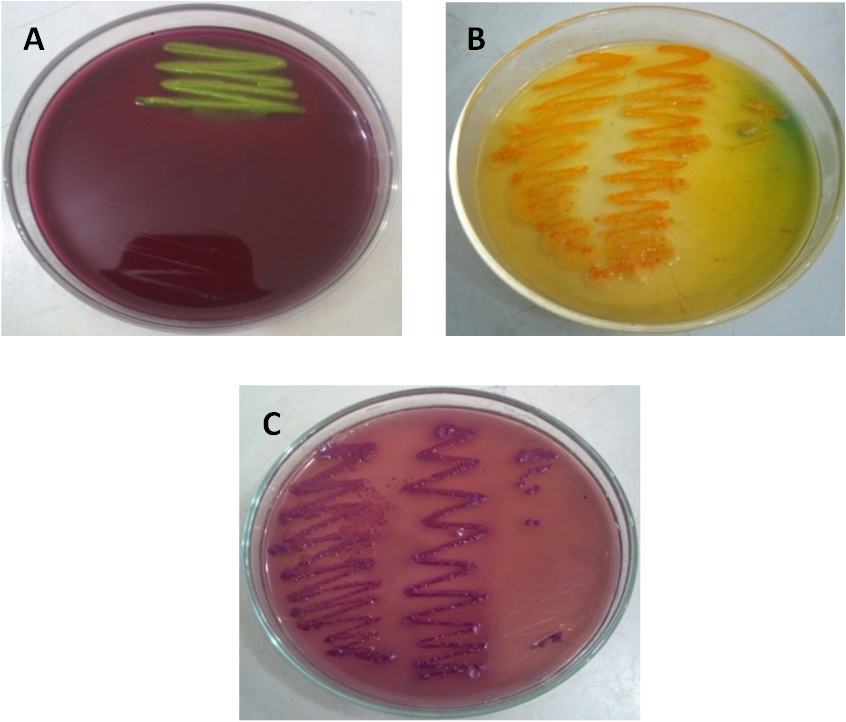
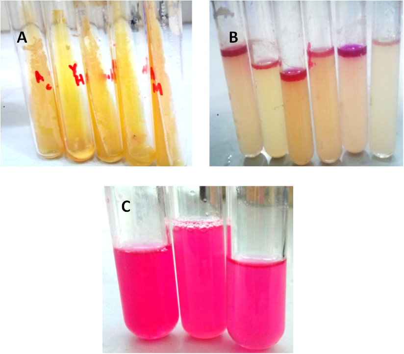
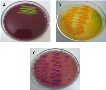
2.3. Isolation and Screening of STECThe samples were speckled on Eosin methylene blue (EMB) agar, TTC agar and violet red bile glucose agar (VRBGA) and kept overnight at 37oC for the isolation of STEC. The colonies with green metallic sheen in case of EMB agar, yellow-orange in case of TTC agar and red colonies on VRBGA were picked and confirmed by TSI agar (triple sugar iron agar) slant (yellow), Indole test (red colored layer at the top of trypton broth), methyl red test (red coloration) and catalase test (bubbling). The samples which showed positive results for E. coli were subsequently used for the detection of shiga toxin gene Stx.
2.4. PCR Detection of Virulence FactorExtracted DNA was isolated from E. coli samples using phenol-chloroform method 18. The isolated DNA was amplified by PCR using Taq DNA polymerase to detect Stx1 and Stx2 genes. The sequence of primers were FP-Stx1 (5ʹ-ACA CTG GAT GAT CTC AGT GG-3ʹ), RP-Stx1 (5ʹ-CTG AAT CCC CCT CCA TTA TG-3ʹ), FP-Stx2 (5ʹ-CCA TGA CAA CGG ACA GCA GTT-3ʹ) and RP-Stx2 (5ʹ-CCT GTC AAC TGA GCA CTT TG-3ʹ). PCR was carried out with 35 cycles including major steps of denaturation, annealing and extension. The whole process was performed in Bio-Rad® thermocycler, USA. The denaturation was carried out at 94°C for 4 minutes. The denaturation was followed by primer annealing which employed 55.2°C for 1 minute for effective binding of primers with the template. The primer extension was done at 72°C for 1 min. The final extension was carried out at a temperature of 72°C. The PCR amplification product was run on 1% agarose gel, stained with ethidium bromide (0.5μg mL-1) and photographed using a gel documentation system (GDS).
2.5. Protein ProfilingThe protein was extracted from the cell culture by taking 3 mL of Luria-Bertani medium (LB) in sterile 50 mL sterile falcon tubes and inoculated with selected colony of STEC. Falcon tubes were kept in shaking incubator at 37°C for three days. Cultures were centrifugated at 6000 rpm for 5 minutes and supernatant was discarded, pellet was washed with 1X PBS buffer and resuspended in lysis buffer. All the samples were sheared in a sonicator for 20 minutes with interval of 20 seconds till solution became clear. The total cell lysate was then centrifuged at 13200 rpm for 30 minutes and supernatant was taken for further examination and stored at 4°C. The proteins extracted were then subjected to 15% SDS-PAGE at 90 volts. The gel was stained with coomassie brilliant blue and bands of shiga toxin were then visualized after destaining the gel 18.
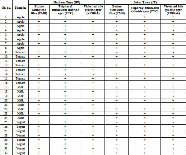
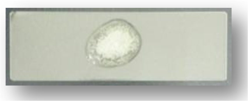
A total of 64 samples were collected from 4 food materials from 2 selected areas of Lahore for the isolation of STEC E. coli. Results indicated that out of 64 food samples 49 revealed the incidence of E. coli strains by conventional microbial methods (Table 1). The existence of E. coli in the food samples was indicated by standard microbiological techniques. (Figure 1A, 1B, 1C).
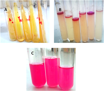
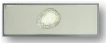
All the 64 strains were also subjected to biochemical analysis among which 49 strains of E. coli were picked as positive colonies from agar when inoculated in TSI slant, trypton broth, methyl red broth and normal saline where the occurrence of E. coli was confirmed by observing the respective change in color (Table 2). The biochemical test with TSI slant was demonstrated in Figure 2A that represented yellow color, CO2 gas emission was noticed at the bottom of the slant however there was no blackening of butt which indicated that there was no H2S gas production. The presence of E. coli grown on Tryptone broth was confirmed by the formation of red layers at the top of Tryptone broth (Figure 2B). Further biochemical analysis signified the occurrence of E. coli by red coloration of the methyl red broth (Figure 2C).
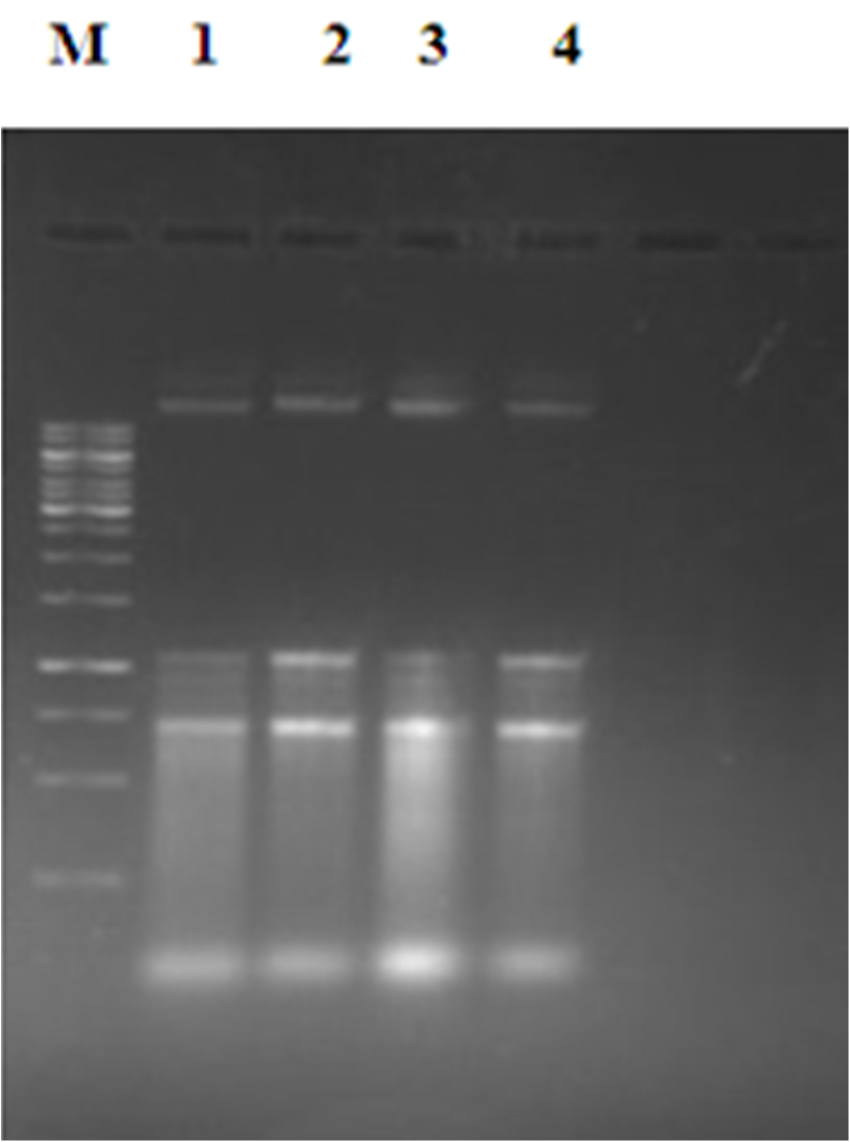
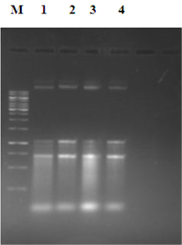
In subsequent studies, DNA was isolated from all (49) strains of E. coli of each food sample and it was separated on 1% agarose to check the quality of extracted DNA and the pertaining results are summarized in Figure 4. All the samples revealed clear and easily detectable bands.
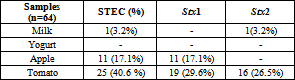
Extracted DNA was utilized to perform PCR to perceive the presence/absence of the bands of Stx genes in the fluorimetric profiles. Out of 49 samples of E. coli, 37 samples confirmed the presence of Stx genes. Amomg all the samples, Stx1 gene was identified in 30 samples while Stx2 was identified in 17 samples (Table 3). The results represented that none of the toxigenic STEC genes was amplified in all the yogurt samples inferring that the E. coli strains isolated from all yogurt samples of all localities were non pathogenic (Table 3).
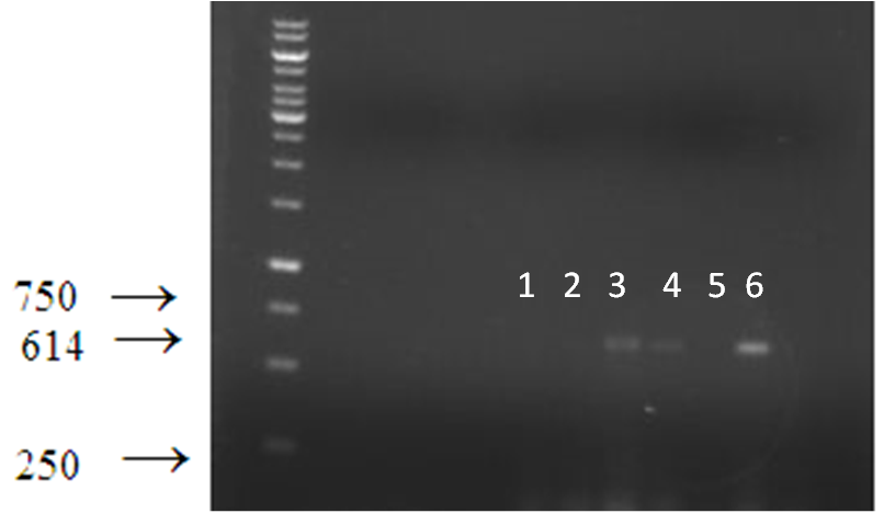
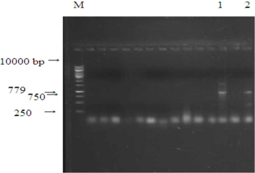
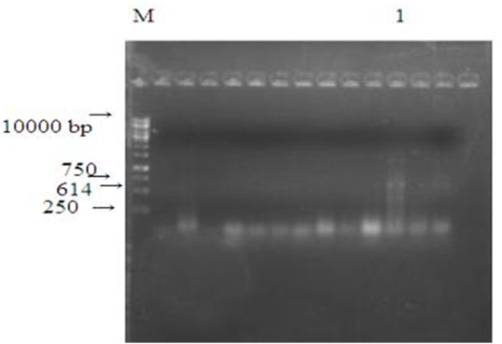
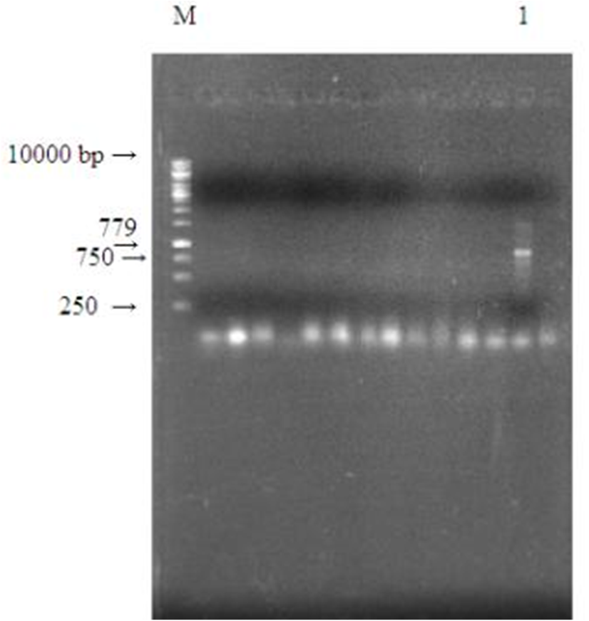
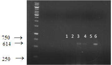
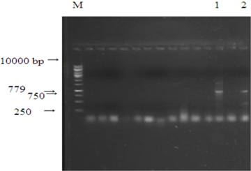
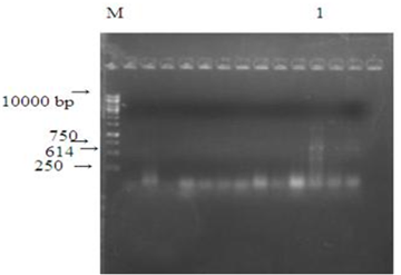
Data regarding the PCR analysis of tomato samples presented in Figure 5 shows that all the samples were not equally capable of amplification of Stx1 gene. The band pattern revealed that Stx1 gene is present in few tomato samples and absent in others. As only samples in lane 3, 4 and 6 exhibited the intense bands at 614bp while no PCR amplification was observed by the samples in lane 1, 2 and 5. However, it is evident from Figure 6 that Stx2 gene was primed at 779bp in only 2 tomato samples while it was completely lacking in all other samples.
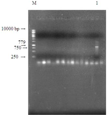
Pattern of DNA fragments amplified for both Stx genes of milk samples depicted that milk samples were quite different from tomato samples. The Stx2 gene was expressed only in 1 milk sample. Whereas this gene was not visible in other milk samples examined. While Stx1 gene was not primed in any sample (Figure 7).
Conversely the PCR analysis conducted for apple samples revealed an entirely different expression profile in comparison to milk samples as only Stx1 gene was detected. Whereas no band of 779bp was visualized in gel pattern confirming that Stx2 gene was found to be absent in all the samples (Figure 8).
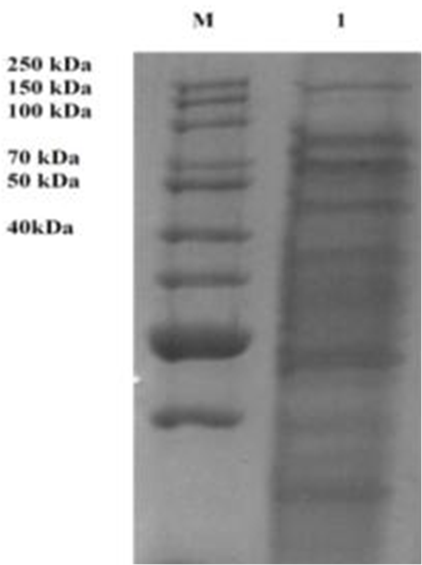
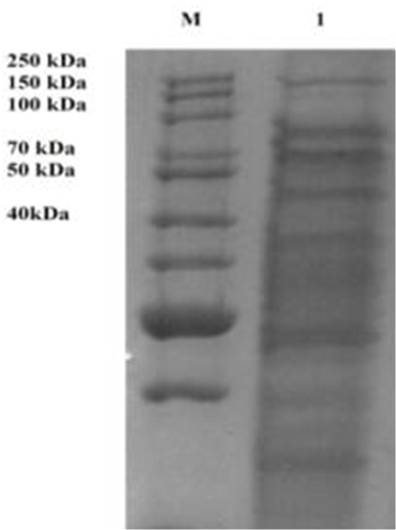
All the E. coli strains (37) possessing STEC genes from all food samples were subjected to protein extraction and the electro–phoretic mobility profiles of recognized molecular weight proteins were matched with marker that exhibited a band which confirmed the presence of shiga toxin protein (Figure 9). The molecular weight assays of these polypeptide protein bands exposed around 70 kDa. These bands (proteins) possibly represent a subunit of the same enzyme protein detected on electrophoresis gel.
STEC is customarily observed as causativte agent of several gastrointestinal diseases and responsible for many foodborne outbreaks. WHO estimated that half of the foodborne diseases are due to STEC 19. They are the most significant agents of diaarhea that are responsible of morbiditty and mortility in children 20. Routine phenotypic methods are used for identification purpose but they are time consuming and less sensitive and they are unable to distinguish diarrheagenic E. coli 21, 22. On the other hand, PCR is the most reliable and sensitive technique for the identification of pathogenic strains 23.
A number of culturing techniques have been established for the isolation of E. coli 24. In current study, E. coli was detected in a large number of samples collected from highly populated areas of Harbanspura and Johar Town. These samples produced characteristics colonies on different minimal media 25.
In the present study the isolation and identification of E. coli from different food samples was carried out by a combination of biochemical and molecular methods.
Earlier, Souza et al. 26 characterized thirty-eight strains of Shiga toxin-producing Escherichia coli (STEC) by describing biochemical properties, enterohaemolysin production and plasmid carriage. An inclusive disparity in the biochemical characteristics was perceived in the STEC, with 14 distinctive biotypes recognized. The common profile, characterized by the occurrence of a single plasmid of ~90 kb, was found in 50% of these strains. This data indicated wide range amongst STEC isolates.
Numerous community strengthening laboratories are using PCR analysis are considered as a reliable technique for the detection of stx1 and stx2 genes for identification and verification of STEC infection 27, 28. Depending on the primers used, these assays can distinguish between stx1 and stx2 29, 30, 31. Presently, in PCR assays, out of 49 E. coli strains Stx1 and Stx2 genes were found in 37 strains (Stx1= tomato, apple and Stx2= tomato and milk). Stx1 and stx2 genes in raw milk sample 32, 33. A number of researchers have scrutinized 593 foodborne STEC isolates for their serotypes and for nine virulence genes (stx1, stx1c, stx1d, stx2, stx2b, stx2e, stx2g, E-hlyand eae), and they perceived a substantial relationship of stx1and stx2 genes with bovine meat and milk products. They also reported a comparison of the characteristics of foodborne STEC with printed data on fecal STEC from food producing animals, and found that virulence profiles and serotypes of STEC from foodstuff exhibited noteworthy similarities with fecal STEC belonging to similar animal species 34, 35.
Furthermore, the toxin nature of STEC protein was predicted by using SDS-PAGE. The electrophoretic analysis of purified proteins revealed the presence of protein bands with MWs of 70 KD demonstrated by SDS-PAGE. Similar results were obtained with a Shiga-like toxin II variant purified from E. coli TB1(pCG6). Sodium dodecyl sulfate polyacrylamide gel electrophoresis of purified toxin depicted three protein bands that traveled with calculated molecular weights of 33,000, 27,500, and 7,500. These bands corresponded to values for the A, A1, and B subunits, respectively, that would be expected on the basis of the nucleotide sequence and comparison with data for Shiga toxin and other Shiga-like toxins [36]. However, Shiga toxin in various food samples was also detected by 34, 35.
Thus the current work reports the novelty of identification and screening of shiga toxigenic E. coli from the food samples. Precise identification of the pathogens causing contamination in food materials is imperative for the management against most devastating pathogens and to control the disease out-break caused by them.
| [1] | Mohammadi K. and Karim G.H., 2009. Study on the growth and survival of Escherichia coli O157:H7 during the manufacture and storage of Iranian white cheese in brine. Iran. J. Vet. Res., 10: 346. | ||
| In article | |||
| [2] | Couturier B.L. 2011. Shiga-Toxigenic Escherichia coli Detection in Stool Samples Screened for Viral Gastroenteritis in Alberta, Canada. J. Microbiol., 49: 574-578. | ||
| In article | View Article PubMed | ||
| [3] | Lim J.W.Y. and C.J. Hovde, 2019. Plasmid O157. A Brief Overview of Escherichia coli O157:H7 and Its Plasmid O157. J. Microbiol. Biotechnol., 20: 5-14. | ||
| In article | View Article PubMed | ||
| [4] | Luna D.K.F.S., 2009. Characterization of virulence factors in Escherichiacoli isolated from diarrheic and healthy calves in Austria shedding various enteropathogenic agents. Vet. Med., 54(1): 1-11. | ||
| In article | View Article | ||
| [5] | Tsutsuki H., K. Ogura, J. Moss and K. Yahiro, 2020. Host response to the subtilase cytotoxin produced by locus of enterocyte effacement-negative Shiga-toxigenic Escherichia coli. Microbiol. Immunol., 64(10): 657-665. | ||
| In article | View Article PubMed | ||
| [6] | Collis R.M., P.J. Biggs, A.C. Midwinter, A.S. Browne, D.A. Wilkinson, H. Irshad and A.L. Cookson, 2020. Genomic epidemiology and carbon metabolism of Escherichia coli serogroup O145 reflect contrasting phylogenies. PloS. one., 15(6): 0235066. | ||
| In article | View Article PubMed | ||
| [7] | Tumer D.B., 2015. Do the A Subunits Contribute to the Differences in the Toxicity of Shiga Toxin 1 and Shiga Toxin 2. Toxin (basel)., 7: 1468. | ||
| In article | View Article PubMed | ||
| [8] | Tanabe R.H.S., M.A. Vieira and N.A.B. Mariano, 2019. Identification and characterization of atypical enteropathogenic and Shiga toxin-producing Escherichia coli isolated from ground beef and poultry breast purchased in Botucatu, Brazil. Braz. J. Microbiol., 50: 1099-1103. | ||
| In article | View Article PubMed | ||
| [9] | Liu D., 2009. Introduction. Molecular Detection of Foodborne Pathogens. 1-3. | ||
| In article | View Article | ||
| [10] | Shahzad K.M., 2013. Isolation and molecular characterization of shiga toxin producing E. Coli O157. J. Ani. Plant. Sci., 23(6): 1618-1621. | ||
| In article | |||
| [11] | Dunn J.R. 2003. Background Shiga-Toxigenic Escherichia coli (STEC).The epidermiology of Shiga-toxigenic Escherichia coli o157:h7 in louisiana dairy cattle, beef cattle, and white-tailed deer. 1. | ||
| In article | |||
| [12] | Krishnaiah C.B., 2010. Detection of Escherichia coli O157:H7 prevalence in foods of animal origin by cultural methods and PCR technique. Vet. Wrld., 3: 14-15. | ||
| In article | View Article | ||
| [13] | Ames L. 2009. Clinical signs. Enterohemorrhagic Escherichia coli Infections. 3. | ||
| In article | |||
| [14] | Laura M.S., 2012. E. coli Alpha Hemolysin and Properties. Biochemistry, InTech., 108-110. | ||
| In article | View Article | ||
| [15] | Menge C., 2020. The Role of Escherichia coli Shiga Toxins in STEC Colonization of Cattle. Toxins., 12(9): 607. | ||
| In article | View Article PubMed | ||
| [16] | Awofisayo-Okuyelu A, J. Brainard, I. Hall and N. McCarthy, 2019. Incubation period of Shiga toxin–producing Escherichia coli. Epidemiol. rev., 41(1): 121-129. | ||
| In article | View Article PubMed | ||
| [17] | Harrison L.M., D.W. Lacher, M.K. Mammel and S.R. Leonard, 2020. Comparative Transcriptomics of Shiga Toxin-Producing and Commensal Escherichia coli and Cytokine Responses in Colonic Epithelial Cell Culture Infections. Fron. Cell. Inf. Microbiol., 10. | ||
| In article | View Article PubMed | ||
| [18] | Sambrook J., Amp and D.W. Russell, 2001. Molecular cloning:a laboratory manual (3 volume set)(Vol. 999): Cold spring harbor laboratory press Cold spring Harbor, New York. | ||
| In article | |||
| [19] | World Health Organization. (2019). Shiga Toxin-producing Escherichia Coli (STEC) and Food: Attribution Characterization and Monitoring (Vol. 19). World Health Organization. | ||
| In article | |||
| [20] | Abd El, S.S.H. Gany, G.G.F. Mahmoud, S.O. Mousa and R.A. Ibrahem, 2020. Characterization of verotoxigenic E. coli and enteropathogenic E. coli isolated from infants with diarrhea in combination with antimicrobial resistance pattern in Minia. Egypt. J. Adv. Biomed. Pharm. Sci., 3(2): 101-109. | ||
| In article | |||
| [21] | Karunasagar H.S., 2008. Detection of Shiga-toxigenic Escherichia coli (STEC) in fresh seafood and meat marketed in Mangalore, India by PCR. J. App. Microbiol., 335. | ||
| In article | |||
| [22] | Bai, X., Fu, S., Zhang, J., Fan, R., Xu, Y., Sun, H., ... & Xiong, Y. (2018). Identification and pathogenomic analysis of an Escherichia coli strain producing a novel Shiga toxin 2 subtype. Scientific reports, 8(1), 1-11. | ||
| In article | View Article PubMed | ||
| [23] | Zhang J., Y. Xu, X. Ling, Y. Zhou, Z. Lin, Z. Huang and B. Kan, 2020. Identification of diarrheagenic Escherichia coli by a new multiplex PCR assay and capillary electrophoresis. Mol. Cell. Prob., 49: 101477. | ||
| In article | View Article PubMed | ||
| [24] | Heuvelink A.E. 2003. Chapter 16 Review of media for the isolation of diarrhoeagenic Escherichia coli. Prog. Ind. Microbiol., 37: 229-247. | ||
| In article | View Article | ||
| [25] | Shinde, D. B., Singhvi, S., Koratkar, S. S., & Saroj, S. D. (2020). Isolation and characterization of Escherichia coli serotype O157: H7 and other verotoxin-producing E. coli in healthy Indian cattle. Veterinary World, 13(10), 2269. | ||
| In article | View Article PubMed | ||
| [26] | Souza M.R.S.M., G. Klassen, F.D. Toni, L.U. Rigo, C. Henkes, C.P. Pigatto, C.B. Dalagassa and C. MTFadel-Picheth, 2010. Biochemical properties, enterohaemolysin production and plasmid carriage of Shiga toxin-producing Escherichia coli strains. Mem. Inst. Oswaldo. Cruz., Rio. De. Janeiro., 105(3): 318-321. | ||
| In article | View Article PubMed | ||
| [27] | Brandal, L. T., Wester, A. L., Lange, H., Løbersli, I., Lindstedt, B. A., Vold, L., & Kapperud, G. (2015). Shiga toxin-producing Escherichia coli infections in Norway, 1992-2012: characterization of isolates and identification of risk factors for haemolytic uremic syndrome. BMC infectious diseases, 15(1), 1-10. | ||
| In article | View Article PubMed | ||
| [28] | Singh, P., Liu, Y., Bosilevac, J. M., & Mustapha, A. (2019). Detection of Shiga toxin-producing Escherichia coli, stx1, stx2 and Salmonella by two high resolution melt curve multiplex real-time PCR. Food Control, 96, 251-259. | ||
| In article | View Article | ||
| [29] | Reischl U., M.T. Youssef, J. Kilwinski, et al., 2002. Real-time fluorescence PCR assays for detection and characterization of Shiga toxin, intimin, and enterohemolysin genes from Shiga toxin--producing Escherichia coli. J. Clin. Microbiol., 40: 2555-65. | ||
| In article | View Article PubMed | ||
| [30] | Pulz M., A. Matussek, M. Monazahian, et al., 2003. Comparison of a Shiga toxin enzyme-linked immunosorbent assay and two types of PCR for detection of Shiga toxin-producing Escherichia coli in human stool specimens. J. Clin. Microbiol., 41: 4671-5. | ||
| In article | View Article PubMed | ||
| [31] | El Sayed Z.M. and H. El-Adrosy, 2007. Diagnosis of Shiga toxin producing Escherichia coli infection, contribution of genetic amplification technique. Microbes. Infect., 9: 200-3. | ||
| In article | View Article PubMed | ||
| [32] | Ombarak, R. A., Hinenoya, A., Awasthi, S. P., Iguchi, A., Shima, A., Elbagory, A. R. M., & Yamasaki, S. (2016). Prevalence and pathogenic potential of Escherichia coli isolates from raw milk and raw milk cheese in Egypt. International Journal of Food Microbiology, 221, 69-76. | ||
| In article | View Article PubMed | ||
| [33] | Okechukwu, E. C., Amuta, E. U., Gberikon, G. M., Chima, N., Yakubu, B., Igwe, J. C., & Njoku, M. (2020). Molecular Identification of Virulence Genes of Escherichia coli Isolated from Cow Milk and Its Products in Abuja, Nigeria. Microbiology Research Journal International, 11-18. | ||
| In article | View Article | ||
| [34] | Martin A. and L. Beutin, 2011. Characteristics of shiga toxin producing Escherichia coli from meat and milk products of different origins and association with food producing animals as main contamination sources. Int. J. Food Microbiol., 146(1): 99-104. | ||
| In article | View Article PubMed | ||
| [35] | Brusa, V., Restovich, V., Galli, L., Teitelbaum, D., Signorini, M., Brasesco, H., ... & Leotta, G. A. (2017). Isolation and characterization of non-O157 Shiga toxin-producing Escherichia coli from beef carcasses, cuts and trimmings of abattoirs in Argentina. PLoS One, 12(8), e0183248. | ||
| In article | View Article PubMed | ||
Published with license by Science and Education Publishing, Copyright © 2022 Usaal Tahir, Mariam Zameer, Nureen Zahra, Maham Mazhar, Ayesha Rafique, Sobia Alyas, Aroosh Shabbir, Arifa Mehreen, Muhammad Imtiaz Shafiq, Modasrah Mazhar and Qaiser Akram
 This work is licensed under a Creative Commons Attribution 4.0 International License. To view a copy of this license, visit
http://creativecommons.org/licenses/by/4.0/
This work is licensed under a Creative Commons Attribution 4.0 International License. To view a copy of this license, visit
http://creativecommons.org/licenses/by/4.0/
| [1] | Mohammadi K. and Karim G.H., 2009. Study on the growth and survival of Escherichia coli O157:H7 during the manufacture and storage of Iranian white cheese in brine. Iran. J. Vet. Res., 10: 346. | ||
| In article | |||
| [2] | Couturier B.L. 2011. Shiga-Toxigenic Escherichia coli Detection in Stool Samples Screened for Viral Gastroenteritis in Alberta, Canada. J. Microbiol., 49: 574-578. | ||
| In article | View Article PubMed | ||
| [3] | Lim J.W.Y. and C.J. Hovde, 2019. Plasmid O157. A Brief Overview of Escherichia coli O157:H7 and Its Plasmid O157. J. Microbiol. Biotechnol., 20: 5-14. | ||
| In article | View Article PubMed | ||
| [4] | Luna D.K.F.S., 2009. Characterization of virulence factors in Escherichiacoli isolated from diarrheic and healthy calves in Austria shedding various enteropathogenic agents. Vet. Med., 54(1): 1-11. | ||
| In article | View Article | ||
| [5] | Tsutsuki H., K. Ogura, J. Moss and K. Yahiro, 2020. Host response to the subtilase cytotoxin produced by locus of enterocyte effacement-negative Shiga-toxigenic Escherichia coli. Microbiol. Immunol., 64(10): 657-665. | ||
| In article | View Article PubMed | ||
| [6] | Collis R.M., P.J. Biggs, A.C. Midwinter, A.S. Browne, D.A. Wilkinson, H. Irshad and A.L. Cookson, 2020. Genomic epidemiology and carbon metabolism of Escherichia coli serogroup O145 reflect contrasting phylogenies. PloS. one., 15(6): 0235066. | ||
| In article | View Article PubMed | ||
| [7] | Tumer D.B., 2015. Do the A Subunits Contribute to the Differences in the Toxicity of Shiga Toxin 1 and Shiga Toxin 2. Toxin (basel)., 7: 1468. | ||
| In article | View Article PubMed | ||
| [8] | Tanabe R.H.S., M.A. Vieira and N.A.B. Mariano, 2019. Identification and characterization of atypical enteropathogenic and Shiga toxin-producing Escherichia coli isolated from ground beef and poultry breast purchased in Botucatu, Brazil. Braz. J. Microbiol., 50: 1099-1103. | ||
| In article | View Article PubMed | ||
| [9] | Liu D., 2009. Introduction. Molecular Detection of Foodborne Pathogens. 1-3. | ||
| In article | View Article | ||
| [10] | Shahzad K.M., 2013. Isolation and molecular characterization of shiga toxin producing E. Coli O157. J. Ani. Plant. Sci., 23(6): 1618-1621. | ||
| In article | |||
| [11] | Dunn J.R. 2003. Background Shiga-Toxigenic Escherichia coli (STEC).The epidermiology of Shiga-toxigenic Escherichia coli o157:h7 in louisiana dairy cattle, beef cattle, and white-tailed deer. 1. | ||
| In article | |||
| [12] | Krishnaiah C.B., 2010. Detection of Escherichia coli O157:H7 prevalence in foods of animal origin by cultural methods and PCR technique. Vet. Wrld., 3: 14-15. | ||
| In article | View Article | ||
| [13] | Ames L. 2009. Clinical signs. Enterohemorrhagic Escherichia coli Infections. 3. | ||
| In article | |||
| [14] | Laura M.S., 2012. E. coli Alpha Hemolysin and Properties. Biochemistry, InTech., 108-110. | ||
| In article | View Article | ||
| [15] | Menge C., 2020. The Role of Escherichia coli Shiga Toxins in STEC Colonization of Cattle. Toxins., 12(9): 607. | ||
| In article | View Article PubMed | ||
| [16] | Awofisayo-Okuyelu A, J. Brainard, I. Hall and N. McCarthy, 2019. Incubation period of Shiga toxin–producing Escherichia coli. Epidemiol. rev., 41(1): 121-129. | ||
| In article | View Article PubMed | ||
| [17] | Harrison L.M., D.W. Lacher, M.K. Mammel and S.R. Leonard, 2020. Comparative Transcriptomics of Shiga Toxin-Producing and Commensal Escherichia coli and Cytokine Responses in Colonic Epithelial Cell Culture Infections. Fron. Cell. Inf. Microbiol., 10. | ||
| In article | View Article PubMed | ||
| [18] | Sambrook J., Amp and D.W. Russell, 2001. Molecular cloning:a laboratory manual (3 volume set)(Vol. 999): Cold spring harbor laboratory press Cold spring Harbor, New York. | ||
| In article | |||
| [19] | World Health Organization. (2019). Shiga Toxin-producing Escherichia Coli (STEC) and Food: Attribution Characterization and Monitoring (Vol. 19). World Health Organization. | ||
| In article | |||
| [20] | Abd El, S.S.H. Gany, G.G.F. Mahmoud, S.O. Mousa and R.A. Ibrahem, 2020. Characterization of verotoxigenic E. coli and enteropathogenic E. coli isolated from infants with diarrhea in combination with antimicrobial resistance pattern in Minia. Egypt. J. Adv. Biomed. Pharm. Sci., 3(2): 101-109. | ||
| In article | |||
| [21] | Karunasagar H.S., 2008. Detection of Shiga-toxigenic Escherichia coli (STEC) in fresh seafood and meat marketed in Mangalore, India by PCR. J. App. Microbiol., 335. | ||
| In article | |||
| [22] | Bai, X., Fu, S., Zhang, J., Fan, R., Xu, Y., Sun, H., ... & Xiong, Y. (2018). Identification and pathogenomic analysis of an Escherichia coli strain producing a novel Shiga toxin 2 subtype. Scientific reports, 8(1), 1-11. | ||
| In article | View Article PubMed | ||
| [23] | Zhang J., Y. Xu, X. Ling, Y. Zhou, Z. Lin, Z. Huang and B. Kan, 2020. Identification of diarrheagenic Escherichia coli by a new multiplex PCR assay and capillary electrophoresis. Mol. Cell. Prob., 49: 101477. | ||
| In article | View Article PubMed | ||
| [24] | Heuvelink A.E. 2003. Chapter 16 Review of media for the isolation of diarrhoeagenic Escherichia coli. Prog. Ind. Microbiol., 37: 229-247. | ||
| In article | View Article | ||
| [25] | Shinde, D. B., Singhvi, S., Koratkar, S. S., & Saroj, S. D. (2020). Isolation and characterization of Escherichia coli serotype O157: H7 and other verotoxin-producing E. coli in healthy Indian cattle. Veterinary World, 13(10), 2269. | ||
| In article | View Article PubMed | ||
| [26] | Souza M.R.S.M., G. Klassen, F.D. Toni, L.U. Rigo, C. Henkes, C.P. Pigatto, C.B. Dalagassa and C. MTFadel-Picheth, 2010. Biochemical properties, enterohaemolysin production and plasmid carriage of Shiga toxin-producing Escherichia coli strains. Mem. Inst. Oswaldo. Cruz., Rio. De. Janeiro., 105(3): 318-321. | ||
| In article | View Article PubMed | ||
| [27] | Brandal, L. T., Wester, A. L., Lange, H., Løbersli, I., Lindstedt, B. A., Vold, L., & Kapperud, G. (2015). Shiga toxin-producing Escherichia coli infections in Norway, 1992-2012: characterization of isolates and identification of risk factors for haemolytic uremic syndrome. BMC infectious diseases, 15(1), 1-10. | ||
| In article | View Article PubMed | ||
| [28] | Singh, P., Liu, Y., Bosilevac, J. M., & Mustapha, A. (2019). Detection of Shiga toxin-producing Escherichia coli, stx1, stx2 and Salmonella by two high resolution melt curve multiplex real-time PCR. Food Control, 96, 251-259. | ||
| In article | View Article | ||
| [29] | Reischl U., M.T. Youssef, J. Kilwinski, et al., 2002. Real-time fluorescence PCR assays for detection and characterization of Shiga toxin, intimin, and enterohemolysin genes from Shiga toxin--producing Escherichia coli. J. Clin. Microbiol., 40: 2555-65. | ||
| In article | View Article PubMed | ||
| [30] | Pulz M., A. Matussek, M. Monazahian, et al., 2003. Comparison of a Shiga toxin enzyme-linked immunosorbent assay and two types of PCR for detection of Shiga toxin-producing Escherichia coli in human stool specimens. J. Clin. Microbiol., 41: 4671-5. | ||
| In article | View Article PubMed | ||
| [31] | El Sayed Z.M. and H. El-Adrosy, 2007. Diagnosis of Shiga toxin producing Escherichia coli infection, contribution of genetic amplification technique. Microbes. Infect., 9: 200-3. | ||
| In article | View Article PubMed | ||
| [32] | Ombarak, R. A., Hinenoya, A., Awasthi, S. P., Iguchi, A., Shima, A., Elbagory, A. R. M., & Yamasaki, S. (2016). Prevalence and pathogenic potential of Escherichia coli isolates from raw milk and raw milk cheese in Egypt. International Journal of Food Microbiology, 221, 69-76. | ||
| In article | View Article PubMed | ||
| [33] | Okechukwu, E. C., Amuta, E. U., Gberikon, G. M., Chima, N., Yakubu, B., Igwe, J. C., & Njoku, M. (2020). Molecular Identification of Virulence Genes of Escherichia coli Isolated from Cow Milk and Its Products in Abuja, Nigeria. Microbiology Research Journal International, 11-18. | ||
| In article | View Article | ||
| [34] | Martin A. and L. Beutin, 2011. Characteristics of shiga toxin producing Escherichia coli from meat and milk products of different origins and association with food producing animals as main contamination sources. Int. J. Food Microbiol., 146(1): 99-104. | ||
| In article | View Article PubMed | ||
| [35] | Brusa, V., Restovich, V., Galli, L., Teitelbaum, D., Signorini, M., Brasesco, H., ... & Leotta, G. A. (2017). Isolation and characterization of non-O157 Shiga toxin-producing Escherichia coli from beef carcasses, cuts and trimmings of abattoirs in Argentina. PLoS One, 12(8), e0183248. | ||
| In article | View Article PubMed | ||