The consequence of the material used during obturation being overextended out of the root canal may cause a reaction of an inflammatory process with a rarefaction area in periapical tissue. The process of healing is not affected by filling materials because the periapical tissues could tolerate it well, but a greater failure frequency is identified in teeth with an overfilled. Case report: This case report depicts an overextended guttapercha material and a different nonsurgical approach to remove this material. Conclusion: This conservative approach is a secure and plausible choice to remove extruded guttapercha in the periapical area. Clinical significance: The failure of endodontic treatment could increase with overextended guttapercha materials. One of the most common approaches to remove this overextended guttapercha cone is a surgical technique. Nonetheless, this nonsurgical technique could be a better alternative treatment technique and could increase success.
Obturation of the root canal system is one of the most critical factors to successful endodontic treatment. It is generally proven that all root canal filling materials should be confined to the root canal system. Nevertheless, in endodontics practice, it is not infrequent for overfilling to happen, specifically in cases of immature, resorbed, or over instrumented root canal apically. It is generally identified that a higher failure rate is found in over instrumented and overfilled teeth 1, 2, 3, 4. The effect of the material overextended out of the root canal system which used during obturation may cause an inflammatory reaction with a rarefaction area in periapical tissue. The process of healing is not affected by filling materials because the periapical tissues could tolerate it well, but a higher rate of failure is identified in overfilled teeth 1, 3.
Despite all the factors discussed, the clinician may often encounter a condition in which a fragment of guttapercha could be placed in the periapical tissues, with a small portion of it inside the root canal system. The clinician may experience this and ask the question of how the guttapercha should be removed in these cases. Thus, an example of this clinical scenario and a nonsurgical situation is described in the following case study.
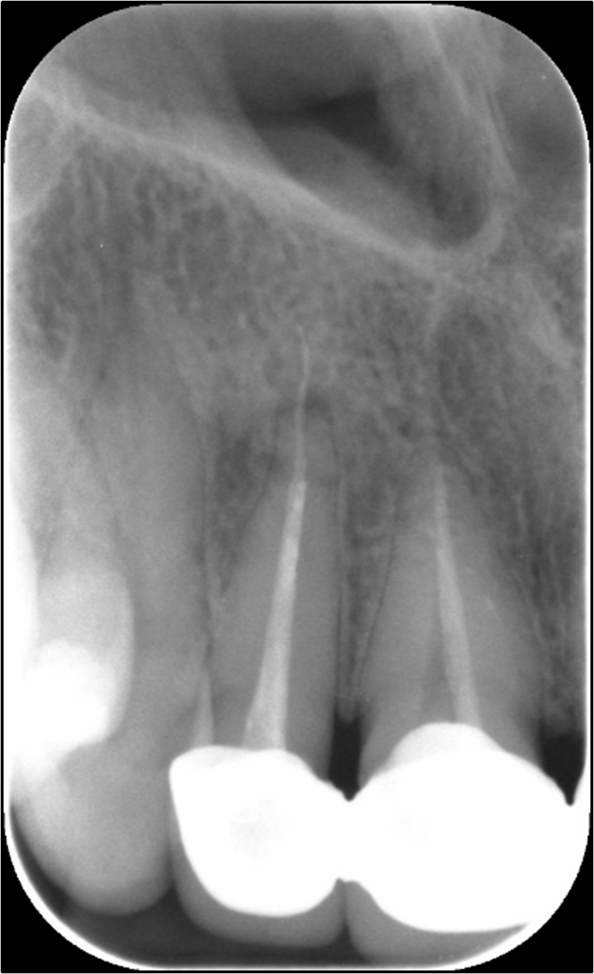
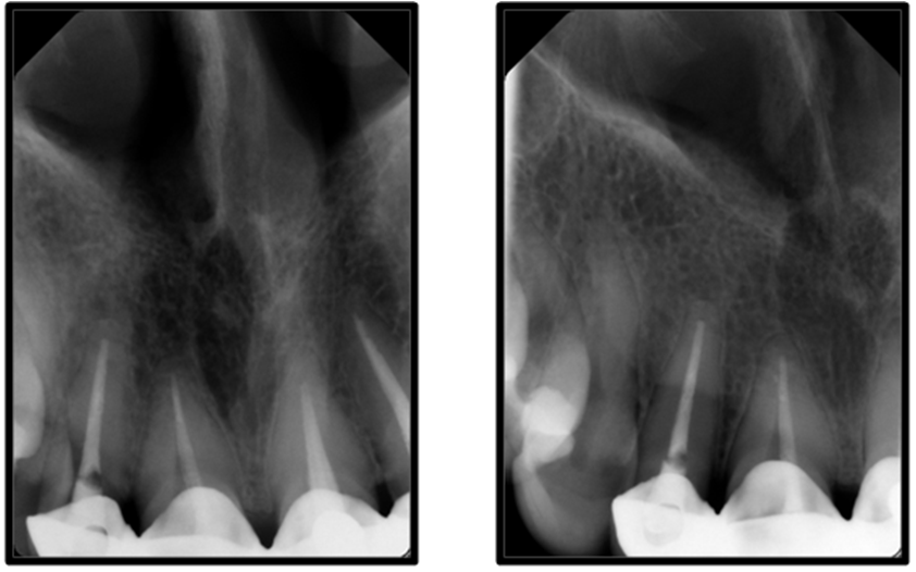
A 40-year-old female was referred to the endodontic department regarding endodontic retreatment of the maxillary lateral right incisor. The medical history was noncontributory and clinical examination showed pain on palpation and tenderness to percussion. The patient was in pain and had done root canal treatment a year earlier ago. She has done crowns after root canal treatments except for maxillary lateral right incisor, which she had done before the root canal treatment. At the time of root canal treatment, the pain was severe but has ceased since then. The patient also describes the former treatment has been done without a rubber dam, and she reported that every visit during previous treatment, she feels pain. A radiograph examination of the tooth showed poor obturation, with overextended gutta percha and slight apical bone destruction (Figure 1). A complete history and meticulous clinical and radiographic examinations confirmed the diagnosis of root filled tooth, symptomatic apical periodontitis of maxillary lateral right incisor. After discussing the possible treatment options, the optimal choice was considered to be a nonsurgical root canal retreatment. The treatment procedure, risks, and benefits have been thoroughly clarified to the patient. The decision was decided of performing a nonsurgical root canal retreatment.
After accomplishing anesthesia, the tooth was isolated, and a rubber dam was used. The access cavity was prepared using Endo access and Endo-z burs (Dentsply, Maillefer, Baillaigues, Switzerland). The root canal retreatment was started with a combination of reciprocation WaveOne GOLD (Dentsply Sirona, York, Pennsylvania, USA) and K-files (Dentsply Maillefer, Ballaigues, Switzerland) to remove the guttapercha from the canal.
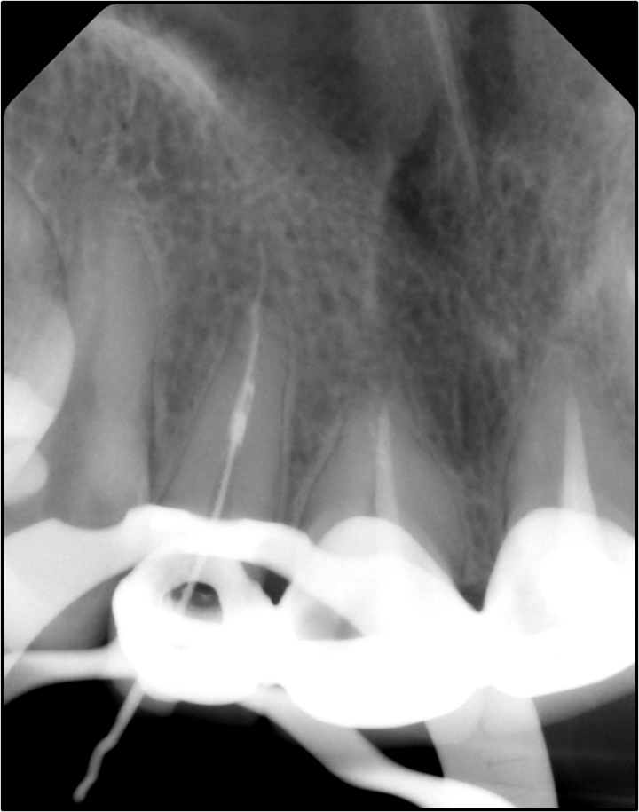
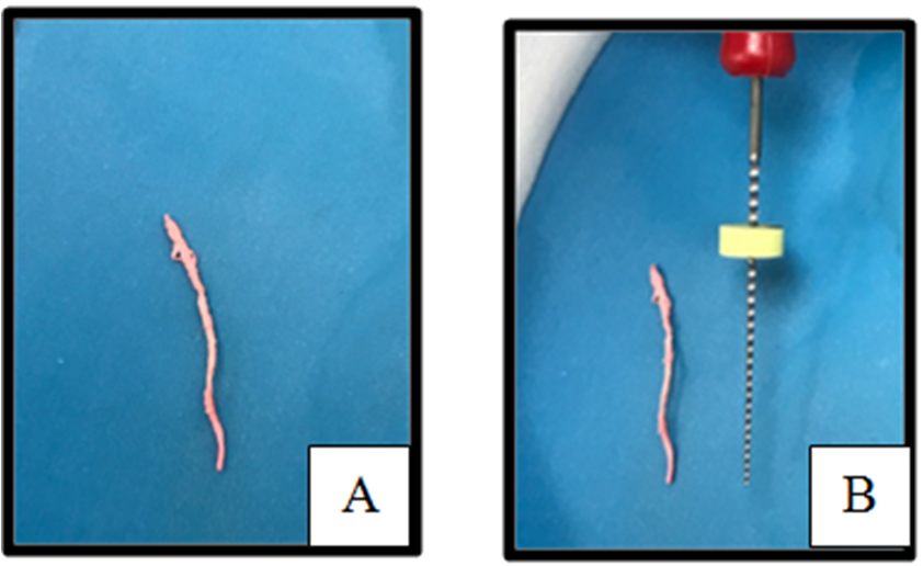
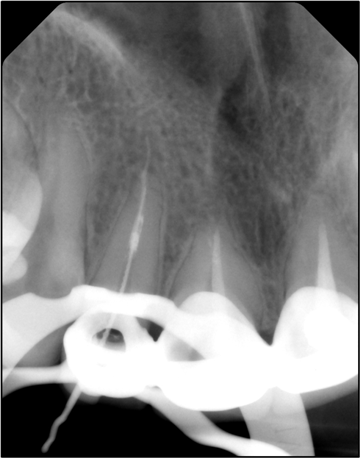
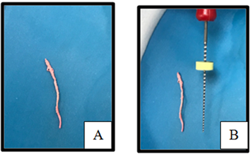
It was perceived that overextended material stayed in the root canal through removing guttapercha, and the radiograph was taken to prove that (Figure 2). Attempts were made to decide if the guttapercha could be removed predictably. The preliminary use of Hedstrom files 10 and 15 disclosed that the instrument could be partly bypassed and that it was loose within the root canal. However, an endodontic explorer (Odous, Belo Horizonte, MG, Brazil) was used to check if this part of the guttapercha could be removed safely. Although, several attempts at removal with hand files and copious irrigation with 1% NaOCl (Septodont, Saint-Maur, France) using a side vented needle (SuYun, Jiangsu, China) solution was unsuccessful. Following this, the apical 3 mm of a 15 K-file was curved. The file was then introduced into the canal, and using careful movements, the guttapercha was pierced and pulled out of the canal (Figure 3A to B).
The gutta percha was removed as one piece (Figure 3A). Based on the radiograph (Figure 2), the working length is determined as 19.5mm. The canal was then cleaned and shaped with WaveOne GOLD 45 to optimal working length and irrigated using 1% NaOCl (Septodont, Saint-Maur, France). The final rinse was made using 17% EDTA (META, Chungbuk, ROK), and the canal was dried using sterile paper points (Dentsply, Maillefer, Baillaigues, Switzerland).
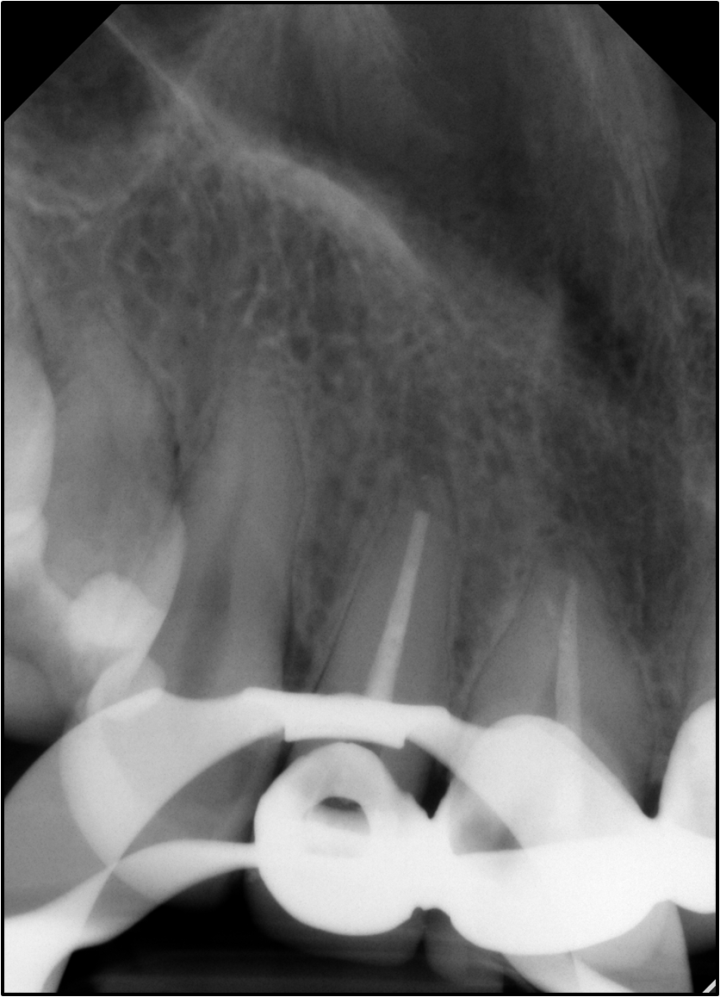
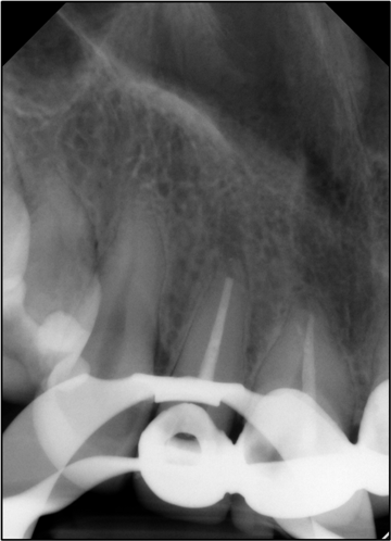
Obturation was done using a warm vertical compaction technique (Figure 4) and matching cone WaveOne GOLD 45. Resin based AH Plus sealer (Dentsply, Maillefer, Baillaigues, Switzerland) was used. A temporary restoration was placed, and the patient was scheduled for permanent restoration after two weeks.
The patient came to the clinic for the 24-follow-up examination and was asymptomatic. The tooth was not sensitive to percussion or palpation. The final permanent composite restoration was placed. Periapical radiographs showed apical tissue was healed (Figure 5).


A case report was addressed in the present work, making it more difficult to remove the apical portion of guttapercha after performing the chemomechanical instrumentation. In addition, a part of guttapercha in the periradicular tissues was identified to be loose. However, guttapercha is an inert material which does not lead to tissue damage 1. It is documented that success rates in guttapercha overfilling are lower than in cases where this material is limited to the root canal system 1, 4, 5, 6. An extended clinical follow-up study was concluded that the highest success rate was achieved when obturation ended 1 mm short of the radiographic apex 5. Another study also showed that the best prognosis was founded for root canals in which the filling material reached within 0 to 2 mm from the radiographic apex 6. For this reason, it was decided to try the removal of extruded guttapercha.
Most previously explained, removing root filling techniques, such as heated pluggers, ultrasonics, rotary files, and solvents, is not safe for removing extrusion guttapercha 7, 8. Compared to the above techniques, the proposed approach is a conservative technique that does not require dentine removal. It is quick and straightforward to perform, does not require direct vision or straight-line access, and can be easily performed.
This conservative approach is a predictable and plausible choice for removing extrusion of guttapercha material in the periapical area. Such information may also add to further endodontic treatments increasing the success in endodontic treatment.
| [1] | Swartz, D.B., A.E. Skidmore, and J.A. Griffin, Jr., Twenty years of endodontic success and failure. J Endod, 1983. 9(5): p. 198-202. | ||
| In article | View Article | ||
| [2] | Bergenholtz, G., et al., Influence of apical overinstrumentation and overfilling on re-treated root canals. J Endod, 1979. 5(10): p. 310-4. | ||
| In article | View Article | ||
| [3] | Seltzer, S., W. Soltanoff, and J. Smith, Biologic aspects of endodontics. V. Periapical tissue reactions to root canal instrumentation beyond the apex and root canal fillings short of and beyond the apex. Oral Surg Oral Med Oral Pathol, 1973. 36(5): p. 725-37. | ||
| In article | View Article | ||
| [4] | Seltzer, S., et al., Biologic aspects of endodontics part III. periapical tissue reactions to root canal instrumentation. 1968. J Endod, 2004. 30(7): p. 491-9; discussion 489-90. | ||
| In article | View Article PubMed | ||
| [5] | Souza, R.A., et al., Apical limit of root canal filling and its relationship with success on endodontic treatment of a mandibular molar: 11-year follow-up. Oral Surg Oral Med Oral Pathol Oral Radiol Endod, 2011. 112(1): p. e48-50. | ||
| In article | View Article PubMed | ||
| [6] | Sjogren, U., et al., Factors affecting the long-term results of endodontic treatment. J Endod, 1990. 16(10): p. 498-504. | ||
| In article | View Article | ||
| [7] | Takahashi, C.M., et al., In vitro evaluation of the effectiveness of ProTaper universal rotary retreatment system for guttapercha removal with or without a solvent. J Endod, 2009. 35(11): p. 1580-3. | ||
| In article | View Article PubMed | ||
| [8] | Schirrmeister, J.F., et al., Efficacy of different rotary instruments for guttapercha removal in root canal retreatment. J Endod, 2006. 32(5): p. 469-72. | ||
| In article | View Article PubMed | ||
Published with license by Science and Education Publishing, Copyright © 2021 Nawaf Almutairi
 This work is licensed under a Creative Commons Attribution 4.0 International License. To view a copy of this license, visit
http://creativecommons.org/licenses/by/4.0/
This work is licensed under a Creative Commons Attribution 4.0 International License. To view a copy of this license, visit
http://creativecommons.org/licenses/by/4.0/
| [1] | Swartz, D.B., A.E. Skidmore, and J.A. Griffin, Jr., Twenty years of endodontic success and failure. J Endod, 1983. 9(5): p. 198-202. | ||
| In article | View Article | ||
| [2] | Bergenholtz, G., et al., Influence of apical overinstrumentation and overfilling on re-treated root canals. J Endod, 1979. 5(10): p. 310-4. | ||
| In article | View Article | ||
| [3] | Seltzer, S., W. Soltanoff, and J. Smith, Biologic aspects of endodontics. V. Periapical tissue reactions to root canal instrumentation beyond the apex and root canal fillings short of and beyond the apex. Oral Surg Oral Med Oral Pathol, 1973. 36(5): p. 725-37. | ||
| In article | View Article | ||
| [4] | Seltzer, S., et al., Biologic aspects of endodontics part III. periapical tissue reactions to root canal instrumentation. 1968. J Endod, 2004. 30(7): p. 491-9; discussion 489-90. | ||
| In article | View Article PubMed | ||
| [5] | Souza, R.A., et al., Apical limit of root canal filling and its relationship with success on endodontic treatment of a mandibular molar: 11-year follow-up. Oral Surg Oral Med Oral Pathol Oral Radiol Endod, 2011. 112(1): p. e48-50. | ||
| In article | View Article PubMed | ||
| [6] | Sjogren, U., et al., Factors affecting the long-term results of endodontic treatment. J Endod, 1990. 16(10): p. 498-504. | ||
| In article | View Article | ||
| [7] | Takahashi, C.M., et al., In vitro evaluation of the effectiveness of ProTaper universal rotary retreatment system for guttapercha removal with or without a solvent. J Endod, 2009. 35(11): p. 1580-3. | ||
| In article | View Article PubMed | ||
| [8] | Schirrmeister, J.F., et al., Efficacy of different rotary instruments for guttapercha removal in root canal retreatment. J Endod, 2006. 32(5): p. 469-72. | ||
| In article | View Article PubMed | ||