The present study was aimed to investigate the anti-diabetic activity of Anemarrhena asphodeloides Bge. Polysaccharides (AAP). Wistar rats were injected with streptozotocin(STZ)to induce diabetes. The APP was prepared at first. After the rats were orally administered for 28 days, related biochemical indicator were determined. AAP was composed of mannose, galacturonic acid, galactose, glucose and xylose in the molar ratio of 12:8.1:2.7:1.3:1 with the average molecular weight of 52.4 kDa. After treatment with AAP (100 and 200 mg·kg-1) for 28 days there was a significant decrease in blood glucose, SGPT, SGOT, ALP, TC, TG, LDL, BUN and significant increase in body weight, fasting serum insulin, pancreatic insulin and HDL. The activities of SOD and CAT in liver and kidneys also increased. The results of the experiments showed that AAP exerted significant anti-diabetic activity.
Diabetes mellitus is a complicated metabolic disease which is associated with abnormalities in protein, fat and glucose metabolism resulting from the defects in insulin secretion, insulin action or both 1. Now it is considered as a world-wide health problem at present leading to cardiovascular disease complications, like retinopathy, nephropathy, stroke, heart attack, etc 2. It is estimated that about 366 million people are likely to be diabetic in 2030 with the prevalence being higher in developed countries than in developing countries 3. The currently synthesized anti-diabetic agents, including sulfonylureas, biguanides and thiazolidinediones, were proven to have side effects and their own other limitations, like high prices, contraindications, etc. Thus, natural plants are constantly being explored with the hope of developing relatively safe antidiabetic traditional folk medicine alone or in combination with other reagents 4. Recent researches showed that plant polysaccharides were proved to have many bioactivities including antioxidant, hypoglycemic and hypolipidemic activities 5. In recent years, the anti-diabetic activity of plant polysaccharides has attracted considerable attention.
Anemarrhena asphodeloides Bge. (family Liliaceae) is mainly distributed in China, Mongolia, Japan and other eastern Asian countries and has been commonly used in traditional medicine in China, Korea and Japan for thousands of years 6. In Traditional Chinese Medicine, Anemarrhena asphodeloides Bge. has been traditionally used to treat diabetes, febrile diseases, lung heat with dry cough and constipation. Pharmacological studies showed that Anemarrhena asphodeloides Bge. possesses anti-platelet aggregation 7, anti-neoplastic 8, antianaphylaxis 9 and anti-dementia activities 10. However, few studies have examined the therapeutic effects of Anemarrhena asphodeloides Bge. Polysaccharides (AAP) on diabetes. The present study was undertaken to evaluate the anti-diabetic, antioxidant and antilipidemic effect of AAP in streptozotocin(STZ) induced diabetes rats.
The rhizomes of Anemarrhena asphodeloides Bge. was collected during the month of October, 2018, from Heibai province of China and identified by Dr. Gao Peng-fei from Dali University.
The assay kits for insulin, total triglyceride (TG), total cholesterol (TC), high density lipoprotein cholesterol (HDL), low density lipoprotein cholesterol(LDL), alkaline phosphatase (ALP), serum glutamic pyruvic transaminase (SGPT), serum glutamic oxaloacetic transaminase(SGOT), superoxide dismutase (SOD), catalase (CAT), and blood urea nitrogen (BUN) were purchased from Jiancheng Bioengineering Institute, Nanjing, Jiangsu Province, China.
2.2. Polysaccharides PreparationThe powdered Anemarrhena asphodeloides Bge. was extracted with 95% ethanol under reflux for 2 h to remove lipophilic and low molecular weight compounds. This step was repeated three times. After filtration, the residue was sun-dried, and then extracted twice with drinking water (mass ratio Anemarrhena asphodeloides Bge.:water was 1:7) refluxing for 2h. The extracting solution was condensed under reduced pressure using rotary evaporator, then the concentrated solution was deproteinated by Sevag method. The water layer was added with 8 volumes of 90% (v/v) ethanol to precipitate the polysaccharides at 4°C for 24 h, and then separated by centrifugation. The crude polysaccharides were collected, washed successively with acetone and ethanol to remove lipids completely, then dissolved in distilled water. The insoluble substances are removed by centrifugation. The supernatant was loaded to a DEAE-52 cellulose column (2.6 cm×40 cm) and eluted with 3 bed volumes distilled water followed by 0.4 mol·L-1 NaCl solution. The eluent was collected, concentrated, dialyzed, and lyophilized for further purification by a Sephacryl S-400 column (5 cm × 50 cm). The polysaccharides (AAP) were collected and lyophilized. AAP was used for further study. The extracting rate of polysaccharide was 2.3%. The content of determination in AAP was 93.1% as determined by phenol-sulfuric acid method, which is mainly comprised of mannose, galacturonic acid, galactose, glucose and xylose in the molar ratio of 12:8.1:2.7:1.3:1 with the average molecular weight of 52.4 kDa. AAP was dissolved in distilled water when administration.
2.3. Animals and TreatmentMale albino Wistar rats, weighing 150-175 g, were maintained in a 12 h day/night cycle (lights on at 06:00 A.M.) at constant room temperature (22-23°C) and humidity (55%±5%) with free access to water and standard laboratory chow. Animals were acclimatized the laboratory conditions for 7 days before starting the experiment. The experiments were conducted in accordance with the P. R. China legislation on the use and care of laboratory animals and US guidelines (NIH publication no. 85-23, revised in 1985) for laboratory animal use and care. Overnight fasted animals were intraperitoneally injected with streptozotocin (STZ) at a dose of 47 mg·kg-1 body weight 11. After 72 h of induction, animals with fasting blood glucose (FBG) levels above 11.1 mmol·L−1 were used to categorize as diabetic animals. Diabetic animals were randomly divided into five groups comprising 12 animals in each group and treated as follows: Group I (Diabetic control), Diabetic rats were treated with drinking water alone; Group II, Diabetic rats were treated with AAP (50 mg·kg-1 b.w.); Group III, Diabetic rats were treated with AAP (100 mg·kg-1 b.w.); Group IV, Diabetic rats were treated with AAP (200 mg·kg-1 b.w.); Group V, Diabetic rats were treated with glibenclamide (1 mg·kg-1 b.w.). Another 12 healthy rats were treated with drinking water as Group VI (Normal control). Treatment was given between 9.00-13.00 hours orally using an intragastric tube once daily for 28 days. Day 0 was designated as the day when rats were confirmed to be diabetic.
2.4. Determination of Fasting Blood Glucose (FBG)Tail vein blood samples of rats after fasting overnight were estimated for fasting blood glucose by glucometer. FBG levels were measured on days 0, 7, 14, 21 and 28 after treatment.
2.5. Determination of Body WeightsThe body weights were monitored on days 0, 7, 14, 21, 28 after treatment and before induction of diabetes.
2.6. Biochemical AnalysisOn the 28th day, blood samples were collected through cardiac puncture and allowed to coagulate at ambient temperature for 30 min. Serum was obtained by centrifugation at 4000 rpm. for 5 min and stored at -20°C, for the analysis of insulin, TG, TC, HDL, LDL, SGPT, SGOT, ALP, and BUN. Assorted biochemical parameters were measured using commercial kits. The liver and kidneys homogenate, prepared in chilled phosphate buffer solution of pH 7.0, was used to estimation the levels and activities of CAT 12 and SOD 13. Pancreas homogenate, prepared in ice-cold acid-alcohol, was stored at -20°C overnight, and finally centrifuged at 4000 rpm at 4°C for 5 minutes. The supernatant was stored at -20 °C for the determination of insulin in the pancreas.
2.7. Statistical AnalysisData were presented as the mean±standard deviation (SD). Statistical analysis of all the data obtained was evaluated using one-way ANOVA followed by Student’s t-test (SPSS 17.0). The values were considered to be significant when P≤0.05.
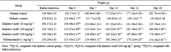
Animals of the same weight range were used in experimental protocol. During the study, the body weight of normal control group was increased naturally, whereas body weight was found to be markedly attenuated in STZ-induced diabetic control group as compared to normal control. After 28 days of treatment with AAP at 100 and 200 mg·kg-1, the body weights were significantly increased by 6.68% and 8.58%, respectively when compared to day 0. The normal control group and glibenclamide group rats showed 13.89% and 12.27% increasing in body weights. The body weight in diabetic+AAP (200 mg·kg-1) group was significantly greater than that of diabetic+AAP (100 mg·kg-1) group at the end of the experimental period but it was markedly lower than that of normal control group or glibenclamide group (Table 1). The effect of AAP administration produced dose-independent effect on body weight in diabetic rats.
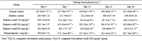
The fasting blood glucose (FBG) of normal control group over the course of 28 days ranged from 103.63±3.75 to 108.48±8.12 g·L-1 and was not notably different from one another. Diabetic control group showed notable increase in the levels of FBG when compared to normal control group. After treatment with AAP at 100 and 200 mg·kg-1 FBG was markedly reduced compared to diabetic control rats. FBG in 100 and 200 mg·kg-1 groups was decreased by 17.8% and 25.7% at the end of experiment. Glibenclamide group also showed significant reduction in FBG to 203.78 g·L-1 (by 29.8%). Diabetic+AAP (200 mg·kg-1) group’s FBG was significantly less than that in diabetic+AAP (100 mg·kg-1) group but it was notably greater than that in normal control group and glibenclamide group from the 7th day to 28th day. AAP showed less potency for antihyperglycemic activity than glibenclamide (Table 2).
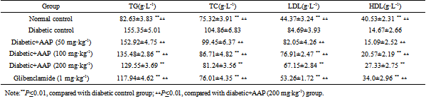
3.3. Effect of AAP on Lipid ProfileTo evaluate the effect of AAP on lipid profile level, TG, TC, LDL and HDL, they were assessed at the end of experiment. There was a significant increase in TC, TG and LDL levels in diabetic control group when compared to normal control group, but the level of HDL significantly decreased. Diabetic control group showed an increase of 88.0% in TG, 39.2% in TC, 90.9% in LDL and a 63.8% decrease in HDL. Diabetic+AAP (100 mg·kg-1), diabetic+AAP (200 mg·kg-1) and glibenclamide groups were significantly different from diabetic control. At the end of treatment diabetic+AAP (200 mg·kg-1) group showed a decrease of 16.6% in TG, 22.5% in TC, 20.7% in LDL and an 86.3% increase in HDL. The levels of TC, TG and LDL in diabetic+AAP (200 mg·kg-1) group were still greater than that of the glibenclamide group, but the level of HDL was less (Table 3).
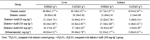
3.4. Effect of AAP on SOD and CATIn diabetic control group a significant decreased activity of SOD and CAT was observed. Liver’s and kidneys’ SOD activity in diabetic control group showed notable decrease by 39.1% and by 39.4%, respectively and liver’s and kidneys’ CAT activity decreased by 23.8 % and by 47.9%, respectively. Diabetic+AAP (100 mg·kg-1), diabetic+AAP (200 mg·kg-1) and glibenclamide groups restored the activity of SOD and CAT than that of diabetic control group markedly. The activity of SOD and CAT in diabetic+AAP (200 mg·kg-1) group was significantly different from that of glibenclamide group and normal control group. After treatment diabetic+AAP(200 mg·kg-1) group showed an increase of 43.0% in liver’s SOD, 40.7% in kidneys’ SOD, 21.5% in liver’s CAT and 60.8% in kidneys’ CAT as compared to diabetic control group. Glibenclamide group also showed significant increase in liver’s SOD, kidneys’ SOD, liver’s CAT, kidneys’ CAT, that is, 58.0%, 54.1%, 29.1%, and 79.3%, respectively. AAP showed less potency for restoring SOD and CAT activity than glibenclamide (Table 4).
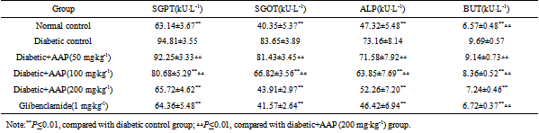
At the end of experiment diabetic control group shown significant increase in the levels of SGPT, SGOT, ALP, and BUT as compared to normal group, that is 50.2%, 107.3%, 54.6%, and 47.5%, respectively. After treatment with 100 and 200 mg·kg-1 of AAP and glibenclamide there was a significant decrease in the activities of SGPT, SGOT and ALP. The content of BUT was also significantly decreased. Diabetic+AAP (200 mg·kg-1) group shown significant attenuation in SGPT, SGOT, ALP, and BUT as compared to diabetic group, that is 30.7%, 47.5%, 28.6%, and 25.3%, respectively, and the levels of SGPT, SGOT, ALP were normalised to that of normal control group’s levels. Glibenclamide group showed a decrease of 32.1% in SGPT, 50.3% in SGOT, 36.6% in ALP, and 30.6% in BUT. There were no significant differences in the levels of SGPT, SGOT, ALP between glibenclamide group and diabetic+AAP (200 mg·kg-1) group (Table 5).
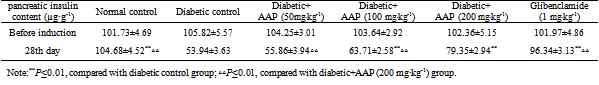
3.6. Effect of AAP on Pancreatic InsulinPancreatic insulin level significantly decreased by 48.5% in diabetic control group as compared to normal control group. After 28 days of treatment, with 100 and 200 mg·kg-1 of AAP and glibenclamide shown significant increase in pancreatic insulin level as compared to diabetic control group, that is, 18.1%, 47.1%, and 78.6%, respectively. There was significant difference between diabetic+AAP (200 mg·kg-1) group and glibenclamide group (Table 6).
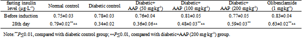
The variation tendency of fasting insulin level was similar to pancreatic insulin. Fasting insulin level was notably decreased (by 57.0%) in diabetic control group as compared to normal control group. On the 28th day, fasting insulin levels in 100 and 200 mg·kg-1 of AAP and glibenclamide groups were significantly increased by 41.2%, 73.5%, and 85.3%, respectively. Fasting insulin level in diabetic+AAP (200 mg·kg-1) group was still significant less than that of glibenclamide group (Table 7).
This present study was designed to investigate the anti-diabetic, antioxidant and antilipidemic activities of AAP in STZ-induced diabetic rats. STZ is commonly used to induce type-I diabetes mellitus in experimental rat models. STZ has potent alkylating property 14 and specifically cytotoxic to the pancreatic β-cell in mammals. Pancreatic islet β-cells preferentially uptake STZ resulting in the destruction of β-cells by necrosis mediated through the release of nitric oxide (NO) liberated from STZ 15.
STZ-induced diabetes is characterized by a notable loss in body weight, which is due to the degradation of structural protein or increased muscle destruction 16. Similar effect was also observed in the current study. When diabetic rats were treated with AAP (100, 200 mg·kg-1), they showed a significant increase in body weight as compared to diabetic control group but the increase was not as much as that in glibenclamide treated group. The ability to protect body weight loss signifies its protective effect in inhibiting the degradation of structural protein and muscle destruction by reduce hyperglycemia.
Lipid plays an important role in the pathogenesis of diabetic complications. The reduced level of serum HDL cholesterol and elevated level of serum cholesterol, poses to be a risk factor for developing cardiovascular disease like coronary heart disease. The abnormal high level of serum lipid mainly due to increased mobilization of peripheral free fatty acids. Insulin deficiency, insulin resistance or insulin inhibits the hormone sensitive lipase may be the pathogenesis 17. This present study showed that diabetic rats has abnormal lipid profile as earlier report 18, whereas the AAP treated groups (100, 200 mg·kg-1) showed significant improvement in the lipid level comparable diabetic control group but the improvement was inferior to glibenclamide treated group. After 28 days treatment, AAP reduced TG, TC, LDL and increased HDL. Thus, AAP could have a potential to reduce cardiovascular complications associated with diabetes.
Pervious study indicated that the antioxidant treatment could provide protection to pancreatic islet β-cells against glucose toxicity in diabetic mice and was beneficial for treating diabetes. SOD and CAT are enzymatic antioxidants. SOD is capable of reducing the superoxide radical into hydrogen peroxide (H2O2). CAT catalyzes the breakdown of H2O2 and protects the tissues from the damage caused by reactive hydroxyl radicals 19. In diabetes mellitus, high glucose can decrease the activity of SOD and CAT by glycating these proteins thus inducing oxidative stress, which in turn, causes lipid peroxidation 20. The SOD and CAT activities in liver and kidneys of diabetic rats were remarkably increased. So AAP as a antioxygen may directly or indirectly protect pancreatic islet β-cells in the duration of high blood glucose. This was evidenced by increased fasting insulin and pancreatic insulin levels after treatment with AAP.
SGPT, SGOT and ALP are cellular enzymes which are reliable markers indicating the presence of injury in the liver. When cellular injury happens, the enzymes are released from the cytosol into the blood stream, suggesting liver damage 21. There was increase in liver enzymes in serum of diabetic rats like previous report 22. AAP (100 200 mg·kg-1) significantly reduced the serum SGPT, SGOT and ALP levels which showed the protective effect of liver functioning in reversing the organ damage due to diabetes that was clearly observed by elevated levels of SGPT, SGOT and ALP in diabetic control group.
The results of the experiments showed that AAP exerted significant anti-diabetic, antioxidant and antilipidemic activities in STZ-induced diabetic rats which provides scientific proof for the use in traditional medicine.
| [1] | Malchoff C D. Diagnosis and Classification of Diabetes Mellitus[J]. Diabetes Care, 1991, 11:625-629. | ||
| In article | |||
| [2] | Uma A, Ahmed Q U, Muhammad B Y, et al. Antihyperglycemic activity of the leaves of Tetracera scandens Linn. Merr. (Dilleniaceae) in alloxan induced diabeticrats[J]. Journal of Ethnopharmacology, 2010, 131(1): 140-145. | ||
| In article | View Article PubMed | ||
| [3] | Rathmann W, Giani G. Global prevalence of diabetes: estimates for the year 2000 and projections for 2030[J]. Diabetes Care, 2004, 27(5): 1047-1053. | ||
| In article | View Article PubMed | ||
| [4] | Wondmagegn T, Ephrem E, Kaleab A. Evaluation of the effects of 80% methanolic leaf extract ofCaylusea abyssinica (fresen.)fisch. & Mey. on glucose handling in normal, glucose loaded and diabetic rodents[J]. BMC Complementary and Alternative Medicine, 2012, 12(1): 151-151. | ||
| In article | View Article PubMed | ||
| [5] | Jia X, Yang J, Wang Z, et al. Polysaccharides from Laminaria japonica show hypoglycemic and hypolipidemic activities in mice with experimentally induced diabetes[J]. Experimental Biology and Medicine, 2014, 239(12): 1663-1670. | ||
| In article | View Article PubMed | ||
| [6] | Tang Z S, Li G L, Yang J, et al. Anemarrhena asphodeloides Non-Steroidal Saponin Components Alter the Pharmacokinetic Profile of Its Steroidal Saponins in Rat[J]. Molecules, 2015, 20(7): 11777-11792. | ||
| In article | View Article PubMed | ||
| [7] | Akihiko T, Mutsumi T, Junich K. Spirostanols Obtained by Cyclization of Pseudosaponin Derivatives and Comparison of Anti-Platelet Agglutination Activities of Spirostanol Glycosides[J]. Eur J Med Chem, European Journal of Medicinal Chemistry, 2000, 35(5): 511-527. | ||
| In article | View Article | ||
| [8] | Kang Y J, Chung H J, Nam J W, et al. Cytotoxic and antineoplastic activity of timosaponin A-III for human colon cancer cells[J]. Journal of Natural Products, 2011, 74(4): 701-706. | ||
| In article | View Article PubMed | ||
| [9] | Lee B, Trinh H T, Jung K, et al. Inhibitory effects of steroidal timosaponins isolated from the rhizomes of Anemarrhena asphodeloides against passive cutaneous anaphylaxis reaction and pruritus[J]. Immunopharmacol Immunotoxicol, 2010, 32(3): 357-363. | ||
| In article | View Article PubMed | ||
| [10] | Deng Y, Ma B P, Xu Q P, et al. Effect and mechanism of effective component in Zhimu on ability of learning and memory in vascular dementia rats[J]. Chinese Pharmacological Bulletin, 2005, 21(7): 830-833. | ||
| In article | |||
| [11] | Sharma I, Aaradhya M, Kodikonda M, et al. Antihyperglycemic, antihyperlipidemic and antioxidant activity of phenolic rich extract of Brassica oleraceae var gongylodes on streptozotocin induced Wistar rats[J]. SpringerPlus, 2015, 4(1): 212. | ||
| In article | View Article PubMed | ||
| [12] | Sinha A K. Colorimetric assay of catalase[J]. Analytical Biochemistry, 1972, 47(2): 389-394. | ||
| In article | View Article | ||
| [13] | Kakkar P, Das B, Viswanathan P N. A modified spectrophotometric assay of superoxide dismutase[J]. Indian Journal of Biochemistry and Biophysics, 1984, 21(2): 130-132. | ||
| In article | |||
| [14] | Szkudelski T. The mechanism of alloxan and streptozotocin action in B cells of the rat pancreas[J]. Physiological Research, 2001, 50(6): 537-546. | ||
| In article | |||
| [15] | Prasath G S, Subramanian S. Fisetin. A bioflavonoid ameliorates hyperglycemia in STZ-induced experimental diabetes in rats[J]. International Journal of Pharmaceutical Sciences Review and Research, 2011, 6(1): 68-74. | ||
| In article | |||
| [16] | Salahuddin M, Jalalpure S S. Antidiabetic activity of aqueous fruit extract of Cucumis trigonus, Roxb. in streptozotocin-induced-diabetic rats[J]. Journal of Ethnopharmacology, 2010, 127(2): 565. | ||
| In article | View Article PubMed | ||
| [17] | Singh N S, Geetha M, Amudha P, et al. Evaluation of anti-diabetic activity of methanol extract of Flacourtia jangomas (Lour) in streptozotocin induced diabetic rats[J]. International Journal of Pharma and Bio Sciences, 2010, 20(8): 1105-1114. | ||
| In article | |||
| [18] | Sunila C, Ignacimuthua S, Agastianb P. Antidiabetic effect of Symplocos cochinchinensis (Lour.) S. Moore. in type 2 diabetic rats[J]. J Ethnopharmacol, 2011, 134(2): 298-304. | ||
| In article | View Article PubMed | ||
| [19] | Eliza J, Daisy P, Ignacimuthu S. Antioxidant activity of costunolide and eremanthin isolated from Costus speciosus (Koen ex. Retz) Sm[J]. Chemico-Biological Interactions, 2010, 188(3): 467-472. | ||
| In article | View Article PubMed | ||
| [20] | Kennedy A L, Lyons T J. Glycation, oxidation, and lipoxidation in the development of diabetic complications[J]. Metabolism-clinical and Experimental, 1997, 46(1): 14-21. | ||
| In article | View Article | ||
| [21] | Ozer J, Ratner M, Shaw M, et al. The current state of serum biomarkers of hepatotoxicity[J]. Toxicology, 2008, 245(3): 194-205. | ||
| In article | View Article PubMed | ||
| [22] | Stephen I S, Sunil C, Duraipandiyan V, et al. Antidiabetic and antioxidant activities of Toddalia asiatica (L.) Lam. leaves in streptozotocin induced diabetic rats[J]. Journal of Ethnopharmacology, 2012, 143(2): 515-523. | ||
| In article | View Article PubMed | ||
Published with license by Science and Education Publishing, Copyright © 2021 YIN Ai-wu and GAO Peng-fei
![]() This work is licensed under a Creative Commons Attribution 4.0 International License. To view a copy of this license, visit
http://creativecommons.org/licenses/by/4.0/
This work is licensed under a Creative Commons Attribution 4.0 International License. To view a copy of this license, visit
http://creativecommons.org/licenses/by/4.0/
| [1] | Malchoff C D. Diagnosis and Classification of Diabetes Mellitus[J]. Diabetes Care, 1991, 11:625-629. | ||
| In article | |||
| [2] | Uma A, Ahmed Q U, Muhammad B Y, et al. Antihyperglycemic activity of the leaves of Tetracera scandens Linn. Merr. (Dilleniaceae) in alloxan induced diabeticrats[J]. Journal of Ethnopharmacology, 2010, 131(1): 140-145. | ||
| In article | View Article PubMed | ||
| [3] | Rathmann W, Giani G. Global prevalence of diabetes: estimates for the year 2000 and projections for 2030[J]. Diabetes Care, 2004, 27(5): 1047-1053. | ||
| In article | View Article PubMed | ||
| [4] | Wondmagegn T, Ephrem E, Kaleab A. Evaluation of the effects of 80% methanolic leaf extract ofCaylusea abyssinica (fresen.)fisch. & Mey. on glucose handling in normal, glucose loaded and diabetic rodents[J]. BMC Complementary and Alternative Medicine, 2012, 12(1): 151-151. | ||
| In article | View Article PubMed | ||
| [5] | Jia X, Yang J, Wang Z, et al. Polysaccharides from Laminaria japonica show hypoglycemic and hypolipidemic activities in mice with experimentally induced diabetes[J]. Experimental Biology and Medicine, 2014, 239(12): 1663-1670. | ||
| In article | View Article PubMed | ||
| [6] | Tang Z S, Li G L, Yang J, et al. Anemarrhena asphodeloides Non-Steroidal Saponin Components Alter the Pharmacokinetic Profile of Its Steroidal Saponins in Rat[J]. Molecules, 2015, 20(7): 11777-11792. | ||
| In article | View Article PubMed | ||
| [7] | Akihiko T, Mutsumi T, Junich K. Spirostanols Obtained by Cyclization of Pseudosaponin Derivatives and Comparison of Anti-Platelet Agglutination Activities of Spirostanol Glycosides[J]. Eur J Med Chem, European Journal of Medicinal Chemistry, 2000, 35(5): 511-527. | ||
| In article | View Article | ||
| [8] | Kang Y J, Chung H J, Nam J W, et al. Cytotoxic and antineoplastic activity of timosaponin A-III for human colon cancer cells[J]. Journal of Natural Products, 2011, 74(4): 701-706. | ||
| In article | View Article PubMed | ||
| [9] | Lee B, Trinh H T, Jung K, et al. Inhibitory effects of steroidal timosaponins isolated from the rhizomes of Anemarrhena asphodeloides against passive cutaneous anaphylaxis reaction and pruritus[J]. Immunopharmacol Immunotoxicol, 2010, 32(3): 357-363. | ||
| In article | View Article PubMed | ||
| [10] | Deng Y, Ma B P, Xu Q P, et al. Effect and mechanism of effective component in Zhimu on ability of learning and memory in vascular dementia rats[J]. Chinese Pharmacological Bulletin, 2005, 21(7): 830-833. | ||
| In article | |||
| [11] | Sharma I, Aaradhya M, Kodikonda M, et al. Antihyperglycemic, antihyperlipidemic and antioxidant activity of phenolic rich extract of Brassica oleraceae var gongylodes on streptozotocin induced Wistar rats[J]. SpringerPlus, 2015, 4(1): 212. | ||
| In article | View Article PubMed | ||
| [12] | Sinha A K. Colorimetric assay of catalase[J]. Analytical Biochemistry, 1972, 47(2): 389-394. | ||
| In article | View Article | ||
| [13] | Kakkar P, Das B, Viswanathan P N. A modified spectrophotometric assay of superoxide dismutase[J]. Indian Journal of Biochemistry and Biophysics, 1984, 21(2): 130-132. | ||
| In article | |||
| [14] | Szkudelski T. The mechanism of alloxan and streptozotocin action in B cells of the rat pancreas[J]. Physiological Research, 2001, 50(6): 537-546. | ||
| In article | |||
| [15] | Prasath G S, Subramanian S. Fisetin. A bioflavonoid ameliorates hyperglycemia in STZ-induced experimental diabetes in rats[J]. International Journal of Pharmaceutical Sciences Review and Research, 2011, 6(1): 68-74. | ||
| In article | |||
| [16] | Salahuddin M, Jalalpure S S. Antidiabetic activity of aqueous fruit extract of Cucumis trigonus, Roxb. in streptozotocin-induced-diabetic rats[J]. Journal of Ethnopharmacology, 2010, 127(2): 565. | ||
| In article | View Article PubMed | ||
| [17] | Singh N S, Geetha M, Amudha P, et al. Evaluation of anti-diabetic activity of methanol extract of Flacourtia jangomas (Lour) in streptozotocin induced diabetic rats[J]. International Journal of Pharma and Bio Sciences, 2010, 20(8): 1105-1114. | ||
| In article | |||
| [18] | Sunila C, Ignacimuthua S, Agastianb P. Antidiabetic effect of Symplocos cochinchinensis (Lour.) S. Moore. in type 2 diabetic rats[J]. J Ethnopharmacol, 2011, 134(2): 298-304. | ||
| In article | View Article PubMed | ||
| [19] | Eliza J, Daisy P, Ignacimuthu S. Antioxidant activity of costunolide and eremanthin isolated from Costus speciosus (Koen ex. Retz) Sm[J]. Chemico-Biological Interactions, 2010, 188(3): 467-472. | ||
| In article | View Article PubMed | ||
| [20] | Kennedy A L, Lyons T J. Glycation, oxidation, and lipoxidation in the development of diabetic complications[J]. Metabolism-clinical and Experimental, 1997, 46(1): 14-21. | ||
| In article | View Article | ||
| [21] | Ozer J, Ratner M, Shaw M, et al. The current state of serum biomarkers of hepatotoxicity[J]. Toxicology, 2008, 245(3): 194-205. | ||
| In article | View Article PubMed | ||
| [22] | Stephen I S, Sunil C, Duraipandiyan V, et al. Antidiabetic and antioxidant activities of Toddalia asiatica (L.) Lam. leaves in streptozotocin induced diabetic rats[J]. Journal of Ethnopharmacology, 2012, 143(2): 515-523. | ||
| In article | View Article PubMed | ||