Calciphylaxis is a rare condition in which calcification of medium blood vessels in the skin results in the purpura and painful, necrotic skin ulcers. Although most commonly seen in adults with end-stage kidney disease and long-term dialysis use, reports of nonuremic calciphylaxis exist. We present the case of a 29-year old woman with recently diagnosed end-stage liver disease who presented with an acute kidney injury and painful lower extremity purpura and ulcers with biopsy-confirmed calciphylaxis. The patient had been receiving dialysis for less than one month. Treatment with sodium thiosulfate was provided but was unsuccessful. The patient ultimately pursued palliative care.
Calciphylaxis, also known as calcific uremic arteriolopathy (CUA), is a rare clinical syndrome in which purpura and necrotic skin ulcers develop secondary to vascular calcifications. 1, 2 It often presents as poorly demarcated, painful retiform purpura in areas with high fat content such as the trunk, abdomen, or thighs. 3 These lesions eventually transition into ulcers with necrosis. As the name implies, it is most often seen in the context of uremia and chronic renal failure, appearing in approximately 4% of patients with end-stage renal disease (ESRD) on hemodialysis. The classic risk factors associated with the development of calciphylaxis include: elevated levels of calcium, phosphate, and parathyroid hormone (PTH), diabetes, obesity, hypoalbuminemia, female sex, warfarin use, and protein C or S deficiency. 4 Despite the strong association between chronic renal disease and calciphylaxis, cases of non-uremic calciphylaxis have also been reported. Risk factors for non-uremic calciphylaxis include: malignancy, autoimmune disease, diabetes, alcoholic liver disease, glucocorticoid or warfarin use. 5
We present a rare case of calciphylaxis in a 29-year-old Caucasian female that had been recently diagnosed with alcohol-induced end-stage liver disease and an acute kidney injury with dialysis treatments for less than one month.
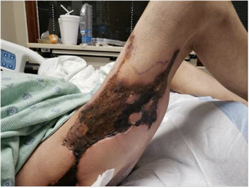
A 29 year-old woman was admitted to complaining of painful lower extremity lesions. Her only past medical history was a four-month history of alcohol-induced end-stage liver disease complicated by refractory ascites and recurrent hepatic encephalopathy. A week prior to her presentation, she had been hospitalized at an outside institution for hepatic encephalopathy and was found to have an acute kidney injury for which she had received two dialysis treatments. Due to the extent of her injury, her kidney function was still abnormal with a creatine of 2.53 (normal range: 0.7-1.3) and a blood urea nitrogen of 23 mg/dL (normal range: 6-23), which prompted our medical team to begin dialysis. Upon physical examination, there were painful, firm, indurated, stellate-shaped, purpuric patches and plaques with focal overlying necrosis on the left upper and posterior thigh and hip and the right thigh (Figure 1 - Figure 3).
Due to concern for calciphylaxis, a skin biopsy and computed tomography (CT) scan of the abdomen, pelvis, and legs was performed. CT scan demonstrated diffuse arterial calcification in the right and left thigh, supporting a diagnosis of calciphylaxis. Initial biopsy was non-diagnostic, but repeat biopsy demonstrated abundant soft tissue calcification including calcification and thrombosis of various small and medium sized blood vessels (Figure 4). Abnormal labs included phosphorus and ionized calcium levels which were low at levels of 2.1 mg/dL (normal range: 2.5 – 4.5 mg/dL) and 1.12 mg/dL (normal range: 4.4 – 5.2 mg/dL) respectively; albumin was low 2.2 g/dL (normal range: 3.4 – 5.4 g/dL), and total protein levels were 4.2 g/dL (normal range: 6.0 – 8.3 g/dL). The PTH level was normal at 48.1pg/ml(normal range: 11– 51 pg/mL). Sodium thiosulfate 25 mg IV was given during dialysis treatments and wound care with daily betadine swabs on the affected areas were performed.
The patient did not have other predisposing factors such as use of warfarin, calcium or vitamin d supplements, presence of phospholipid antibodies, and protein C and S deficiency.
Although 8 sodium thiosulfate treatments were administered during dialysis treatments, there were no improvements in the appearance or symptoms associated with the lesions. Due to pain and comorbidities, patient elected for palliative care and to not pursue further dialysis.


Calciphylaxis is a rare disease with an estimated 1-year survival rate between 20 - 50%. 3 The typical demographic of patients who develop calciphylaxis are older female patients, with the average age of being between 50-70 years old. 7 When examining a cohort of 30 patients with confirmed calciphylaxis, Panchel et al. (2020) found that 73% of patients were female and 90% were Caucasian. 6 Cases of young adults and children with calciphylaxis have been reported but are rare and usually occur in the setting of chronic renal failure such as ESRD. 10, 11
The vast majority of patients diagnosed with this condition have chronic renal disease and are dialysis-dependent. In fact, calciphylaxis has been diagnosed in almost 4% of all dialysis patients. 9 As it is most often associated with patients who have undergone dialysis treatments for more than a year, the presentation of calciphylaxis in our patient who was a younger woman with an acute kidney injury which had required dialysis for less than one month is unusual. 8
To our knowledge, there has only been one reported case of calciphylaxis after an acute kidney injury in addition to alcohol induced cirrhosis however the patient was 40 years old compared to this patient at 29 years old. 12
Additionally, it is important to note that despite high clinical suspicion for calciphylaxis, the initial biopsy performed for this patient was not diagnostic. It is not uncommon for initial biopsies to be negative when evaluating for calciphylaxis. 9, 13 Therefore, if a patient’s presentation suggests calciphylaxis, it is important to continue a work-up by obtaining CT scans or additional biopsies, and providers may even consider initiating sodium thiosulfate while awaiting final results.
We report calciphylaxis in an atypical clinical context of a young woman with end-stage liver disease and acute kidney injury who had only recently started dialysis. As there have been few case reports demonstrating the presence of calciphylaxis patients under the age of 30 without chronic kidney disease, we present this case to raise clinician’s awareness of calciphylaxis in nonuremic and younger patients and promote work-up for these patients with characteristic painful retiform purpura.
| [1] | Coates T, Kirkland GS, Dymock RB, et al. Cutaneous necrosis from calcific uremic arteriolopathy. Am J Kidney Dis. 1998; 32(3): 384-391. | ||
| In article | View Article PubMed | ||
| [2] | Selye H. Calciphylaxis. University of Chicago Press; 1962. | ||
| In article | |||
| [3] | Marques SA, Mendaçolli TJ, Marques MEA, Kakuda AC, Abbade LPF. Calciphylaxis: a rare but potentially fatal event of chronic kidney disease. Case report. An Bras Dermatol. 2013; 88(6 SUPPL.1): 44-47. | ||
| In article | View Article PubMed | ||
| [4] | Nigwekar SU, Kroshinksy D, Nazarian RM, et al. Calciphylaxis: Risk Factors, Diagnosis, and Treatment. | ||
| In article | |||
| [5] | Nigwekar SU, Wolf M, Sterns RH, Hix JK. Calciphylaxis from nonuremic causes: A systematic review. Clin J Am Soc Nephrol. 2008; 3(4): 1139-1143. | ||
| In article | View Article PubMed | ||
| [6] | Panchal S, Holtermann K, Trivedi N, Regunath H, Yerram P. Calciphylaxis: An analysis of concomitant factors, treatment effectiveness and prognosis in 30 patients. Int J Nephrol Renovasc Dis. 2020; 13: 65-71. | ||
| In article | View Article PubMed | ||
| [7] | McCarthy JT, el-Azhary RA, Patzelt MT, et al. Survival, Risk Factors, and Effect of Treatment in 101 Patients With Calciphylaxis. Mayo Clin Proc. 2016; 91(10): 1384-1394. | ||
| In article | View Article PubMed | ||
| [8] | Budisavijevic MN, Cheek D, Ploth DW. Calciphylaxis in Chronic Renal Failure. J Am Soc Nephrol. 1996; 7: 978-982. | ||
| In article | View Article PubMed | ||
| [9] | Fine A, Zacharias J. Calciphylaxis is usually non-ulcerating: Risk factors, outcome and therapy. Kidney Int. 2002; 61(6): 2210-2217. | ||
| In article | View Article PubMed | ||
| [10] | Stârcea M, Gavrilovici C, Elsayed A, et al. A case report of pediatric calciphylaxis-a rare and potentially fatal under diagnosed condition. Med (United States). 2018; 97(27). | ||
| In article | View Article PubMed | ||
| [11] | Timmis A, Morgan H. Calciphylaxis in a paediatric patient. BMJ Case Rep. 2010; 2010: bcr0520102989. | ||
| In article | View Article PubMed | ||
| [12] | Wichienkuer P, Naugler W, Wusirika R. Calciphylaxis in a patient with acute kidney injury and alcoholic cirrhosis. Clin Nephrol. 2011; 76(6): 499-503. | ||
| In article | View Article PubMed | ||
| [13] | Stavros K, Motiwala R, Zhou L, Sejdiu F, Shin S. Calciphylaxis in a dialysis patient diagnosed by muscle biopsy. J Clin Neuromuscul Dis. 2014; 15(3): 108-111. | ||
| In article | View Article PubMed | ||
Published with license by Science and Education Publishing, Copyright © 2021 Rachel Russell, Walid Omer, Nagat Mudabal and Karolyn A. Wanat
![]() This work is licensed under a Creative Commons Attribution 4.0 International License. To view a copy of this license, visit
http://creativecommons.org/licenses/by/4.0/
This work is licensed under a Creative Commons Attribution 4.0 International License. To view a copy of this license, visit
http://creativecommons.org/licenses/by/4.0/
| [1] | Coates T, Kirkland GS, Dymock RB, et al. Cutaneous necrosis from calcific uremic arteriolopathy. Am J Kidney Dis. 1998; 32(3): 384-391. | ||
| In article | View Article PubMed | ||
| [2] | Selye H. Calciphylaxis. University of Chicago Press; 1962. | ||
| In article | |||
| [3] | Marques SA, Mendaçolli TJ, Marques MEA, Kakuda AC, Abbade LPF. Calciphylaxis: a rare but potentially fatal event of chronic kidney disease. Case report. An Bras Dermatol. 2013; 88(6 SUPPL.1): 44-47. | ||
| In article | View Article PubMed | ||
| [4] | Nigwekar SU, Kroshinksy D, Nazarian RM, et al. Calciphylaxis: Risk Factors, Diagnosis, and Treatment. | ||
| In article | |||
| [5] | Nigwekar SU, Wolf M, Sterns RH, Hix JK. Calciphylaxis from nonuremic causes: A systematic review. Clin J Am Soc Nephrol. 2008; 3(4): 1139-1143. | ||
| In article | View Article PubMed | ||
| [6] | Panchal S, Holtermann K, Trivedi N, Regunath H, Yerram P. Calciphylaxis: An analysis of concomitant factors, treatment effectiveness and prognosis in 30 patients. Int J Nephrol Renovasc Dis. 2020; 13: 65-71. | ||
| In article | View Article PubMed | ||
| [7] | McCarthy JT, el-Azhary RA, Patzelt MT, et al. Survival, Risk Factors, and Effect of Treatment in 101 Patients With Calciphylaxis. Mayo Clin Proc. 2016; 91(10): 1384-1394. | ||
| In article | View Article PubMed | ||
| [8] | Budisavijevic MN, Cheek D, Ploth DW. Calciphylaxis in Chronic Renal Failure. J Am Soc Nephrol. 1996; 7: 978-982. | ||
| In article | View Article PubMed | ||
| [9] | Fine A, Zacharias J. Calciphylaxis is usually non-ulcerating: Risk factors, outcome and therapy. Kidney Int. 2002; 61(6): 2210-2217. | ||
| In article | View Article PubMed | ||
| [10] | Stârcea M, Gavrilovici C, Elsayed A, et al. A case report of pediatric calciphylaxis-a rare and potentially fatal under diagnosed condition. Med (United States). 2018; 97(27). | ||
| In article | View Article PubMed | ||
| [11] | Timmis A, Morgan H. Calciphylaxis in a paediatric patient. BMJ Case Rep. 2010; 2010: bcr0520102989. | ||
| In article | View Article PubMed | ||
| [12] | Wichienkuer P, Naugler W, Wusirika R. Calciphylaxis in a patient with acute kidney injury and alcoholic cirrhosis. Clin Nephrol. 2011; 76(6): 499-503. | ||
| In article | View Article PubMed | ||
| [13] | Stavros K, Motiwala R, Zhou L, Sejdiu F, Shin S. Calciphylaxis in a dialysis patient diagnosed by muscle biopsy. J Clin Neuromuscul Dis. 2014; 15(3): 108-111. | ||
| In article | View Article PubMed | ||


