Bioactive restorative materials have transformed restorative dentistry by combining structural restoration with therapeutic functions that promote tissue repair and oral health. Unlike conventional restorative materials, these materials actively interact with the surrounding dental tissues through ion release, remineralization, antibacterial activity, and stimulation of dentin–pulp regeneration. This review provides a comprehensive overview of the principles of bioactivity, classification of bioactive restorative materials, including glass ionomer cements, calcium silicate-based materials, bioactive composite resins, and bioactive ceramics, their mechanisms of action, clinical applications, advantages, limitations, and future developments. Current evidence suggests that bioactive restorative materials improve restoration longevity, reduce the risk of secondary caries, and support minimally invasive and regenerative treatment approaches. However, challenges related to long-term clinical evidence, mechanical properties, and cost remain. Future advances in nanotechnology, smart and self-healing materials, artificial intelligence-assisted material selection, and personalized restorative dentistry are expected to further enhance the clinical performance and therapeutic potential of these materials.
The primary objective of restorative dentistry is to restore the form, function, and esthetics of teeth affected by caries, trauma, developmental defects, or wear while preserving as much healthy tooth structure as possible. Over the past century, restorative dental materials have undergone significant evolution, progressing from metallic restorations such as dental amalgam and cast gold to tooth-colored materials including composite resins, glass ionomer cements (GICs), ceramics, and resin-based hybrid systems. This evolution has been driven by increasing patient expectations for esthetic outcomes, advances in material science, and a growing emphasis on minimally invasive and biologically oriented dental care. 1
Despite their widespread clinical success, conventional restorative materials possess inherent limitations. Composite resins, although highly esthetic and mechanically favorable, are susceptible to polymerization shrinkage, marginal leakage, secondary caries, and degradation over time. 2 Similarly, dental amalgam, while durable and cost-effective, lacks adhesive properties and has raised environmental and esthetic concerns. 2 Traditional restorative materials are generally considered bioinert, serving primarily as passive replacements for lost tooth structure without actively participating in the biological processes of the oral environment. Consequently, restoration failure due to recurrent caries, marginal deterioration, and restoration fracture remains a significant clinical challenge. 3
In response to these limitations, the concept of bioactivity has emerged as a transformative approach in restorative dentistry. A bioactive material is broadly defined as a material capable of eliciting a favorable biological response at the interface between the material and surrounding tissues, thereby promoting tissue repair, regeneration, or remineralization. 4 Unlike conventional restorative materials, bioactive materials interact dynamically with the oral environment through mechanisms such as ion release, apatite formation, antibacterial activity, pH modulation, and stimulation of cellular responses. 5 These properties enable bioactive materials to contribute not only to structural restoration but also to the maintenance and enhancement of tooth vitality and oral health.
Bioactive restorative materials encompass a diverse range of products, including glass ionomer cements, resin-modified glass ionomers, calcium silicate-based materials such as mineral trioxide aggregate (MTA) and Biodentine, bioactive glass-containing materials, giomers, and newer bioactive composite systems. 6 Many of these materials release calcium, phosphate, fluoride, or other therapeutic ions that promote remineralization of demineralized dental tissues and may reduce the risk of secondary caries. Furthermore, certain bioactive materials have demonstrated the ability to stimulate dentin bridge formation, support pulp healing, and encourage regenerative processes within the dentin-pulp complex. 7
The increasing emphasis on preventive, minimally invasive, and regenerative dentistry has heightened interest in bioactive restorative materials. Their potential to combine restorative function with therapeutic benefits represents a paradigm shift from passive restoration toward biologically active treatment strategies. 8 As research in material science continues to advance, bioactive restorative materials are expected to play an increasingly important role in contemporary dental practice, offering new opportunities for improving restoration longevity, preserving tooth vitality, and enhancing patient outcomes. Therefore, this review aims to provide a comprehensive overview of the principles, classification, mechanisms of action, clinical applications, and future perspectives of bioactive restorative materials in modern dentistry.
This narrative review was conducted through a comprehensive literature search to identify published evidence on bioactive restorative materials used in contemporary dentistry. Electronic databases including PubMed/MEDLINE, Scopus, Web of Science, and Google Scholar were searched using combinations of the keywords bioactive restorative materials, bioactive dental materials, glass ionomer cement, resin-modified glass ionomer, mineral trioxide aggregate, Biodentine, bioactive composites, giomers, bioactive glass, calcium silicate, hydroxyapatite, calcium phosphate, remineralization, and restorative dentistry.
Original research articles, clinical trials, systematic reviews, meta-analyses, narrative reviews, and relevant in vitro and in vivo studies published in English were considered for inclusion. Conference abstracts, editorials, letters to the editor, non-English publications, duplicate records, and studies lacking sufficient methodological detail or relevance to restorative dentistry were excluded. Additional relevant studies were identified through manual screening of the reference lists of selected articles. The retrieved literature was critically evaluated and synthesized to provide a comprehensive overview of the principles, classification, mechanisms of action, clinical applications, advantages, limitations, and future perspectives of bioactive restorative materials in modern dentistry.
The concept of bioactivity was first introduced by Hench during the development of bioactive glass materials for orthopedic applications. A bioactive material is generally defined as a material capable of eliciting a specific biological response at the interface between the material and surrounding tissues, resulting in the formation of a bond between the material and the host tissue. 9 In dentistry, bioactive materials extend beyond their conventional restorative role by actively interacting with the oral environment to promote tissue repair, remineralization, and regeneration.
Unlike bioinert materials, which remain relatively passive after placement, bioactive restorative materials can release therapeutic ions, modulate the local pH, inhibit bacterial growth, stimulate mineral deposition, and enhance cellular activity within dental tissues. 10 These dynamic interactions enable the material to contribute to the maintenance of tooth vitality and restoration longevity. Consequently, bioactivity has emerged as a key characteristic in the development of next-generation restorative materials aimed at addressing both structural and biological aspects of dental disease.
Although definitions vary slightly among researchers, a dental restorative material is generally considered bioactive when it can induce a measurable biological effect that contributes to the preservation, repair, or regeneration of dental tissues without causing adverse local or systemic reactions. 11
3.2. Requirements of Bioactive Restorative MaterialsFor a restorative material to be considered genuinely bioactive, it should possess several essential characteristics. First, the material must exhibit excellent biocompatibility, ensuring that it does not induce cytotoxic, inflammatory, or adverse tissue responses following placement 12. Biocompatibility is particularly important in deep restorations where direct or indirect contact with the pulp-dentin complex may occur.
Second, bioactive materials should be capable of releasing therapeutic ions such as calcium, phosphate, fluoride, silicate, or strontium ions. These ions play crucial roles in promoting remineralization, enhancing mineral deposition, and maintaining the integrity of surrounding dental tissues 13.
Third, the material should demonstrate adequate mechanical properties, including sufficient compressive strength, wear resistance, fracture toughness, and dimensional stability to withstand functional stresses within the oral cavity. The biological benefits of a material must not compromise its clinical durability. Additionally, bioactive restorative materials should possess antibacterial or anti-biofilm properties to reduce microbial colonization and minimize the risk of secondary caries. Long-term ion release, chemical stability, resistance to degradation, and the ability to form a stable interface with tooth structure are also desirable features. Ideally, these materials should support natural healing processes while maintaining restorative function throughout their service life. 13
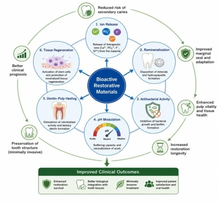
3.3. Mechanisms of Action of Bioactive Restorative MaterialsThe clinical effectiveness of bioactive restorative materials is largely attributed to their ability to interact with the surrounding biological environment through multiple mechanisms. These mechanisms often occur simultaneously and contribute collectively to tissue preservation and regeneration.
Ion release is considered the fundamental mechanism underlying bioactivity. Many bioactive restorative materials continuously release calcium, phosphate, fluoride, silicate, sodium, and other ions into the surrounding environment 14. These ions influence local biochemical processes and facilitate mineral deposition at the tooth-restoration interface. Calcium and phosphate ions contribute directly to hydroxyapatite formation, whereas fluoride ions enhance enamel resistance to acid dissolution and promote remineralization. The release of hydroxyl ions may increase local pH levels, creating an environment less favorable for cariogenic bacteria and supporting tissue healing. 15
The extent and duration of ion release depend on material composition, environmental conditions, and exposure to oral fluids. Sustained ion release is particularly important for maintaining long-term therapeutic effects and supporting continuous mineral exchange between the restoration and dental tissues.
Remineralization refers to the restoration of mineral content within demineralized enamel or dentin. Bioactive restorative materials facilitate this process through the release of calcium, phosphate, and fluoride ions that precipitate as apatite crystals on tooth surfaces 16. When exposed to saliva or dentinal fluid, many bioactive materials form a hydroxycarbonate apatite layer that closely resembles the mineral phase of natural tooth structure. This newly formed mineral layer can occlude dentinal tubules, strengthen weakened tissues, and improve the seal between the restoration and the tooth. 17
The remineralization potential of bioactive materials is particularly valuable in minimally invasive dentistry, where preservation of affected but remineralizable dentin is preferred over extensive tissue removal. Through continuous mineral replenishment, these materials contribute to the prevention of recurrent caries and prolong restoration longevity.
Secondary caries remains one of the leading causes of restoration failure. Therefore, the antibacterial properties of bioactive restorative materials represent a significant clinical advantage. Several mechanisms contribute to their antimicrobial activity, including ion release, pH elevation, fluoride release, and inhibition of bacterial adhesion. 18
Calcium silicate-based materials such as mineral trioxide aggregate and Biodentine produce alkaline environments with pH values exceeding 11, which can suppress the growth of cariogenic microorganisms. Similarly, fluoride-releasing materials reduce bacterial metabolism and acid production while enhancing enamel resistance to demineralization. 19 Certain contemporary bioactive materials also incorporate antimicrobial nanoparticles, bioactive glass particles, or quaternary ammonium compounds to further enhance antibacterial performance. By limiting bacterial colonization and biofilm formation, these materials help reduce the risk of recurrent decay around restorations. 19
One of the most distinctive characteristics of bioactive restorative materials is their ability to interact with the dentin-pulp complex. Traditional restorative materials typically provide only a physical barrier, whereas bioactive materials can stimulate biological responses within underlying tissues 20.
Calcium silicate-based materials release calcium ions that activate cellular signaling pathways involved in odontoblastic differentiation and mineralized tissue formation. These signals promote the formation of tertiary or reparative dentin, which serves as a natural protective barrier against external stimuli and bacterial invasion 21. Furthermore, bioactive materials have been shown to enhance pulp cell proliferation, migration, and differentiation while maintaining favorable conditions for tissue healing. These properties make them particularly valuable in vital pulp therapy procedures such as direct pulp capping, indirect pulp treatment, and pulpotomy.
Recent advances in biomaterials research have expanded the role of bioactive restorative materials beyond repair toward true tissue regeneration. Regeneration involves the restoration of both structure and function through activation of cellular and molecular pathways responsible for tissue development and healing. 22
Many bioactive materials serve as scaffolds capable of supporting stem cell attachment, proliferation, and differentiation. Calcium silicate cements and bioactive glasses can stimulate the release of growth factors from dentin matrices, promoting angiogenesis, mineralization, and tissue regeneration. 23 The regenerative potential of these materials aligns with contemporary concepts of minimally invasive and biologically driven dentistry. As material science continues to evolve, future bioactive restorative systems may integrate smart biomaterials, nanotechnology, and regenerative medicine approaches to further enhance tissue repair and restoration outcomes.
Collectively, these mechanisms demonstrate that bioactive restorative materials function not merely as passive replacements for lost tooth structure but as active therapeutic agents capable of influencing biological processes within the oral environment. Their ability to release beneficial ions, promote remineralization, inhibit bacterial activity, interact with the dentin-pulp complex, and support tissue regeneration forms the scientific foundation for their growing role in modern restorative dentistry. Figure 1
Bioactive restorative materials comprise a diverse group of biomaterials designed to interact beneficially with dental tissues through ion release, remineralization, antibacterial activity, and regenerative potential. Based on their chemical composition and primary mechanism of action, these materials can be broadly classified into four major categories: glass ionomer cements, calcium silicate-based materials, bioactive composite resins, and bioactive ceramics and glasses. Each category possesses unique physicochemical properties and clinical indications that contribute to the advancement of biologically driven restorative dentistry.
4.1. Glass Ionomer Cements (GICs)Glass ionomer cements (GICs) were introduced by Wilson and Kent in the early 1970s and remain among the earliest restorative materials recognized for their bioactive properties. 29
These materials are formed through an acid-base reaction between a fluoroaluminosilicate glass powder and aqueous polyalkenoic acids, resulting in a chemically bonded restoration capable of releasing therapeutic fluoride ions over extended periods. 24
Conventional GICs exhibit several properties that distinguish them from traditional restorative materials. Their ability to chemically bond to enamel and dentin eliminates the need for extensive cavity preparation and adhesive systems. Furthermore, continuous fluoride release contributes to enamel remineralization and reduces the incidence of secondary caries, making these materials particularly valuable for pediatric dentistry, atraumatic restorative treatment (ART), cervical lesions, and restorations in high-caries-risk patients. 25
The bioactivity of conventional GICs is primarily attributed to sustained fluoride release, calcium and phosphate ion exchange, and their capacity to absorb fluoride from external sources and subsequently re-release it, a phenomenon commonly referred to as the "fluoride recharge" effect. In addition, the formation of an ion-exchange layer at the tooth-restoration interface contributes to a durable chemical bond and improved marginal sealing. 26
Despite these biological advantages, conventional GICs possess relatively low flexural strength, poor fracture resistance, moisture sensitivity during initial setting, and inferior wear resistance compared with resin composites, thereby limiting their use in stress-bearing posterior restorations. 26
Resin-modified glass ionomer cements (RMGICs) were developed to overcome many of the mechanical shortcomings associated with conventional GICs. These materials combine the traditional acid-base reaction with resin polymerization, typically through incorporation of hydrophilic monomers such as 2-hydroxyethyl methacrylate (HEMA). 27
Compared with conventional GICs, RMGICs exhibit improved mechanical strength, enhanced esthetics, greater wear resistance, reduced moisture sensitivity, and immediate light-activated setting. Importantly, they retain many of the biological properties of conventional GICs, including fluoride release, chemical adhesion to tooth structure, and remineralization potential. 27
However, the presence of resin components may reduce overall ion release compared with conventional GICs, while polymerization shrinkage and potential cytotoxicity associated with residual HEMA monomers remain concerns. Nevertheless, RMGICs continue to serve as versatile restorative materials for cervical lesions, pediatric restorations, liners, bases, and intermediate restorations. 27
4.2. Calcium Silicate-Based MaterialsCalcium silicate-based materials represent one of the most biologically active classes of restorative and reparative dental materials. Their bioactivity results primarily from the release of calcium hydroxide during hydration, leading to an alkaline environment, antibacterial effects, hydroxyapatite formation, and stimulation of dentin regeneration. 28
Mineral trioxide aggregate (MTA), introduced by Torabinejad and colleagues in the 1990s, revolutionized vital pulp therapy and endodontic repair procedures due to its exceptional biocompatibility and regenerative potential. MTA consists primarily of tricalcium silicate, dicalcium silicate, tricalcium aluminate, and radiopacifying agents such as bismuth oxide. Upon hydration, calcium hydroxide is released, increasing local pH and promoting the deposition of hydroxyapatite crystals at the material-tooth interface. Numerous clinical and histological studies have demonstrated that MTA stimulates odontoblastic differentiation, tertiary dentin formation, angiogenesis, and pulp healing. Consequently, it has become the gold standard for direct pulp capping, pulpotomy, apexification, perforation repair, and root-end filling procedures. 29
Despite its excellent biological performance, MTA exhibits several disadvantages, including prolonged setting time, difficult handling characteristics, relatively high cost, tooth discoloration associated with bismuth oxide, and limited compressive strength during early setting. 29, 30
Biodentine is a newer calcium silicate-based material developed to address many of the limitations associated with MTA. It contains highly purified tricalcium silicate, calcium carbonate, zirconium oxide, and water-soluble polymers that accelerate setting and improve handling characteristics.
Compared with MTA, Biodentine demonstrates shorter setting time, superior mechanical properties, improved handling, greater compressive strength, and reduced risk of tooth discoloration. It also releases calcium ions capable of stimulating mineralization, dentin bridge formation, and pulp healing. Due to these favorable properties, Biodentine is widely used for direct and indirect pulp capping, pulpotomy, dentin replacement beneath restorations, perforation repair, and management of deep carious lesions. Several clinical studies have reported comparable or superior outcomes to MTA in vital pulp therapy. 31, 32
Bio-C Repair represents a newer generation of premixed calcium silicate-based bioceramic repair materials. Unlike traditional powder-liquid systems, Bio-C Repair is supplied as a ready-to-use paste, reducing operator variability and improving clinical convenience. 33
These materials exhibit excellent sealing ability, dimensional stability, high alkalinity, sustained calcium ion release, and favorable cytocompatibility with human dental pulp stem cells. Experimental studies have demonstrated their capacity to induce mineralized tissue formation and support regenerative healing processes comparable to those observed with MTA and Biodentine. Although early laboratory and clinical evidence is promising, additional long-term randomized clinical trials are required to establish their durability and clinical performance across various restorative and endodontic applications. 34
4.3. Bioactive Composite ResinsConventional resin composites provide excellent esthetics and mechanical properties but generally lack intrinsic biological activity. Recent developments in material science have led to the incorporation of bioactive fillers capable of releasing therapeutic ions while preserving the favorable physical characteristics of composite restorations. 35
Giomers are hybrid restorative materials containing prereacted glass ionomer (PRG) fillers embedded within a resin matrix. These fillers are produced by reacting fluoroaluminosilicate glass with polyacrylic acid prior to incorporation into the composite resin. 36
Giomers combine the esthetics and polishability of resin composites with the fluoride release and recharge capabilities of glass ionomer technology. In addition to fluoride release, they demonstrate moderate remineralization potential, reduced plaque accumulation, and improved resistance to recurrent caries. Clinically, giomers are widely used for Class III, IV, and V restorations, pediatric restorations, fissure sealants, and preventive resin restorations. 36
ACTIVA BioACTIVE Restorative is a relatively new restorative material that combines a resilient resin matrix with bioactive ionic fillers and modified glass ionomer technology. The manufacturer claims that the material releases and recharges calcium, phosphate, and fluoride ions while exhibiting physical properties comparable to resin composites. 37
Laboratory investigations have demonstrated favorable fracture resistance, ion release, marginal adaptation, and reduced microleakage. Furthermore, ACTIVA has shown the ability to form apatite-like mineral deposits on its surface under simulated oral conditions. However, independent long-term clinical evidence remains limited, and further randomized controlled clinical trials are necessary to validate the manufacturer's claims regarding superior bioactivity and restoration longevity. 38
Recent research has focused on developing next-generation bioactive composites incorporating nanoparticles, bioactive glass fillers, amorphous calcium phosphate (ACP), nano-hydroxyapatite, calcium silicate particles, antibacterial monomers, and self-healing technologies. These materials aim to provide controlled ion release, enhanced remineralization, antibacterial activity, and improved resistance to polymerization shrinkage while maintaining excellent esthetic and mechanical performance. Although many remain experimental, they represent a promising direction toward multifunctional restorative materials capable of actively preventing restoration failure. 39
4.4. Bioactive Ceramics and GlassesBioactive ceramics constitute another important class of restorative biomaterials characterized by their ability to chemically bond with mineralized tissues through apatite formation. These materials are increasingly incorporated into restorative, endodontic, and regenerative dental applications.
Bioactive glass, originally developed by Hench as 45S5 Bioglass®, consists primarily of silica, calcium oxide, sodium oxide, and phosphorus pentoxide. Upon exposure to physiological fluids, the material undergoes controlled dissolution, releasing calcium and phosphate ions that facilitate hydroxycarbonate apatite formation on its surface. 40
This apatite layer promotes strong bonding with hard tissues, enhances remineralization, occludes dentinal tubules, and exhibits antibacterial activity due to local pH elevation. Consequently, bioactive glass has found applications in restorative materials, air-polishing powders, desensitizing agents, periodontal regeneration, bone grafts, and implant coatings. 40
Hydroxyapatite (HA) is chemically similar to the inorganic mineral phase of enamel and dentin, making it highly biocompatible and osteoconductive. Synthetic hydroxyapatite has been incorporated into restorative materials, adhesives, toothpastes, and preventive products because of its excellent remineralization potential. Nano-hydroxyapatite particles possess increased surface area and enhanced interaction with enamel crystals, allowing effective repair of early enamel lesions and reduction of dentin hypersensitivity through tubule occlusion. 41
Calcium phosphate-based materials include amorphous calcium phosphate (ACP), tricalcium phosphate (TCP), octacalcium phosphate, and other calcium phosphate derivatives capable of releasing calcium and phosphate ions under acidic conditions. Among these, ACP has received considerable attention because of its ability to maintain supersaturation of calcium and phosphate ions, thereby promoting rapid remineralization of early carious lesions. Calcium phosphate nanoparticles have also been incorporated into composite resins and adhesive systems to provide continuous mineral release while maintaining satisfactory mechanical performance. Ongoing research continues to optimize these materials by improving ion-release kinetics, mechanical strength, and long-term clinical stability. Their integration into restorative systems represents an important step toward developing multifunctional biomaterials capable of simultaneously restoring tooth structure and promoting biological repair. 42, 43 [Table 1]
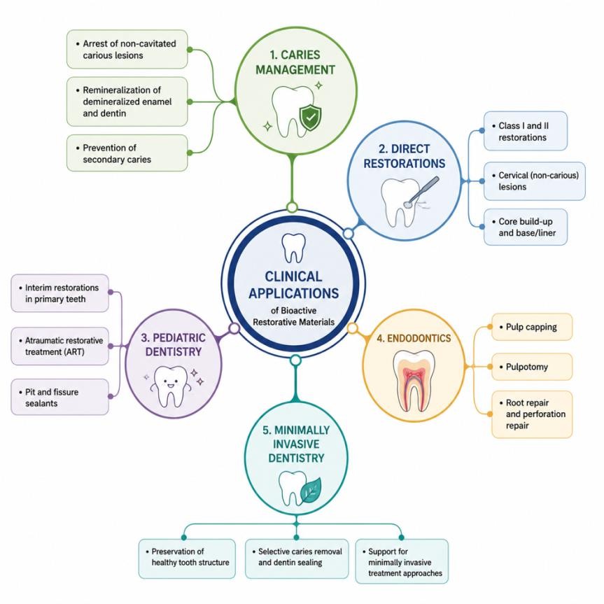
Bioactive restorative materials have gained widespread clinical acceptance because of their ability to restore tooth structure while simultaneously promoting biological repair and reducing disease progression. Their unique properties, including ion release, remineralization, antibacterial activity, and stimulation of hard tissue formation, make them suitable for a variety of clinical applications.
5.1. Caries ManagementBioactive restorative materials play a significant role in contemporary caries management by promoting remineralization of demineralized tooth structure and reducing the risk of recurrent caries. Continuous release of calcium, phosphate, and fluoride ions facilitates hydroxyapatite formation and strengthens adjacent enamel and dentin. Furthermore, their antibacterial properties help suppress cariogenic microorganisms, thereby contributing to the prevention of secondary caries around restoration margins. In deep carious lesions, calcium silicate-based materials such as Biodentine and mineral trioxide aggregate (MTA) promote dentin bridge formation and pulp healing, supporting minimally invasive management strategies. 44
5.2. Direct RestorationsSeveral bioactive restorative materials, including glass ionomer cements, resin-modified glass ionomers, giomers, and bioactive composite resins, are routinely used for direct restorations. These materials are particularly suitable for Class I–V restorations in patients with moderate to high caries risk because of their fluoride release and remineralization potential. Their ability to chemically bond to tooth structure and maintain marginal integrity contributes to improved restoration longevity and reduced incidence of secondary caries. 45
5.3. Pediatric DentistryBioactive restorative materials are extensively used in pediatric dentistry due to their biocompatibility, fluoride release, and ease of clinical application. Glass ionomer cements remain the material of choice for atraumatic restorative treatment (ART), particularly in community-based and resource-limited settings. Additionally, these materials are effective in the management of early childhood caries, where their cariostatic properties and chemical adhesion to tooth structure help preserve primary teeth while minimizing the need for extensive cavity preparation. 46
5.4. Endodontic ApplicationsCalcium silicate-based bioactive materials have transformed vital pulp therapy and endodontic treatment. Materials such as MTA and Biodentine are widely used for direct and indirect pulp capping, pulpotomy, apexification, perforation repair, and root-end filling procedures. Their ability to release calcium ions, induce hard tissue formation, and provide an excellent biological seal promotes pulp vitality, tissue regeneration, and long-term treatment success. 47
5.5. Minimally Invasive DentistryThe philosophy of minimally invasive dentistry emphasizes preservation of healthy tooth structure and biological repair rather than extensive tissue removal. Bioactive restorative materials support this approach by enabling selective caries removal and facilitating remineralization of affected dentin. Their regenerative and antibacterial properties allow clinicians to preserve tooth vitality while achieving durable restorations, making them integral components of modern conservative dental practice. 48 Figure 2
Bioactive restorative materials have gained considerable attention because they offer both therapeutic and restorative benefits, distinguishing them from conventional restorative materials. Their ability to promote remineralization, release bioactive ions, and support tissue healing has expanded their clinical applications in restorative, pediatric, and endodontic dentistry. However, despite these advantages, several limitations related to mechanical properties, cost, esthetics, and long-term clinical evidence continue to influence their widespread adoption. 49, 50, 51, 52 Table 2
The field of bioactive restorative materials is rapidly evolving with advances in biomaterials science, nanotechnology, artificial intelligence (AI), and regenerative medicine. Future research aims to develop restorative materials that not only replace lost tooth structure but also actively monitor, repair, and regenerate dental tissues, ultimately improving long-term clinical outcomes.
7.1. Nanotechnology-Enhanced Bioactive MaterialsNanotechnology has emerged as a promising approach to improve the biological and mechanical properties of restorative materials. Incorporation of nanoparticles such as nano-hydroxyapatite, bioactive glass nanoparticles, calcium phosphate nanoparticles, and silver nanoparticles enhances ion release, remineralization, antibacterial activity, and mechanical strength. Owing to their high surface-area-to-volume ratio, nanoparticles facilitate greater interaction with dental tissues, resulting in improved mineral deposition and restoration longevity. Future developments are expected to focus on controlled ion-release systems and multifunctional nanocomposites with enhanced durability and therapeutic efficacy. 53
7.2. Smart Restorative MaterialsSmart restorative materials are designed to respond dynamically to changes in the oral environment. These materials can detect variations in pH, temperature, moisture, or bacterial activity and subsequently release therapeutic ions or antimicrobial agents when needed. Such "responsive" behavior allows targeted remineralization during acidic challenges and enhanced protection against recurrent caries. Continued research into stimuli-responsive biomaterials may significantly improve the longevity and clinical performance of restorative treatments. 54, 55
7.3. Self-Healing MaterialsOne of the most exciting innovations in restorative dentistry is the development of self-healing materials capable of repairing microcracks before they progress to restoration failure. These materials incorporate microcapsules, healing agents, or dynamic polymer networks that are activated when structural damage occurs. Self-healing technology has the potential to increase restoration lifespan, reduce replacement frequency, and minimize treatment costs while preserving tooth structure. Although still largely experimental, this technology represents a promising direction for future restorative materials. 56
7.4. AI-Guided Material SelectionArtificial intelligence is increasingly being integrated into clinical decision-making in dentistry. AI-based systems may assist clinicians in selecting the most appropriate restorative material by analyzing patient-specific factors such as caries risk, occlusal loading, tooth location, esthetic requirements, and medical history. Machine learning algorithms can also predict restoration longevity and treatment outcomes using large clinical datasets, thereby facilitating evidence-based and personalized treatment planning. As digital dentistry continues to advance, AI is expected to play an important role in optimizing restorative material selection and improving clinical outcomes. 57
7.5. Personalized Restorative DentistryThe future of restorative dentistry is shifting toward personalized treatment approaches tailored to individual patient characteristics. Advances in genomics, salivary diagnostics, oral microbiome analysis, and digital technologies may enable clinicians to select restorative materials based on each patient's biological profile, caries susceptibility, and regenerative capacity. Combined with chairside digital workflows and three-dimensional (3D) printing technologies, personalized restorative dentistry has the potential to improve treatment predictability, restoration longevity, and patient satisfaction. The integration of bioactive materials with regenerative therapies and precision dentistry is expected to redefine restorative dental care in the coming decades. 58
Overall, future bioactive restorative materials are likely to evolve from passive restorative agents into multifunctional therapeutic systems capable of preventing disease, promoting tissue regeneration, and adapting to the dynamic oral environment. Continued interdisciplinary research involving material scientists, engineers, clinicians, and artificial intelligence experts will be essential for translating these innovations into routine clinical practice.
Bioactive restorative materials have transformed modern restorative dentistry by combining structural restoration with therapeutic functions such as ion release, remineralization, antibacterial activity, and tissue regeneration. Current evidence suggests that materials including glass ionomer cements, calcium silicate-based materials, bioactive composite resins, and bioactive ceramics can improve restoration longevity while preserving tooth vitality.
Clinically, these materials are particularly beneficial in minimally invasive dentistry, caries management, pediatric dentistry, and vital pulp therapy due to their biological compatibility and regenerative potential. However, their selection should be based on the clinical indication and the available scientific evidence.
Future research should focus on long-term clinical studies, enhancement of mechanical properties, and the development of advanced technologies such as nanotechnology-based materials, smart and self-healing restoratives, and AI-assisted personalized treatment approaches. These innovations are expected to further expand the role of bioactive restorative materials in contemporary dental practice.
| [1] | Khurana S, Bhullar KK, Handa A, Dhami TK, Kaur G. Incidence of restoration failure and its etiology: A retrospective study. J Conserv Dent Endod. 2025 Dec; 28(12): 1228-1233. | ||
| In article | View Article PubMed | ||
| [2] | Ferracane JL. Resin composite--state of the art. Dent Mater. 2011 Jan; 27(1): 29-38. | ||
| In article | View Article PubMed | ||
| [3] | Ferracane JL. A Historical Perspective on Dental Composite Restorative Materials. J Funct Biomater. 2024 Jun 25; 15(7): 173. | ||
| In article | View Article PubMed | ||
| [4] | Hench LL. The story of Bioglass. J Mater Sci Mater Med. 2006 Nov; 17(11): 967-78. | ||
| In article | View Article PubMed | ||
| [5] | Jones JR. Reprint of: Review of bioactive glass: From Hench to hybrids. Acta Biomater. 2015 Sep; 23 Suppl: S53-82. | ||
| In article | View Article PubMed | ||
| [6] | Almulhim KS, Syed MR, Alqahtani N, Alamoudi M, Khan M, Ahmed SZ, Khan AS. Bioactive Inorganic Materials for Dental Applications: A Narrative Review. Materials (Basel). 2022 Oct 2; 15(19): 6864. | ||
| In article | View Article PubMed | ||
| [7] | Abozaid D, Azab A, Bahnsawy MA, Eldebawy M, Ayad A, Soomro R, Elwakeel E, Mohamed MA. Bioactive restorative materials in dentistry: a comprehensive review of mechanisms, clinical applications, and future directions. Odontology. 2026 Apr; 114(2): 349-377. | ||
| In article | View Article PubMed | ||
| [8] | Elnawam H, Abdelmougod M, Mobarak A, Hussein M, Aboualmakarem H, Girgis M, El Backly R. Regenerative Endodontics and Minimally Invasive Dentistry: Intertwining Paths Crossing Over Into Clinical Translation. Front Bioeng Biotechnol. 2022 Feb 8; 10: 837639. | ||
| In article | View Article PubMed | ||
| [9] | Hench LL. Biomaterials: a forecast for the future. Biomaterials. 1998 Aug; 19(16): 1419-23. | ||
| In article | View Article PubMed | ||
| [10] | Vallittu PK, Boccaccini AR, Hupa L, Watts DC. Bioactive dental materials-Do they exist and what does bioactivity mean? Dent Mater. 2018 May; 34(5): 693-694. | ||
| In article | View Article PubMed | ||
| [11] | Pires PM, Rosa TDC, Ribeiro-Lages MB, Duarte ML, Cople Maia L, Neves AA, Sauro S. Bioactive Restorative Materials Applied over Coronal Dentine-A Bibliometric and Critical Review. Bioengineering (Basel). 2023 Jun 19; 10(6): 731. | ||
| In article | View Article PubMed | ||
| [12] | St John KR. Biocompatibility of dental materials. Dent Clin North Am. 2007 Jul; 51(3): 747-60, viii. | ||
| In article | View Article PubMed | ||
| [13] | Spagnuolo G. Bioactive Dental Materials: The Current Status. Materials (Basel). 2022 Mar 9; 15(6): 2016. | ||
| In article | View Article PubMed | ||
| [14] | Marovic, D.; Par, M.; Posavec, K.; Marić, I.; Štajdohar, D.; Muradbegović, A.; Tauböck, T.T.; Attin, T.; Tarle, Z. Long-Term Assessment of Contemporary Ion-Releasing Restorative Dental Materials. Materials 2022, 15, 4042. | ||
| In article | View Article PubMed | ||
| [15] | Guimarães BM, Prati C, Duarte MAH, Bramante CM, Gandolfi MG. Physicochemical properties of calcium silicate-based formulations MTA Repair HP and MTA Vitalcem. J Appl Oral Sci. 2018 Apr 5; 26: e2017115. | ||
| In article | View Article | ||
| [16] | Daruich PM, Brizuela M. Remineralization of Initial Carious Lesions. [Updated 2023 Apr 17]. In: StatPearls [Internet]. Treasure Island (FL): StatPearls Publishing; 2026 Jan-. Available from: https: //www.ncbi.nlm.nih.gov/books/NBK573067/ | ||
| In article | |||
| [17] | Zafar K, Jamal S, Ghafoor R. Bio-active cements-Mineral Trioxide Aggregate based calcium silicate materials: a narrative review. J Pak Med Assoc. 2020 Mar; 70(3): 497-504. | ||
| In article | View Article PubMed | ||
| [18] | Zhang K, Zhang N, Weir MD, Reynolds MA, Bai Y, Xu HHK. Bioactive Dental Composites and Bonding Agents Having Remineralizing and Antibacterial Characteristics. Dent Clin North Am. 2017 Oct; 61(4): 669-687. | ||
| In article | View Article PubMed | ||
| [19] | Wiegand A, Buchalla W, Attin T. Review on fluoride-releasing restorative materials--fluoride release and uptake characteristics, antibacterial activity and influence on caries formation. Dent Mater. 2007 Mar; 23(3): 343-62. | ||
| In article | View Article PubMed | ||
| [20] | Goldberg M, Six N, Decup F, Lasfargues JJ, Salih E, Tompkins K, Veis A. Bioactive molecules and the future of pulp therapy. Am J Dent. 2003 Feb; 16(1): 66-76. PMID: 12744417. | ||
| In article | |||
| [21] | Amir, M.; Jeevithan, L.; Barkat, M.; Fatima, S.H.; Khan, M.; Israr, S.; Naseer, F.; Fayyaz, S.; Elango, J.; Wu, W.; et al. Advances in Regenerative Dentistry: A Systematic Review of Harnessing Wnt/β-Catenin in Dentin-Pulp Regeneration. Cells 2024, 13, 1153. | ||
| In article | View Article PubMed | ||
| [22] | Murray PE, Garcia-Godoy F, Hargreaves KM. Regenerative endodontics: a review of current status and a call for action. J Endod. 2007 Apr; 33(4): 377-90.. | ||
| In article | View Article PubMed | ||
| [23] | Galler KM, Hartgerink JD, Cavender AC, Schmalz G, D'Souza RN. A customized self-assembling peptide hydrogel for dental pulp tissue engineering. Tissue Eng Part A. 2012 Jan; 18(1-2): 176-84. | ||
| In article | View Article PubMed | ||
| [24] | Sikka N, Brizuela M. Glass Ionomer Cement. [Updated 2024 Mar 4]. In: StatPearls [Internet]. Treasure Island (FL): StatPearls Publishing; 2026 Jan-. Available from: https: //www.ncbi.nlm.nih.gov/books/NBK582145/ | ||
| In article | |||
| [25] | Lohbauer U. Dental Glass Ionomer Cements as Permanent Filling Materials? — Properties, Limitations Future Trends. Materials (Basel). 2009 Dec 28; 3(1): 76–96. | ||
| In article | View Article PubMed | ||
| [26] | Sidhu SK, Nicholson JW. A Review of Glass-Ionomer Cements for Clinical Dentistry. J Funct Biomater. 2016 Jun 28; 7(3): 16. | ||
| In article | View Article PubMed | ||
| [27] | Ghilotti J, Mayorga P, Sanz JL, Forner L, Llena C. Remineralizing Ability of Resin Modified Glass Ionomers (RMGICs): A Systematic Review. J Funct Biomater. 2023 Aug 11; 14(8): 421. | ||
| In article | View Article PubMed | ||
| [28] | Talabani RM, Garib BT, Masaeli R. Bioactivity and Physicochemical Properties of Three Calcium Silicate-Based Cements: An In Vitro Study. Biomed Res Int. 2020 May 22; 2020: 9576930. | ||
| In article | View Article PubMed | ||
| [29] | Parirokh M, Torabinejad M, Dummer PMH. Mineral trioxide aggregate and other bioactive endodontic cements: an updated overview - part I: vital pulp therapy. Int Endod J. 2018 Feb; 51(2): 177-205. | ||
| In article | View Article PubMed | ||
| [30] | Torabinejad M, Parirokh M, Dummer PMH. Mineral trioxide aggregate and other bioactive endodontic cements: an updated overview - part II: other clinical applications and complications. Int Endod J. 2018 Mar; 51(3): 284-317. | ||
| In article | View Article PubMed | ||
| [31] | Kaur M, Singh H, Dhillon JS, Batra M, Saini M. MTA versus Biodentine: Review of Literature with a Comparative Analysis. J Clin Diagn Res. 2017 Aug; 11(8): ZG01-ZG05. | ||
| In article | View Article PubMed | ||
| [32] | Malkondu Ö, Karapinar Kazandağ M, Kazazoğlu E. A review on biodentine, a contemporary dentine replacement and repair material. Biomed Res Int. 2014; 2014: 160951. | ||
| In article | View Article PubMed | ||
| [33] | Campi LB, Rodrigues EM, Torres FFE, Reis JMDSN, Guerreiro-Tanomaru JM, Tanomaru-Filho M. Physicochemical properties, cytotoxicity and bioactivity of a ready-to-use bioceramic repair material. Braz Dent J. 2023 Jan-Feb; 34(1): 29-38. | ||
| In article | View Article PubMed | ||
| [34] | Kenchappa M, Gupta S, Gupta P, Sharma P. Dentine in a capsule: clinical case reports. J Indian Soc Pedod Prev Dent. 2015 Jul-Sep; 33(3): 250-4. | ||
| In article | View Article PubMed | ||
| [35] | Lopes S, Lopes PC, Fidalgo-Pereira R, Flores-Fraile J, Veiga N, Gomes ATPC. Antimicrobial potential of bioactive resin composites in caries management: a systematic review of in vitro studies. Front Oral Health. 2025 Oct 28; 6: 1625977. | ||
| In article | View Article PubMed | ||
| [36] | Rusnac ME, Gasparik C, Irimie AI, Grecu AG, Mesaroş AŞ, Dudea D. Giomers in dentistry - at the boundary between dental composites and glass-ionomers. Med Pharm Rep. 2019 Apr; 92(2): 123-128. | ||
| In article | View Article PubMed | ||
| [37] | Popa M, Dinu S, Luca MM, Bumbu BA, Maghet E, Bita RG. Clinical and Laboratory Performance of ACTIVA BioACTIVE Restorative in Primary Teeth: A Systematic Review of Pediatric Evidence. J Clin Med. 2026 Jan 4; 15(1): 373. | ||
| In article | View Article PubMed | ||
| [38] | Karabucak B, Li D, Lim J, Iqbal M. Vital pulp therapy with mineral trioxide aggregate. Dent Traumatol. 2005 Aug; 21(4): 240-3. | ||
| In article | View Article PubMed | ||
| [39] | Chen L, Shen H, Suh BI. Bioactive dental restorative materials: a review. Am J Dent. 2013 Aug; 26(4): 219-27. PMID: 24693633. | ||
| In article | |||
| [40] | Baino F, Hamzehlou S, Kargozar S. Bioactive Glasses: Where Are We and Where Are We Going? J Funct Biomater. 2018 Mar 19; 9(1): 25. | ||
| In article | View Article PubMed | ||
| [41] | Chen L, Al-Bayatee S, Khurshid Z, Shavandi A, Brunton P, Ratnayake J. Hydroxyapatite in Oral Care Products-A Review. Materials (Basel). 2021 Aug 27; 14(17): 4865. | ||
| In article | View Article PubMed | ||
| [42] | Zhang L, Weir MD, Chow LC, Antonucci JM, Chen J, Xu HH. Novel rechargeable calcium phosphate dental nanocomposite. Dent Mater. 2016 Feb; 32(2): 285-93. | ||
| In article | View Article PubMed | ||
| [43] | Jeong J, Kim JH, Shim JH, Hwang NS, Heo CY. Bioactive calcium phosphate materials and applications in bone regeneration. Biomater Res. 2019 Jan 14; 23: 4. | ||
| In article | View Article PubMed | ||
| [44] | Sajdeya A, Milgram J, Kot-Limon N, Abramovitz I, Sharir A. Cellular Dynamics and Dentine Bridge Formation Following Direct Pulp Capping With MTA and Biodentine in a Murine Model. Int Endod J. 2026 Jun 26.. | ||
| In article | View Article PubMed | ||
| [45] | Condò R, Cerroni L, Pasquantonio G, Mancini M, Pecora A, Convertino A, Mussi V, Rinaldi A, Maiolo L. A Deep Morphological Characterization and Comparison of Different Dental Restorative Materials. Biomed Res Int. 2017; 2017: 7346317. | ||
| In article | View Article PubMed | ||
| [46] | Yengopal V, Harneker SY, Patel N, Siegfried N. Dental fillings for the treatment of caries in the primary dentition. Cochrane Database Syst Rev. 2009 Apr 15; (2): CD004483. | ||
| In article | View Article PubMed | ||
| [47] | Marks LA, Faict N, Welbury RR. Literature review: Restorations of class II cavities in the primary dentition with compomers. Eur Arch Paediatr Dent. 2010 Jun; 11(3): 109-14. | ||
| In article | View Article PubMed | ||
| [48] | Tassery H, Sauro S, Slimani A. Bioactive and Ion-releasing materials in minimum intervention dentistry: a clinical pathway from prevention to restorative treatment. Front Dent Med. 2026 Apr 8; 7: 1739208. | ||
| In article | View Article PubMed | ||
| [49] | Al-Kaff AA, Alshehri AZ, Alasmari RA, Alsubaie N, Aldaws A, Althaqeel A, Alshehri RS, Alawaji YM. Minimally Invasive Techniques for Managing Dental Caries inChildren: Efficacy, Applications, and Future Directions. Cureus. 2025 Jul 7; 17(7): e87450. | ||
| In article | View Article PubMed | ||
| [50] | BaniHani A, Santamaría RM, Hu S, Maden M, Albadri S. Minimal intervention dentistry for managing carious lesions into dentine in primary teeth: an umbrella review. Eur Arch Paediatr Dent. 2022 Oct; 23(5): 667-693. | ||
| In article | View Article PubMed | ||
| [51] | Philip N, Suneja B. The revolutionary evolution in carious lesion management. J Conserv Dent. 2023 May-Jun; 26(3): 249-257. | ||
| In article | View Article PubMed | ||
| [52] | Innes N, Schwendicke F, Frencken J. An Agreed Terminology for Carious Tissue Removal. Monogr Oral Sci. 2018; 27: 155-161. | ||
| In article | View Article PubMed | ||
| [53] | Sauro S, Carvalho RM, Ferracane J. The rise of advanced bioactive restorative materials: Are they redefining operative dentistry? Dent Mater. 2025 Nov; 41(11): 1411-1429. | ||
| In article | View Article PubMed | ||
| [54] | Montoya C, Roldan L, Yu M, Valliani S, Ta C, Yang M, Orrego S. Smart dental materials for antimicrobial applications. Bioact Mater. 2022 Dec 9; 24: 1-19. | ||
| In article | View Article PubMed | ||
| [55] | Montoya C, Du Y, Gianforcaro AL, Orrego S, Yang M, Lelkes PI. On the road to smart biomaterials for bone research: definitions, concepts, advances, and outlook. Bone Res. 2021 Feb 11; 9(1): 12. | ||
| In article | View Article PubMed | ||
| [56] | Khan AA. Self-healing dental biomaterials: bioinspired pathways to sustainable dentistry. Biomater Investig Dent. 2025 Dec 29; 12: 45229. | ||
| In article | View Article | ||
| [57] | Arjumand B. The Application of artificial intelligence in restorative Dentistry: A narrative review of current research. Saudi Dent J. 2024 Jun; 36(6): 835-840. | ||
| In article | View Article PubMed | ||
| [58] | Sharma A, Tuteja A, Patel M, Tahir H, Daddanala A, Pustake A. Personalized dentistry: Enhancing outcomes through patient-centered innovation. Bioinformation. 2026 Apr 30; 22(4): 2470-2475. | ||
| In article | View Article PubMed | ||
Published with license by Science and Education Publishing, Copyright © 2026 Dr. Mariam Muneer, Dr Urmi Reshamdalal, Dr. Vikram Shad, Dr. Ekta Patel, Dr. Zulekha Yasin, Dr. Ayesha Aijaz and Dr. Sandeep Singh
 This work is licensed under a Creative Commons Attribution 4.0 International License. To view a copy of this license, visit
http://creativecommons.org/licenses/by/4.0/
This work is licensed under a Creative Commons Attribution 4.0 International License. To view a copy of this license, visit
http://creativecommons.org/licenses/by/4.0/
| [1] | Khurana S, Bhullar KK, Handa A, Dhami TK, Kaur G. Incidence of restoration failure and its etiology: A retrospective study. J Conserv Dent Endod. 2025 Dec; 28(12): 1228-1233. | ||
| In article | View Article PubMed | ||
| [2] | Ferracane JL. Resin composite--state of the art. Dent Mater. 2011 Jan; 27(1): 29-38. | ||
| In article | View Article PubMed | ||
| [3] | Ferracane JL. A Historical Perspective on Dental Composite Restorative Materials. J Funct Biomater. 2024 Jun 25; 15(7): 173. | ||
| In article | View Article PubMed | ||
| [4] | Hench LL. The story of Bioglass. J Mater Sci Mater Med. 2006 Nov; 17(11): 967-78. | ||
| In article | View Article PubMed | ||
| [5] | Jones JR. Reprint of: Review of bioactive glass: From Hench to hybrids. Acta Biomater. 2015 Sep; 23 Suppl: S53-82. | ||
| In article | View Article PubMed | ||
| [6] | Almulhim KS, Syed MR, Alqahtani N, Alamoudi M, Khan M, Ahmed SZ, Khan AS. Bioactive Inorganic Materials for Dental Applications: A Narrative Review. Materials (Basel). 2022 Oct 2; 15(19): 6864. | ||
| In article | View Article PubMed | ||
| [7] | Abozaid D, Azab A, Bahnsawy MA, Eldebawy M, Ayad A, Soomro R, Elwakeel E, Mohamed MA. Bioactive restorative materials in dentistry: a comprehensive review of mechanisms, clinical applications, and future directions. Odontology. 2026 Apr; 114(2): 349-377. | ||
| In article | View Article PubMed | ||
| [8] | Elnawam H, Abdelmougod M, Mobarak A, Hussein M, Aboualmakarem H, Girgis M, El Backly R. Regenerative Endodontics and Minimally Invasive Dentistry: Intertwining Paths Crossing Over Into Clinical Translation. Front Bioeng Biotechnol. 2022 Feb 8; 10: 837639. | ||
| In article | View Article PubMed | ||
| [9] | Hench LL. Biomaterials: a forecast for the future. Biomaterials. 1998 Aug; 19(16): 1419-23. | ||
| In article | View Article PubMed | ||
| [10] | Vallittu PK, Boccaccini AR, Hupa L, Watts DC. Bioactive dental materials-Do they exist and what does bioactivity mean? Dent Mater. 2018 May; 34(5): 693-694. | ||
| In article | View Article PubMed | ||
| [11] | Pires PM, Rosa TDC, Ribeiro-Lages MB, Duarte ML, Cople Maia L, Neves AA, Sauro S. Bioactive Restorative Materials Applied over Coronal Dentine-A Bibliometric and Critical Review. Bioengineering (Basel). 2023 Jun 19; 10(6): 731. | ||
| In article | View Article PubMed | ||
| [12] | St John KR. Biocompatibility of dental materials. Dent Clin North Am. 2007 Jul; 51(3): 747-60, viii. | ||
| In article | View Article PubMed | ||
| [13] | Spagnuolo G. Bioactive Dental Materials: The Current Status. Materials (Basel). 2022 Mar 9; 15(6): 2016. | ||
| In article | View Article PubMed | ||
| [14] | Marovic, D.; Par, M.; Posavec, K.; Marić, I.; Štajdohar, D.; Muradbegović, A.; Tauböck, T.T.; Attin, T.; Tarle, Z. Long-Term Assessment of Contemporary Ion-Releasing Restorative Dental Materials. Materials 2022, 15, 4042. | ||
| In article | View Article PubMed | ||
| [15] | Guimarães BM, Prati C, Duarte MAH, Bramante CM, Gandolfi MG. Physicochemical properties of calcium silicate-based formulations MTA Repair HP and MTA Vitalcem. J Appl Oral Sci. 2018 Apr 5; 26: e2017115. | ||
| In article | View Article | ||
| [16] | Daruich PM, Brizuela M. Remineralization of Initial Carious Lesions. [Updated 2023 Apr 17]. In: StatPearls [Internet]. Treasure Island (FL): StatPearls Publishing; 2026 Jan-. Available from: https: //www.ncbi.nlm.nih.gov/books/NBK573067/ | ||
| In article | |||
| [17] | Zafar K, Jamal S, Ghafoor R. Bio-active cements-Mineral Trioxide Aggregate based calcium silicate materials: a narrative review. J Pak Med Assoc. 2020 Mar; 70(3): 497-504. | ||
| In article | View Article PubMed | ||
| [18] | Zhang K, Zhang N, Weir MD, Reynolds MA, Bai Y, Xu HHK. Bioactive Dental Composites and Bonding Agents Having Remineralizing and Antibacterial Characteristics. Dent Clin North Am. 2017 Oct; 61(4): 669-687. | ||
| In article | View Article PubMed | ||
| [19] | Wiegand A, Buchalla W, Attin T. Review on fluoride-releasing restorative materials--fluoride release and uptake characteristics, antibacterial activity and influence on caries formation. Dent Mater. 2007 Mar; 23(3): 343-62. | ||
| In article | View Article PubMed | ||
| [20] | Goldberg M, Six N, Decup F, Lasfargues JJ, Salih E, Tompkins K, Veis A. Bioactive molecules and the future of pulp therapy. Am J Dent. 2003 Feb; 16(1): 66-76. PMID: 12744417. | ||
| In article | |||
| [21] | Amir, M.; Jeevithan, L.; Barkat, M.; Fatima, S.H.; Khan, M.; Israr, S.; Naseer, F.; Fayyaz, S.; Elango, J.; Wu, W.; et al. Advances in Regenerative Dentistry: A Systematic Review of Harnessing Wnt/β-Catenin in Dentin-Pulp Regeneration. Cells 2024, 13, 1153. | ||
| In article | View Article PubMed | ||
| [22] | Murray PE, Garcia-Godoy F, Hargreaves KM. Regenerative endodontics: a review of current status and a call for action. J Endod. 2007 Apr; 33(4): 377-90.. | ||
| In article | View Article PubMed | ||
| [23] | Galler KM, Hartgerink JD, Cavender AC, Schmalz G, D'Souza RN. A customized self-assembling peptide hydrogel for dental pulp tissue engineering. Tissue Eng Part A. 2012 Jan; 18(1-2): 176-84. | ||
| In article | View Article PubMed | ||
| [24] | Sikka N, Brizuela M. Glass Ionomer Cement. [Updated 2024 Mar 4]. In: StatPearls [Internet]. Treasure Island (FL): StatPearls Publishing; 2026 Jan-. Available from: https: //www.ncbi.nlm.nih.gov/books/NBK582145/ | ||
| In article | |||
| [25] | Lohbauer U. Dental Glass Ionomer Cements as Permanent Filling Materials? — Properties, Limitations Future Trends. Materials (Basel). 2009 Dec 28; 3(1): 76–96. | ||
| In article | View Article PubMed | ||
| [26] | Sidhu SK, Nicholson JW. A Review of Glass-Ionomer Cements for Clinical Dentistry. J Funct Biomater. 2016 Jun 28; 7(3): 16. | ||
| In article | View Article PubMed | ||
| [27] | Ghilotti J, Mayorga P, Sanz JL, Forner L, Llena C. Remineralizing Ability of Resin Modified Glass Ionomers (RMGICs): A Systematic Review. J Funct Biomater. 2023 Aug 11; 14(8): 421. | ||
| In article | View Article PubMed | ||
| [28] | Talabani RM, Garib BT, Masaeli R. Bioactivity and Physicochemical Properties of Three Calcium Silicate-Based Cements: An In Vitro Study. Biomed Res Int. 2020 May 22; 2020: 9576930. | ||
| In article | View Article PubMed | ||
| [29] | Parirokh M, Torabinejad M, Dummer PMH. Mineral trioxide aggregate and other bioactive endodontic cements: an updated overview - part I: vital pulp therapy. Int Endod J. 2018 Feb; 51(2): 177-205. | ||
| In article | View Article PubMed | ||
| [30] | Torabinejad M, Parirokh M, Dummer PMH. Mineral trioxide aggregate and other bioactive endodontic cements: an updated overview - part II: other clinical applications and complications. Int Endod J. 2018 Mar; 51(3): 284-317. | ||
| In article | View Article PubMed | ||
| [31] | Kaur M, Singh H, Dhillon JS, Batra M, Saini M. MTA versus Biodentine: Review of Literature with a Comparative Analysis. J Clin Diagn Res. 2017 Aug; 11(8): ZG01-ZG05. | ||
| In article | View Article PubMed | ||
| [32] | Malkondu Ö, Karapinar Kazandağ M, Kazazoğlu E. A review on biodentine, a contemporary dentine replacement and repair material. Biomed Res Int. 2014; 2014: 160951. | ||
| In article | View Article PubMed | ||
| [33] | Campi LB, Rodrigues EM, Torres FFE, Reis JMDSN, Guerreiro-Tanomaru JM, Tanomaru-Filho M. Physicochemical properties, cytotoxicity and bioactivity of a ready-to-use bioceramic repair material. Braz Dent J. 2023 Jan-Feb; 34(1): 29-38. | ||
| In article | View Article PubMed | ||
| [34] | Kenchappa M, Gupta S, Gupta P, Sharma P. Dentine in a capsule: clinical case reports. J Indian Soc Pedod Prev Dent. 2015 Jul-Sep; 33(3): 250-4. | ||
| In article | View Article PubMed | ||
| [35] | Lopes S, Lopes PC, Fidalgo-Pereira R, Flores-Fraile J, Veiga N, Gomes ATPC. Antimicrobial potential of bioactive resin composites in caries management: a systematic review of in vitro studies. Front Oral Health. 2025 Oct 28; 6: 1625977. | ||
| In article | View Article PubMed | ||
| [36] | Rusnac ME, Gasparik C, Irimie AI, Grecu AG, Mesaroş AŞ, Dudea D. Giomers in dentistry - at the boundary between dental composites and glass-ionomers. Med Pharm Rep. 2019 Apr; 92(2): 123-128. | ||
| In article | View Article PubMed | ||
| [37] | Popa M, Dinu S, Luca MM, Bumbu BA, Maghet E, Bita RG. Clinical and Laboratory Performance of ACTIVA BioACTIVE Restorative in Primary Teeth: A Systematic Review of Pediatric Evidence. J Clin Med. 2026 Jan 4; 15(1): 373. | ||
| In article | View Article PubMed | ||
| [38] | Karabucak B, Li D, Lim J, Iqbal M. Vital pulp therapy with mineral trioxide aggregate. Dent Traumatol. 2005 Aug; 21(4): 240-3. | ||
| In article | View Article PubMed | ||
| [39] | Chen L, Shen H, Suh BI. Bioactive dental restorative materials: a review. Am J Dent. 2013 Aug; 26(4): 219-27. PMID: 24693633. | ||
| In article | |||
| [40] | Baino F, Hamzehlou S, Kargozar S. Bioactive Glasses: Where Are We and Where Are We Going? J Funct Biomater. 2018 Mar 19; 9(1): 25. | ||
| In article | View Article PubMed | ||
| [41] | Chen L, Al-Bayatee S, Khurshid Z, Shavandi A, Brunton P, Ratnayake J. Hydroxyapatite in Oral Care Products-A Review. Materials (Basel). 2021 Aug 27; 14(17): 4865. | ||
| In article | View Article PubMed | ||
| [42] | Zhang L, Weir MD, Chow LC, Antonucci JM, Chen J, Xu HH. Novel rechargeable calcium phosphate dental nanocomposite. Dent Mater. 2016 Feb; 32(2): 285-93. | ||
| In article | View Article PubMed | ||
| [43] | Jeong J, Kim JH, Shim JH, Hwang NS, Heo CY. Bioactive calcium phosphate materials and applications in bone regeneration. Biomater Res. 2019 Jan 14; 23: 4. | ||
| In article | View Article PubMed | ||
| [44] | Sajdeya A, Milgram J, Kot-Limon N, Abramovitz I, Sharir A. Cellular Dynamics and Dentine Bridge Formation Following Direct Pulp Capping With MTA and Biodentine in a Murine Model. Int Endod J. 2026 Jun 26.. | ||
| In article | View Article PubMed | ||
| [45] | Condò R, Cerroni L, Pasquantonio G, Mancini M, Pecora A, Convertino A, Mussi V, Rinaldi A, Maiolo L. A Deep Morphological Characterization and Comparison of Different Dental Restorative Materials. Biomed Res Int. 2017; 2017: 7346317. | ||
| In article | View Article PubMed | ||
| [46] | Yengopal V, Harneker SY, Patel N, Siegfried N. Dental fillings for the treatment of caries in the primary dentition. Cochrane Database Syst Rev. 2009 Apr 15; (2): CD004483. | ||
| In article | View Article PubMed | ||
| [47] | Marks LA, Faict N, Welbury RR. Literature review: Restorations of class II cavities in the primary dentition with compomers. Eur Arch Paediatr Dent. 2010 Jun; 11(3): 109-14. | ||
| In article | View Article PubMed | ||
| [48] | Tassery H, Sauro S, Slimani A. Bioactive and Ion-releasing materials in minimum intervention dentistry: a clinical pathway from prevention to restorative treatment. Front Dent Med. 2026 Apr 8; 7: 1739208. | ||
| In article | View Article PubMed | ||
| [49] | Al-Kaff AA, Alshehri AZ, Alasmari RA, Alsubaie N, Aldaws A, Althaqeel A, Alshehri RS, Alawaji YM. Minimally Invasive Techniques for Managing Dental Caries inChildren: Efficacy, Applications, and Future Directions. Cureus. 2025 Jul 7; 17(7): e87450. | ||
| In article | View Article PubMed | ||
| [50] | BaniHani A, Santamaría RM, Hu S, Maden M, Albadri S. Minimal intervention dentistry for managing carious lesions into dentine in primary teeth: an umbrella review. Eur Arch Paediatr Dent. 2022 Oct; 23(5): 667-693. | ||
| In article | View Article PubMed | ||
| [51] | Philip N, Suneja B. The revolutionary evolution in carious lesion management. J Conserv Dent. 2023 May-Jun; 26(3): 249-257. | ||
| In article | View Article PubMed | ||
| [52] | Innes N, Schwendicke F, Frencken J. An Agreed Terminology for Carious Tissue Removal. Monogr Oral Sci. 2018; 27: 155-161. | ||
| In article | View Article PubMed | ||
| [53] | Sauro S, Carvalho RM, Ferracane J. The rise of advanced bioactive restorative materials: Are they redefining operative dentistry? Dent Mater. 2025 Nov; 41(11): 1411-1429. | ||
| In article | View Article PubMed | ||
| [54] | Montoya C, Roldan L, Yu M, Valliani S, Ta C, Yang M, Orrego S. Smart dental materials for antimicrobial applications. Bioact Mater. 2022 Dec 9; 24: 1-19. | ||
| In article | View Article PubMed | ||
| [55] | Montoya C, Du Y, Gianforcaro AL, Orrego S, Yang M, Lelkes PI. On the road to smart biomaterials for bone research: definitions, concepts, advances, and outlook. Bone Res. 2021 Feb 11; 9(1): 12. | ||
| In article | View Article PubMed | ||
| [56] | Khan AA. Self-healing dental biomaterials: bioinspired pathways to sustainable dentistry. Biomater Investig Dent. 2025 Dec 29; 12: 45229. | ||
| In article | View Article | ||
| [57] | Arjumand B. The Application of artificial intelligence in restorative Dentistry: A narrative review of current research. Saudi Dent J. 2024 Jun; 36(6): 835-840. | ||
| In article | View Article PubMed | ||
| [58] | Sharma A, Tuteja A, Patel M, Tahir H, Daddanala A, Pustake A. Personalized dentistry: Enhancing outcomes through patient-centered innovation. Bioinformation. 2026 Apr 30; 22(4): 2470-2475. | ||
| In article | View Article PubMed | ||