sciepub.com
 Quick Submission

Cholesterol Granuloma of Sphenoid Sinus: Report of a Rare Case
Samasuk Thammachantha1, , Pichest Metarugcheep2, Yodkhwan Wattanasen2
1Department of Pathology, Prasat Neurological Institute, 312 Rajvithi road, Thung phayathai, Rajthewi, Bangkok, Thailand
2Department of Neuroradiology, Prasat Neurological Institute, 312 Rajvithi road, Thung phayathai, Rajthewi, Bangkok, Thailand
Abstract
Cholesterol granuloma is chronic inflammatory lesion of head and neck region, caused by bleeding of mucosal vessels of paranasal sinus. Histologically, the lesion shows aggregate of cholesterol crystals surrounded by foreign-body giant cell reaction. The symptoms vary depending on location of an expansile mass and Magnetic resonance imaging (MRI) are helpful in distinguish this disease from mucocoele. We present a case of cholesterol granuloma in the right sphenoid sinus of a 52-year-old man, with the oculomotor nerve dysfunction but sparing pupillary reflex.
Keywords: cholesterol granuloma, sphenoid sinus, magnetic resonance imaging, mucocoele
Copyright © 2017 Science and Education Publishing. All Rights Reserved.Cite this article:
- Samasuk Thammachantha, Pichest Metarugcheep, Yodkhwan Wattanasen. Cholesterol Granuloma of Sphenoid Sinus: Report of a Rare Case. American Journal of Medical Case Reports. Vol. 5, No. 2, 2017, pp 35-37. http://pubs.sciepub.com/ajmcr/5/2/3
- Thammachantha, Samasuk, Pichest Metarugcheep, and Yodkhwan Wattanasen. "Cholesterol Granuloma of Sphenoid Sinus: Report of a Rare Case." American Journal of Medical Case Reports 5.2 (2017): 35-37.
- Thammachantha, S. , Metarugcheep, P. , & Wattanasen, Y. (2017). Cholesterol Granuloma of Sphenoid Sinus: Report of a Rare Case. American Journal of Medical Case Reports, 5(2), 35-37.
- Thammachantha, Samasuk, Pichest Metarugcheep, and Yodkhwan Wattanasen. "Cholesterol Granuloma of Sphenoid Sinus: Report of a Rare Case." American Journal of Medical Case Reports 5, no. 2 (2017): 35-37.
| Import into BibTeX | Import into EndNote | Import into RefMan | Import into RefWorks |
At a glance: Figures
1. Introduction
Cholesterol granuloma (CG) is chronic inflammatory lesion which has cholesterol cleft with multinucleated giant cell, surrounded by fibrous tissue [1]. This disease can present with headache and cranial nerve dysfunction. The hypothesis claims that leakage of occult blood by denuded mucosa is involved [2]. The incidence is 0.6:1 million [3]. This disease usually occurs in middle ear, mastoid and paranasal sinus. But the incidence of sphenoid sinus is extremely rare [4]. We report the case of CG in the sphenoid sinus, which presented with cranial nerve dysfunction.
2. Case Report
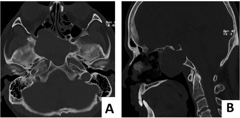
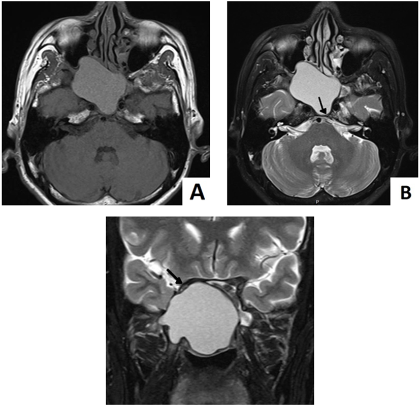
A 52-year-old-man, who was a farmer, presented with gradual onset of ptosis of right eyelid for 3 weeks. No history of double vision or headache. Physical examination showed loss of pinprick sensation at V2 distribution of right trigeminal nerve (cranial nerve V, CN 5). No abnormality of eye movement. Other cranial nerve examinations are within normal limit. Pupillary reflex, visual field (VF) and visual acuity (VA) are not remarkable (VA = 20/20 both). He was referred to our hospital for the mass lesion, suspected of tumor. CT scan revealed a cyst with bone remodeling of petrous part of temporal bone (Figure 1). Magnetic resonance imaging (MRI) showed cystic lesion in the right sphenoid sinus, size 5.5 x 4 x 4.5 cm. On T1 weighted (T1WI) and T2 weighted images (T2WI), this lesion showed high signal intensity in both, and also had pressure effect to base of skull, included right orbital apex. The hypointense rim in T2WI is also observed (Figure 2).

 Download as

 Download as
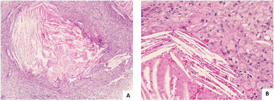
The endoscopic endonasal technique was done, and show mucous content with turbid yellow fluid. Tissue was sent to pathological laboratory and processed. The histological findings revealed the numerous cholesterol cleft with dense macrophages engulfed the lipid content. Multinucleated giant cells are observed with granulation tissue and fibrosis (Figure 3). Patient has been admitted in hospital for 12 days. During first 3 days after operation, he still had a ptosis. However, on the fourth day after operation, his symptom are recovered, he opened his right eye almost as a normal person. Nose irrigation and cleansing are advised by the hospital staffs. He was sent back home without any complications. The patient is asymptomatic during 1-month follow up, with neurosurgeon and ophthalmologist. No ptosis and no obstruction of nasal passage. 5 months after operation, the MRI evaluation show mild residual content in the right sphenoid sinus with no longer pressure effect to the right orbit.
 Download as
3. Discussion
CG is usually seen in middle ear, but is extremely rare among paranasal sinuses [2, 5]. This patient has unilateral cystic mass with compressive symptoms, which supplied by an oculomotor nerve (CN 3) and V2 distribution of CN 5. These are the first presenting manifestation by enlarging lesions.
Radiographically, the findings of CG and mucocoele on CT are the same, which are difficult to differentiate this both entities [6]. Kang suggested that CG shows the isodensity of expansile mass with scalloping bone on CT, however CG must be differentiated from mucocoele and cystic schwannoma [2]. While MRI shows hyperdensity in both T1WI and T2WI, which are characteristic findings of CG. The intense signaling in T1WI caused by the paramagnetic effect of methemoglobin, while hyperintensity in the T2WI caused by granulation tissue in paranasal sinus [5]. Ahmed postulated that differentiation CG from other expansile T1W hyperintense lesions might be difficult unless there is bony erosion [7]. Imaging differential diagnosis could be mucocele, transphenoid meningocele and arachnoid cyst. Basically, the mucocoele has hypointense content in T1WI, unless high protein content which could be hyperintense T1WI, hypointense T2WI. The content of meningocoele and arachnoid cyst is same to CSF signal intensity which the paranasal sinus arachnoid cyst is extremely rare. For the meningocoele, bony defect is the key finding. Moreover, the hypointense rim of the cyst on T2WI, which are due to breakdown products of blood, supporting the diagnosis of CG [8].
This patient had right CN 3 palsy and numbness on right cheek of face (V2 distribution of CN 5). We suspected that the mass should compress the right orbital apex. Because of sparing of pupillary reflex, which is supplied by the inferior division of CN 3. This mass should disturb the superior division of CN 3, and produce the weakness of right levator palpabrae muscle. V2 of CN 5 passes the foreman rotundum and supplies the cheek area of face. According to the CT and MRI, we found that the mass compressed the right optic nerve inferiorly but no visual loss detected in this case. We believed that the optic nerve is a bit big and located beneath the basal brain. So the optic nerve can compensate if there is occupying mass nearby. In the other hand, the orbit and apex are the limited areas. The CN 3, 4, 6, which pass the orbital apex to the eyeball, are easily effected by the compression. This symptoms can explained by the location of mass in imaging techniques.
Pathophysiologically, etiologies of this disease are hemorrhage and impaired drainage, due to obstruction of ventilation of air cells. The vacuum effect makes rupture of mucosal vessels and bleeding. Once the red blood cells breakdown, this event intrigues phogocytosis of macrophages. Finally, they engulf the cholesterol [2, 9].
Due to absence of epithelial lining, total excision of the cyst wall of CG is not necessary [5]. Miyamura also claimed that a goal of treatment is not to remove the cyst, but to make the drainage and provide the ventilation. However, neurosurgeon might prefer the craniotomy but otolaryngologists may prefer an otologic or transmastoid approach [10]. Provide ventilation by nasal cleaning is recommended. Recurrence is rare [11].
Conflict of Interest
The authors state that there is no conflict of interest.
Ethical Approval
All procedures performed in studies were in accordance with the ethical standards of the institutional research committee. And ethical approval is obtained before the research is conducted.
Informed Consent
Informed consent was obtained from participant, and the participant has been informed clearly about article publication.
References
| [1] | Manojlovic S. Cholesterol Granuloma, Middle Ear. In: Volavšek M, editor. Head and Neck Pathology. Springer: 2016. pp. 68-69. | ||
 In article In article | |||
| [2] | Kang H, Kim JK, Kim Y. Association of cholesterol granuloma and aspergillosis in the sphenoid sinus. Korean J Radiol 2008; 9(Suppl): S30-3. | ||
| In article | |||
| [3] | Jaberoo MC, Hassan A, Pulido MA, Saleh HA. Endoscopic endonasal approaches to management of cholesterol granuloma of the petrous apex. Skull base 2010; 20: 375-9. | ||
| In article | View Article PubMed PubMed | ||
| [4] | Ramani P, Murugesan K , Chandrasekar T, Anuja N. Cholesterol granuloma of maxillary sinus. Int J Oral Max Surg 2006; 35(11): 1063-5. | ||
| In article | View Article PubMed | ||
| [5] | Kim DH, Lee SK, Kim CH, Cho HJ. Radiologically unusual presentation of cholesterol granuloma in the sphenoid sinus. Korean J Otorhinolaryngol-Head Neck Surg 2015; 58(9): 641-5. | ||
| In article | View Article | ||
| [6] | Jackler RK, Parker DA. Radiographic differential diagnosis of petrous apex lesions. Am J Otol 1992; 13(6): 561-74. | ||
| In article | |||
| [7] | Ahmed M, Joseph E, Mani S, Chacko G, Rajshekhar V. Cholesterol granuloma of the sphenoid sinus: An unusual sphenoid sinus lesion. Neurology India 2012; 60(3): 332-3. | ||
| In article | View Article PubMed | ||
| [8] | Greeberg JJ, Oot RF, Wismer GL, Davis KR, Goodman ML, Weber AE, et al. Cholesterol granuloma of the petrous apex: MR and CT evaluation. AJNR Am J Neuroradiol 1988; 9: 1205-14. | ||
| In article | |||
| [9] | Sarioglu S, Pabuççuoglu U, Arzu Topal N. Cholesterol granuloma and aspergilloma of the maxillary sinus. Eur Arch Otorhinolaryngol 2001; 258: 74-6. | ||
| In article | View Article PubMed | ||
| [10] | Miyamura S, Yamaguchi S, Tominaga A, Kinoshita Y, Takeda M, Usui S, et al. A case of petrous apex cholesterol granuloma successfully treated with endoscopic endonasal surgery. Hiroshima J Med Sci. 2014; 63(4): 39-42. | ||
| In article | |||
| [11] | Prasad PV, Satyanarayana MA, Susritha K, Bala TK. Cholesterol granuloma in middle ear and mastoid. A case report. IJSR 2015; 4(5): 125-6. | ||
| In article | |||
 CiteULike
 Mendeley
 StumbleUpon
 Delicious
 Facebook
 Twitter
 LinkedIn
. Sagittal CT scan showed clivus bone remodeling with intact sellar floor (B))
 and axial T2WI (B). The hypointense rim is noted in B (arrow). The cystic lesion compressed the right optic nerve (arrow) (C:coronal T2WI))
)