Ameloblastoma (AM) is a benign but locally infiltrative epithelial odontogenic neoplasm. An 86-year-old woman presented with swelling on the left palate gum. CT images as well as panoramic X-ray photograph displayed a multiocular radiolucent lesion extending from the alveolar process to the plate. Pathological diagnosis of the resected tumor tissue was AM arisen from the anterior maxillary bone. AM can present with a follicular, plexiform, acanthomatous, basal cell, granular or desmoplastic subtype. Present case was diagnosed as a mixed pattern of acanthomatous subtype and basal cell subtype. In this study, tumor cells of the basal cell type had a clear continuity to the islands of acanthomatous subtype suggesting that the cells of basal type could differentiate to those of acanthomatous islands. To the best of our knowledge, presence of the unique combination of acanthomatous subtype and basal cell subtype, and the mutual correlation with two subtypes of AM have not yet been described.
Ameloblastoma (AM) is the most common benign tumor of the jaws, comprising 1% of all cysts and tumors of the jaws and approximately 10% of odontogenic tumors 1. Broca in 1866 first described AM suggesting that it arises from epithelial remnants of the developing root sheath 2. However, the exact genesis of AM still largely remains unknown. AM is a benign but locally infiltrative epithelial odontogenic neoplasm. AMs located in the maxillary region are rare compared to those arising in the mandible, the latter being five times more frequent. Indeed, maxillary AMs account for 15% of all AMs. Most of them arise posterior to the premolars and only 2% of them arise from anterior hard plate 3, 4, 5, 6. According to available data, maxillary AMs can occur in all ages but later than mandible AMs. No sex preference has been shown 7.
As for histological subtype of AMs, 6 subtypes are advocated 7. Follicular subtype is the most common subtype. This type consists of neoplastic cellular nests with central loosening of short spindled epithelial cells resembling the stellate reticulum by palisading columnar cells. Plexiform subtype shows a lace like architecture consisting of lamina like strands which show two distinct forms: one where the strands are formed by two layers of basaloid cells and a second form where the strands are thicker and looser having a more squamous appearance. Acanthomatous subtype has squamous differentiation centrally in islands but maintains the reverse polarization of the nuclei in peripheral columnar cells. Basal cell subtype comprises islands and strands of basaloid cells with scant cytoplasm and peripheral palisading, reminiscent of basal cell carcinoma. Granular cell subtype is characterized by granular change where all or some of the neoplastic cells of the stellate reticulum gain abundant deeply granular cytoplasm. Desmoplatic subtype is characterized by intense stromal desmoplasia which compresses the neoplastic islands of the epithelium. It is known that many AMs contain more than one histological subtype. The most common histological patterns seen in the maxillary bone are the follicular and plexiform subtypes 1. The purpose of this study is to examine the histopathological significance of basal cells in ameloblastoma based on the pathological findings of this case.
An 86-year-old woman presented with swelling on the left palatal gum and visited a dentist. She was given a diagnosis of subperiosteal abscess by the dentist, and incision with drainage was performed. The dentist suspected an infected odontogenic cyst and referred the patient to the Department of Dental and Oral Surgery of Ogaki Tokushukai Hospital. At the first examination, was noticed swelling of the left cheek toward left nasal wing. Although there was a mild tenderness of the cheek, no movement disorder or sensory impairment around the cheek was present. She had been noticed diabetes mellitus (Type 2). Clinical laboratory findings at the consultation were as follow; white blood cell count: 8200/μL, CRP: 0.77mg/dL, blood sugar level: 462mg/dL, HbAlc(NGSP): 17.0, BUN: 52.1mg/dL, creatinin value: 2.28mg/dL, eGFR:16.3.
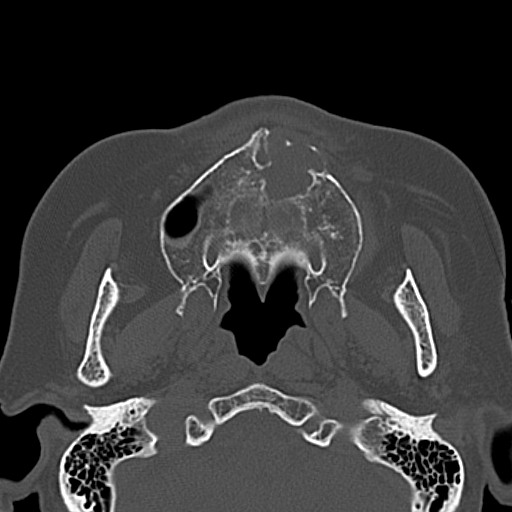
On panoramic X-ray photograph, a clear multilocular radiolucency was apparent in the frontal maxillary bone. In CT images, a multilocular infiltrative lesion extending from alveolar process to the plate, and from maxillary left central incision to the first premolar region was recognized (Figure 1). Result of the biopsy, however, was only a keratinized material with calcification and necrotic tissue with leukocyte infiltration, and clear cystic lesions or neoplasms were not identified. Four months later, operation for resection of the maxillary neoplasm was performed under general anesthesia. The resected tumor tissue had multilocular structures by bone septum and lacked communication with the nasal cavity. The tissues were removed together with palatine periosteum.
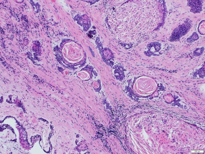
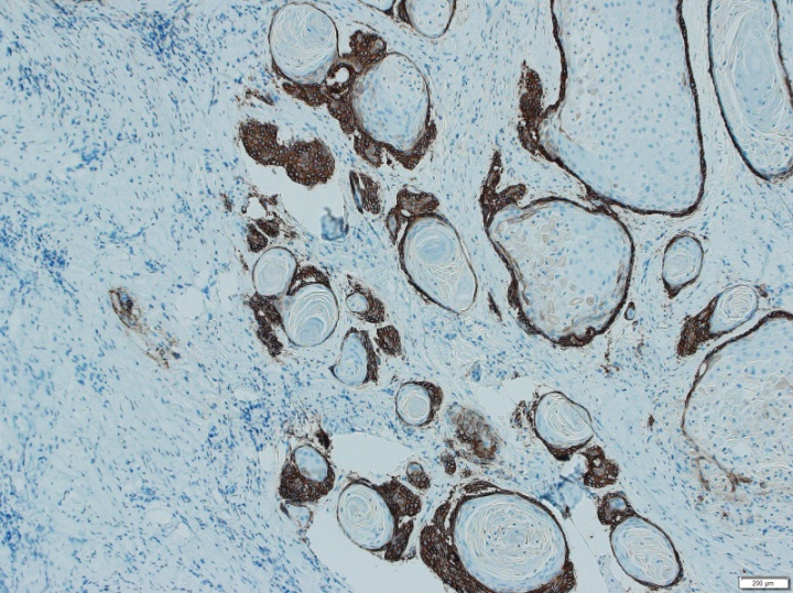
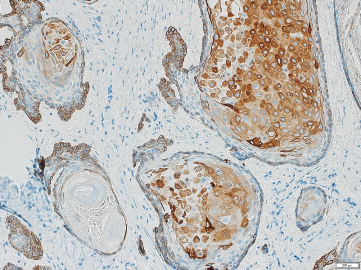
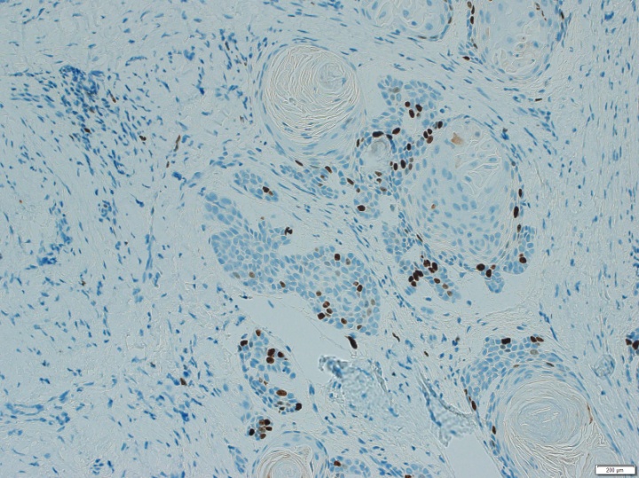
Histologically, one of representative characteristics was presence of pale staining acanthomatous islands of various sizes with squamous differentiation (Figure 2). The islands accompanied formation of keratin filled cavities. Peripheral epithelial cells in the islands had hyperchromatic nuclei. Second characteristic feature was existence of darker-staining islands and strands of basaloid cells with scant cytoplasm, reminiscent of basal cell carcinoma. Such islands and strands had spiky outlines toward into a densely collagenous stroma (Figure 2). Thus, the neoplasm was diagnosed as an ameloblastoma, conventional with mixed pattern of acanthomatous subtype and basal cell subtype. Immunohistochemistry, podoplanin, a lymphatic endometrium marker and p63 was strongly positive in the mass of basal cells (Figure 3). However, both markers were only positive in the peripheral cells of the islands. Reversely, CK19 being a good marker for benign odontogenic tumors, showed clearly positive expression in the central area of acanthomatous islands (Figure 4). Labeling index of Ki-67 of the basal cells was about 10%. However, for the acanthomatous islands, scanty labeled cells were located only in the outer membrane (Figure 5).
The patient has been stable without recurrence of the neoplasm, however, she died of a heart failure 2 years after the operation.


It is known that as many as 87% of AMs arise from the mandible, predominantly in the posterior molar area 8 9. Maxillary AMs are usually posterior sited 7. In this case, AM was developed in the anterior site. Therefore, Maxillary AMs in the anterior site are regarded as exceedingly rare.
AMs can present with follicular, plexiform, acanthomatous, basal cell, granular and desmoplastic subtypes. It is not uncommon to have a combination of two or more patterns in a tumor. Present case seems to be a mixed type of acanthomatous subtype and basal cell subtype. Such combination must be also rare. Andressa-Incert Filizzola 10 reported clinicopathological features of 70 cases of AMs. In the review, 57 solid AMs included: follicular (26%), plexiform (18%), follicular + acanthomatous (14%), follicular + plexiform (6%), follicular + plexiform + acanthomatous (6%), follicular + granular cell (4%), plexiform + angiomatous (3%), follicular + angiomatous (3%), follicular + acanthomatous (3%), follicular + acanthomatous + basaloid (1%). As shown in the research, AM of basal cell subtype is quite unusual. Meanwhile, it is also known that AM exhibits different differentiation and histological changes infrequently. For instance, plexiform AM has acanthomaous changes 11 or basaloid differentiation 12.
In the present study, labeling index of Ki-67 of the tumor mass of basal cell type was higher than of acanthomatous subtype indicating that the basaloid cells in the basal cell subtype has increased activity of cell proliferation comparing to those of acanthomatous subtype. Importantly, the tumor cells of basal cell subtype had clear continuity to the islands of acanthomatous subtype. This implies that the basaloid cells could differentiate to those of acanthomaous islands.
Immunohistochemical expression of podoplanin and p63 was present in the tumor cells of basal cell type and in peripheral cells of acanthomatous islands. On the contrary, expression of CK19 was strongly positive in the central area of the acanthomatous islands and was weak or negative in the peripheral area of acanthomatous islands and basaloid cells. These results are agreement with previous studies 13, 14. Podoplanin is presumed to promote collective cell migration and invasiveness in AMs 13. Thus, the fact that the expression of the mucin-type transmembrane glycoprotein is limited in the basal cells or peripheral cells in the acanthomatous islands is interesting.
Vickers 15 who studied early histopathologic features of ameloblastoma using 10 examples of cystic jaw lesions (dental cysts) described the possible delineation as follows; 1) hyperchromatism of basal cell nuclei of the epithelium lining the cystic cavities 2) palisading and polarization of basal cell nuclei of the epithelium lining the cystic cavities 3) cytoplasmic vacuolization of, especially, basal cells of cystic lining. Furthermore, Pincock 16 illustrated a cyst with developing AM in a patient with multiple jaw cyst and other stigmata of the multiple nevoid basal cell carcinoma syndrome. Accordingly, it is suggested that basal cells have potentials to differentiate to other cell types during the development of AM and to malignant transformation.
Basal cell AM is a rare variant of AM with very few cases reported until date. Furthermore, basal cell subtype of AM has been shown only in recent classification of AM 7. As shown in the present case, tumor cells of the basal cell subtype have potential to infiltrate surrounding connective tissues like the basal cell carcinoma. Accordingly, significance of basal cells in the basal subtype of AM needs to be studied with more cases. So far as we are concerned, presence of AM with unique combination of acanthomatous and basal cell subtypes, and the mutual correlation between the two subtypes have not yet been described.
| [1] | Evangelou Z, Zarachi A, Dumollard JM, Peoc'h M, Komnos I, Kastanioudakis I, Karpathiou G, “Maxillary Ameloblastoma: A Review With Clinical, Histological and Prognostic Data of a Rare Tumor”, In Vivo, 34(5), 2249-2258, Sep, 2020. | ||
| In article | View Article PubMed | ||
| [2] | Broca P, Recherches sur un nouveau groupe de tumeurs désignées sous le nom d’odontomes (ed.1867), HACHETTE BNF, 2017. | ||
| In article | |||
| [3] | Small IA, Waldron CA, “Ameloblastomas of the jaws”, Oral Surg Oral Med Oral Pathol, 8(3), 281-297, Mar, 1955. | ||
| In article | View Article PubMed | ||
| [4] | Robinson HBG, “Histologic study of the ameloblastoma”. Arch Pathol, 23, 664–673, 1937. | ||
| In article | |||
| [5] | Reichart PA、Philipsen HP, Sonner S, “Ameloblastoma: biological profile of 3677 cases”, Eur J Cancer Part B Oral Oncol, 31B(2), 86-99, Mar, 1995. | ||
| In article | View Article PubMed | ||
| [6] | Sehdev MK, Huvos AG, Strong EW, Gerold FP, Willis GW, “Proceedings: Ameloblastoma of maxilla and mandible”, Cancer, 33(2), 324-333, Feb, 1974. | ||
| In article | View Article PubMed | ||
| [7] | Vered M, Wright JM, “Update from the 5th Edition of the World Health Organization Classification of Head and Neck Tumors: Odontogenic and Maxillofacial Bone Tumours”, Head Neck Pathol, 16(1), 63-75, Mar, 2022. | ||
| In article | View Article PubMed | ||
| [8] | Hendra FN, Cann EV, Helder MN, Ruslin M, Visscher J, Forouzanfar T, Vet H-C-W, “Global incidence and profile of ameloblastoma: A systematic review and meta-analysis”, Oral Dis, 26(1), 12-21, Jan, 2020. | ||
| In article | View Article PubMed | ||
| [9] | Zlotogorski-Hurvitz A, Tekkesin MS, Passador-Santos F, Montalli VCM, Salo T, Mauramo M, Kats L, Buchner A, Vered M, “Conceptual changes in ameloblastoma: Suggested re-classification of a "veteran" tumor”, Oral Dis, 28(3), 703-710, Apr, 2022. | ||
| In article | View Article PubMed | ||
| [10] | Filizzola A-I, Bartholomeu-dos-Santos T-C-R, Pires F-R, “Ameloblastomas: clinicopathological features from 70 cases diagnosed in a single oral pathology service in an 8-year period”, Med Oral Patol Oral Cir Bucal, 19(6), e556-e561, Nov, 2014. | ||
| In article | View Article PubMed | ||
| [11] | Tomar A, “Plexiform Ameloblastoma with Acanthomatous Changes: A Rare Case Report”, Ind J Otolaryng Head Neck Surg, 76(6), 6028-3601, Sep, 2024. | ||
| In article | View Article PubMed | ||
| [12] | Ghattamaneni S, Guttikonda VR, Kumari MG, Kumar DR, “Maxillary plexiform ameloblastoma showing basaloid differentiation: Report of a rare case with review of literature”, Ind J Dental Res, 26(6), 633-636, Nov, 2015. | ||
| In article | View Article PubMed | ||
| [13] | Gonzalez-Alva P, Tanaka A, Oku Y, Miyazaki Y, Okamoto E, Fujinami M, Yoshida N, Kikuchi K, Ide F, Sakashita H, Kusama K, “Enhanced expression of podoplanin in ameloblastomas” J Oral Pathol Med, 39(1), 103-109, Jan, 2010. | ||
| In article | View Article PubMed | ||
| [14] | Martinez M, Taylor A, Bregni R, Pires F-R, Azanero W, Silva R, Barrios B, Almeida O, “Comparative histological and immunohistochemical study of ameloblastomas and ameloblastic carcinomas”, Med Oral Patol Oral Cir Bucal, 22(3), 324-332, Apr, 2017. | ||
| In article | |||
| [15] | 15.Vickers RA, Gorlin RJ, “Ameloblastoma: Delineation of early histopathologic features of neoplasia”, Cancer, 26(3), 699-710, Sep, 1970. | ||
| In article | View Article PubMed | ||
| [16] | 16.Pincock HP, Bruce KW, “Odontogenic fibroma”, Oral Surg Oral Med Oral Path, 7(3), 307-311, 1954. | ||
| In article | View Article PubMed | ||
Published with license by Science and Education Publishing, Copyright © 2026 Noriko Matsuzawa, Hideki Mori, Yoshihiro Kimura and Mitsuhiro Aoki
 This work is licensed under a Creative Commons Attribution 4.0 International License. To view a copy of this license, visit
http://creativecommons.org/licenses/by/4.0/
This work is licensed under a Creative Commons Attribution 4.0 International License. To view a copy of this license, visit
http://creativecommons.org/licenses/by/4.0/
| [1] | Evangelou Z, Zarachi A, Dumollard JM, Peoc'h M, Komnos I, Kastanioudakis I, Karpathiou G, “Maxillary Ameloblastoma: A Review With Clinical, Histological and Prognostic Data of a Rare Tumor”, In Vivo, 34(5), 2249-2258, Sep, 2020. | ||
| In article | View Article PubMed | ||
| [2] | Broca P, Recherches sur un nouveau groupe de tumeurs désignées sous le nom d’odontomes (ed.1867), HACHETTE BNF, 2017. | ||
| In article | |||
| [3] | Small IA, Waldron CA, “Ameloblastomas of the jaws”, Oral Surg Oral Med Oral Pathol, 8(3), 281-297, Mar, 1955. | ||
| In article | View Article PubMed | ||
| [4] | Robinson HBG, “Histologic study of the ameloblastoma”. Arch Pathol, 23, 664–673, 1937. | ||
| In article | |||
| [5] | Reichart PA、Philipsen HP, Sonner S, “Ameloblastoma: biological profile of 3677 cases”, Eur J Cancer Part B Oral Oncol, 31B(2), 86-99, Mar, 1995. | ||
| In article | View Article PubMed | ||
| [6] | Sehdev MK, Huvos AG, Strong EW, Gerold FP, Willis GW, “Proceedings: Ameloblastoma of maxilla and mandible”, Cancer, 33(2), 324-333, Feb, 1974. | ||
| In article | View Article PubMed | ||
| [7] | Vered M, Wright JM, “Update from the 5th Edition of the World Health Organization Classification of Head and Neck Tumors: Odontogenic and Maxillofacial Bone Tumours”, Head Neck Pathol, 16(1), 63-75, Mar, 2022. | ||
| In article | View Article PubMed | ||
| [8] | Hendra FN, Cann EV, Helder MN, Ruslin M, Visscher J, Forouzanfar T, Vet H-C-W, “Global incidence and profile of ameloblastoma: A systematic review and meta-analysis”, Oral Dis, 26(1), 12-21, Jan, 2020. | ||
| In article | View Article PubMed | ||
| [9] | Zlotogorski-Hurvitz A, Tekkesin MS, Passador-Santos F, Montalli VCM, Salo T, Mauramo M, Kats L, Buchner A, Vered M, “Conceptual changes in ameloblastoma: Suggested re-classification of a "veteran" tumor”, Oral Dis, 28(3), 703-710, Apr, 2022. | ||
| In article | View Article PubMed | ||
| [10] | Filizzola A-I, Bartholomeu-dos-Santos T-C-R, Pires F-R, “Ameloblastomas: clinicopathological features from 70 cases diagnosed in a single oral pathology service in an 8-year period”, Med Oral Patol Oral Cir Bucal, 19(6), e556-e561, Nov, 2014. | ||
| In article | View Article PubMed | ||
| [11] | Tomar A, “Plexiform Ameloblastoma with Acanthomatous Changes: A Rare Case Report”, Ind J Otolaryng Head Neck Surg, 76(6), 6028-3601, Sep, 2024. | ||
| In article | View Article PubMed | ||
| [12] | Ghattamaneni S, Guttikonda VR, Kumari MG, Kumar DR, “Maxillary plexiform ameloblastoma showing basaloid differentiation: Report of a rare case with review of literature”, Ind J Dental Res, 26(6), 633-636, Nov, 2015. | ||
| In article | View Article PubMed | ||
| [13] | Gonzalez-Alva P, Tanaka A, Oku Y, Miyazaki Y, Okamoto E, Fujinami M, Yoshida N, Kikuchi K, Ide F, Sakashita H, Kusama K, “Enhanced expression of podoplanin in ameloblastomas” J Oral Pathol Med, 39(1), 103-109, Jan, 2010. | ||
| In article | View Article PubMed | ||
| [14] | Martinez M, Taylor A, Bregni R, Pires F-R, Azanero W, Silva R, Barrios B, Almeida O, “Comparative histological and immunohistochemical study of ameloblastomas and ameloblastic carcinomas”, Med Oral Patol Oral Cir Bucal, 22(3), 324-332, Apr, 2017. | ||
| In article | |||
| [15] | 15.Vickers RA, Gorlin RJ, “Ameloblastoma: Delineation of early histopathologic features of neoplasia”, Cancer, 26(3), 699-710, Sep, 1970. | ||
| In article | View Article PubMed | ||
| [16] | 16.Pincock HP, Bruce KW, “Odontogenic fibroma”, Oral Surg Oral Med Oral Path, 7(3), 307-311, 1954. | ||
| In article | View Article PubMed | ||