Metastasis of malignant melanoma (MM) to the breast is very rare, accounting for only 1.3% to 2.7% of all melanoma cases. In addition, many are metastases to the skin and subcutaneous tissue of the breast, and metastasis to the breast parenchyma including the mammary gland is even rarer, with a very limited number of reports. A 47-year-old female presented with a lump in the lower-outer quadrant of the right breast noted 6 months prior. Visual examination and palpation, ultrasonography, and mammography were performed. The skin of both breasts showed no abnormal findings and no melanoma. Core needle biopsy (CNB) was performed. The result of CNB was malignant melanoma. Metastasis from other sites was considered more likely than primary malignancy of the breast. A 10 × 12 mm melanoma lesion on the right upper arm had shown progressive growth over 1.5 years. The lesion on the right upper arm was considered the primary lesion. Positron Emision Tomography-Computed Tomography (PET-CT) detected a nodule in the left upper lobe of the lung, and metastasis of MM was suspected. The right breast tumor was resected. Histopathological results were pT3N0M1, Stage IV. BRAF mutation was negative. Nivolumab + ipilimumab was scheduled for further treatment.
Metastasis of malignant melanoma (MM) to the breast is very rare, accounting for only 1.3% to 2.7% of all melanoma cases 1. In addition, most are metastases to the skin and subcutaneous tissue of the breast, and metastasis to the breast parenchyma including the mammary gland is even rarer, with few reports. The skin of both breasts showed no abnormal findings and no melanoma. This case is extremely rare, unusual, and of great clinical significance.
Here, a case of rare metastatic MM of the breast parenchyma diagnosed by preoperative CNB is reported.
A 47-year-old female presented with a lump in the lower lateral region of the right breast noted 6 months prior. There is nothing of note in the patient's medical history or family history. Physical examination, breast ultrasonography, and mammography
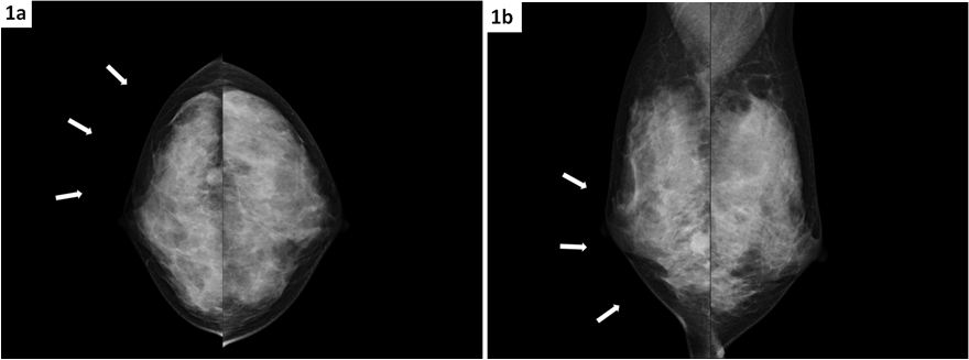
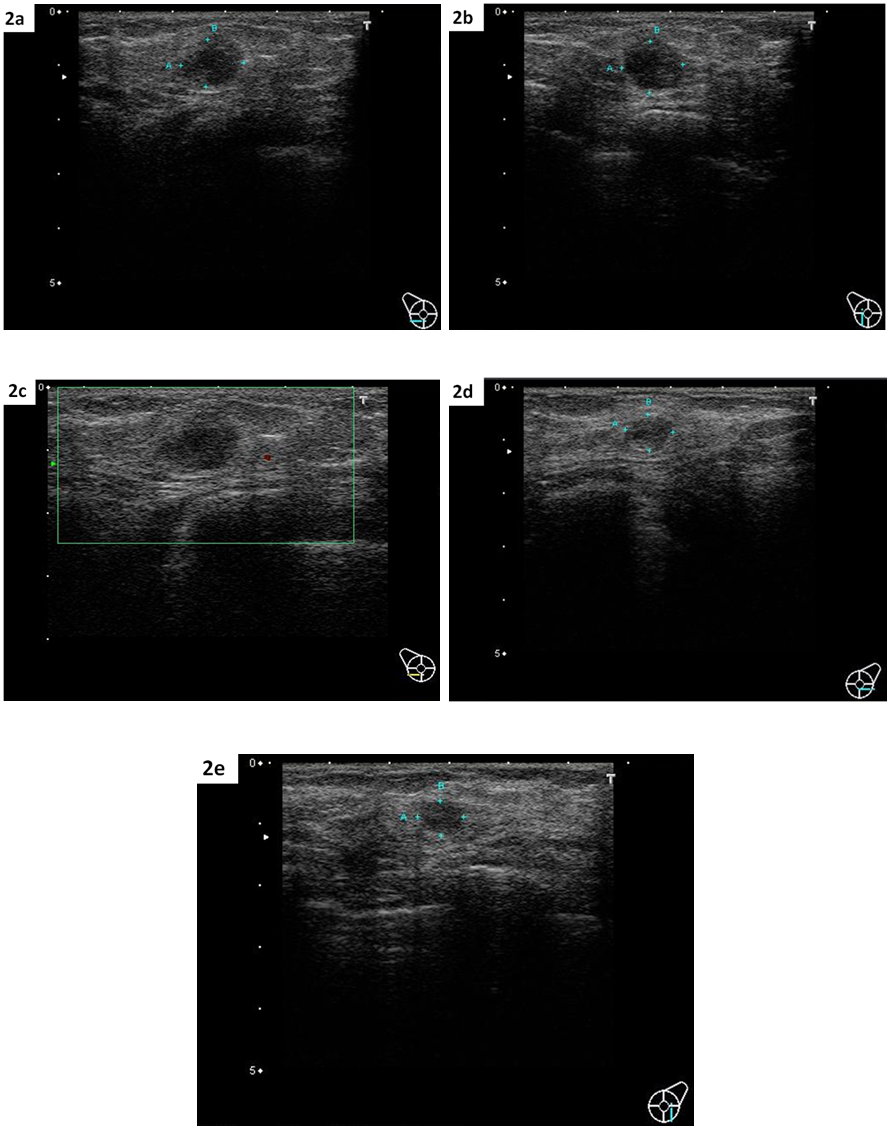
were performed. The skin of both breasts showed no abnormal findings and no melanoma. On MMG taken in the craniocaudal and mediolateral oblique views, localized asymmetric lesions with tumor shadows were observed in the outer and lower quadrants of the right breast, respectively. Thus, the right and left lesions were classified as category 4 and category 1, respectively (Figure 1a, 1b). Breast US revealed a mass shadow measuring 12.0 × 11.7 × 9.5 mm in the lower-outer quadrant of the right breast (Figure 2a, 2b, 2c) and a mass shadow measuring 9.1 × 7.7 × 8.6 mm in the lower-outer quadrant of the left breast. On US, the right and left lesions were classified as category 4 and category 3, respectively (Figure 2d, 2e).
Although the left breast mass was classified as category 1 on MMG, it was classified as category 3 on US; therefore, CNB was performed on both the right and left breast masses.
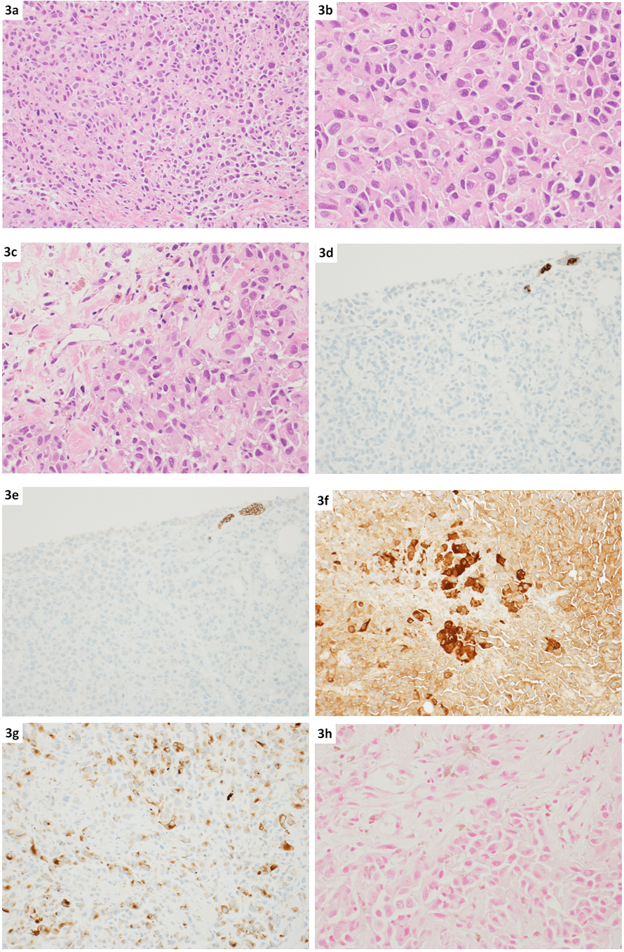
Right CNB results: malignant melanoma. Atypical cells with eccentrically placed nuclei infiltrated in a trabecular and scattered pattern, accompanied by tissue necrosis (HE × 200, × 400, × 400) (Figure 3a, 3b, 3c). Immunohistochemistry (IHC) stain showed that the atypical cells were cytokeratin AE1/3 (−) (cytokeratin AE1/3×200) (Figure 3d), and E-cadherin (−) (E-cadherin×200) (Figure 3e). Due to negativity for cytokeratin, the possibility of a tumor other than carcinoma (e.g., lymphoma) should also be included in the differential diagnosis. Additional IHC staining results showed that the tumor cells exhibited S-100 positivity (S-100, ×200) (Figure 3f), and HMB-45 positivity (HMB-45, ×200) (Figure 3g). The tumor cells completely exhibited negativity for all types of cytokeratin, and negativity for lymphocyte markers. In addition, brown pigment retention was observed in and between tumor cells, which was considered to be melanin pigment due to negativity for iron staining (Berlin Blue,x400) (Figure 3h). Based on these findings, it was considered malignant melanoma. Immunostaining results are summarized as follows: AE1/3 (−), CAM5.2 (−), cytokeratin 7 (−), 34βE12 (−), E-cadherin (−), CD3 (−), CD20 (−), CD30 (−), CD68 (−), CD138 (−), S-100 (+), HMB-45 (+). Metastasis from other sites was considered more likely than primary malignancy of the breast.
Left CNB results showed a phyllodes tumour. The tumor lesion consisted of dilated ducts and edematous connective tissue filling between them. Enlarged nuclei were seen in interstitial cells. Mitotic figures were not obvious. It was considered to be a phyllodes tumor. Although the lesion was not considered aggressively malignant, confirmation by resection of the entire lesion was desirable.
Following a diagnosis of malignant melanoma in the right breast, a systemic melanoma screening was performed, during which a 10 × 12 mm melanoma lesion on the right upper arm—which may be malignant—had shown progressive growth over 1.5 years and a 3 mm lesion on the right cheek—which is thought to be benign—were identified.
The lesion on the right upper arm was dark brown, with an uneven and mottled coloration and indistinct borders; although no ulceration was present, primary malignant melanoma was suspected. Meanwhile, the lesion on the right breast was suspected to be a metastatic malignant lesion.
To date, there has never been a single case in which a patient with melanoma on the right upper arm underwent a medical examination, biopsy, or surgical excision.
A subsequent PET-CT detected a nodule in the left upper lobe of the lung, and metastasis of malignant melanoma was suspected.
A palliative excision was performed on the melanoma lesion on the right upper arm.
The tumor thickness measured 1.1 mm, and the Clark level was IV (invasion into the dermal reticular layer). In addition, mitotic activity was 2–3 per high-power field (HPF); there was no vascular invasion, no neurotropism or nerve invasion, and no microsatellite lesions.
A skin biopsy of the lesion on the right cheek revealed no atypia and showed nevus cells in both the epidermis and dermis.
The tumor in the right breast was resected to ensure that sufficient tumor tissue was available for genetic testing in preparation for future treatment. The surgery involved a resection similar to an enucleation, in which the tumor and a small amount of surrounding tissue were removed together. Histopathological results were pT3N0M1, Stage IV. In addition, BRAF mutation was negative. Therefore, the BRAF inhibitor vemurafenib was not administered, and underwent systemic therapy (a combination of nivolumab and ipilimumab) was scheduled for further treatment.
Regarding the treatment and subsequent course, after starting the nivolumab and ipilimumab combination therapy, the patient completed four cycles, during which the lung metastases remained stable. A follow-up MRI performed 1 year and 3 months after the start of treatment revealed multiple brain metastases; following whole-brain radiation therapy (40 gray), the patient began receiving Pembrolizumab every 3 weeks. A follow-up MRI three months later showed that the metastatic lesions in the brain had shrunk, and no progression has been observed since then.
Two years and one month have passed since the start of initial treatment. Ten months have passed since the initiation of Pembrolizumab, and the patient continues to receive this medication.


MM is a highly malignant skin tumor with excessive proliferation of atypical melanocytes. There is an increasing trend in the incidence of this disease due to changes in daily life, increased exposure to ultraviolet rays associated with ozone layer depletion, and aging of the population. The most common site of MM is the sole of the foot, followed by the lower extremities, face, and trunk. Metastasis of MM is observed in 20% of cases 2. MM usually metastasizes to the lungs, liver, central nervous system, bones and secondary sites of the skin via hematogenous or lymphatic routes 2. However, metastasis of MM to the breast is very rare, accounting for only 1.3% to 2.7% of all melanoma cases 1.
The incidence of mammary MM accounts for less than 0.5% of all breast cancers 3, 4. The average age is 38 to 40 years, and it is more frequently occurred in younger premenopausal women due to the greater vascularity and abundance of glandular tissue 5. MM can be in the breast skin or in the breast tissue and most often occurs in the outer half of the breast 6.
In addition, most are metastases to the skin and subcutaneous tissue of the breast, and metastasis to the breast parenchyma including the mammary gland is even rarer, with few reports 7, 8. MM of the breast does not have specific imaging characteristics and can mimic various primary breast tumors. Clinical tests, MMG, US, and imaging techniques such as magnetic resonance imaging (MRI) often lack specificity. Metastasis in the breast can be misdiagnosed as a benign disease or a primary malignant tumor, and it is usually a rare and unexpected diagnosis in a patient presenting with a breast lump 9. After CNB is performed, pathological examinations play an important role in making a diagnosis. MM is positive for both HMB-45 and S-100 10, 11, 12. IHC staining is important to confirm the diagnosis, particularly combining S100 (a marker sensitive to melanoma) with more specific tumor markers such as Melan-A and HMB-45, and it is important to confirm the absence of cytokeratin staining 13.
The overall survival of patients with metastatic MM ranges from 4.7 to 11 months 14.
Metastatic MM has a poor prognosis, with a survival duration ranging from 4.7 to 11 months. Systemic therapy is very important for patients with metastatic melanoma due to the progressive nature of the disease 15. Therefore, systemic therapy aims not only to relieve symptoms but also to extend and improve quality of life 16.
Nivolumab or pembrolizumab (PD-1 inhibitors), or a combination of nivolumab and ipilimumab (anti-CTLA-4 inhibitor), are most commonly used for the treatment of metastatic malignant melanoma.
Mutations in the V600 codon of the BRAF gene, and less frequently, mutations in the c-KIT gene, are commonly observed and important 17. Based on these factors, the first-line treatment options include immunotherapy with either a PD-1 inhibitor (nivolumab or pembrolizumab) or combination therapy with nivolumab and the anti-CTLA-4 inhibitor ipilimumab 18. This case is BRAF mutation-negative and therefore off-label; however, vemurafenib is indicated for BRAF mutation-positive cases. For patients with BRAF V600 mutations, equivalent options include targeted therapy with either a BRAF inhibitor (vemurafenib, dabrafenib, or encorafenib) alone or in combination with a MEK inhibitor (cobimetinib, trametinib, or binimetinib, respectively) 19, 20.
Metastasis of MM to the breast is extremely rare, and in most cases, metastases are found in the skin or subcutaneous tissue of both breasts.
Given this background, the present case—in which no metastases were found in the skin or subcutaneous tissue, but metastases were instead detected within the breast parenchyma—is extremely rare and unique, and is of great clinical significance.
Here, a case of rare metastatic MM of the breast parenchyma diagnosed by preoperative needle biopsy was reported.
Availability of data and materials: Not applicable
Competing interests Funding: Not applicable
Authors' contributions: Not applicable
Acknowledgements: Not applicable
Consent for publication' statement: Informed consent was obtained from the patient for publication of this case report and any accompanying images.
Conflict of interest statement: Keiichi Takahashi who is the author declares that he has no conflict of interest.
This research did not receive any specific grant from funding agencies in the public, commercial, or not-for-profit sectors.
Informed consent has been obtained for this study involving patient.
Ethics approval and consent to participate: This work does not require ethical considerations or approval. Informed consent to participate was obtained from the patient.
| [1] | J. Cangiarella J, Symmans WF, Cohen JM, Goldenberg A, ShapiroRL, Waisman J. Malignant melanoma metastatic to the breast: a report of seven cases diagnosed by fine-needle aspiration cytology. Cancer 1998;84(3):160-2. | ||
| In article | View Article | ||
| [2] | Samaraee AA, Khout H, Barakat T, Fasih T. Breast metastasis from a melanoma. Ochsner J 2012;12 (2):149-151. | ||
| In article | |||
| [3] | Kurul S, Taş F, Büyükbabani N, Mudun A, Baykal C, Camlica H. Different manifestations of malignant melanoma in the breast: a report of 12 cases and a review of the literature. Jpn J Clin Oncol 2005;35(4):202-206. | ||
| In article | View Article PubMed | ||
| [4] | Drueppel D, Schultheis B, Solass W, Ergonenc H, Tempfer CB. Primary malignant melanoma of the breast: case report and review of the literature. Anticancer Res 2015;35(3): 1709-1713. | ||
| In article | |||
| [5] | Bassia F, Gattia G, Mauria E, Ballardinia B, De Pasd T, Luini A. Breast metastases from cutaneous malignant melanoma. The Breast 2004;13(6):533-5. | ||
| In article | View Article PubMed | ||
| [6] | Ravdel L, Robinson WA, Lewis K, Gonzalez R. Metastatic melanoma in the breast: a report of 27 cases. J Surg Oncol 2006;94(2):101-4. | ||
| In article | View Article PubMed | ||
| [7] | Rassouli M, Voutsadakis LA. Primary Noncutaneous Malignant Melanoma of the Breast. Breast J 2016;22(6):688-91. | ||
| In article | View Article PubMed | ||
| [8] | Alzaraa A, Sharma N. Primary cutaneous melanoma of the breast: A case report. Cases J 2008;1:212. | ||
| In article | View Article PubMed | ||
| [9] | Cholmondeley K, Callan L, Sangle N, D'Souza D. Metastatic cervical adenocarcinoma to the breast: A case report and literature review. Gynecol Oncol Rep 2019;28(29):33-6. | ||
| In article | View Article PubMed | ||
| [10] | Cochran AJ, Wen DR. S-100 Protein as a Marker for Melanocytic and Other Tumours. Pathology 1985;17(2):340-45. | ||
| In article | View Article PubMed | ||
| [11] | Kaufmann O, Koch S, Burghardt J, Audring H, Dietel M. Tyrosinase, melan-A, and KBA62 as markers for the immunohistochemical identification of metastatic amelanotic melanomas on paraffin sections. Mod Pathol 1998;11(8):740-6. | ||
| In article | |||
| [12] | Bishop PW, Menasce LP, Yates AJ, Win NA, Banerjee SS. An immunophenotypic survey of malignant melanomas. Histopathology 1993;23(2):159-66. | ||
| In article | View Article PubMed | ||
| [13] | Bacchi CE, Wludarski SC, Ambaye AB, Lamovec J, Salviato T, Falconieri G. Metastatic melanoma presenting as an isolated breast tumor: a study of 20 cases with emphasis on several primary mimickers. Arch Pathol Lab Med 2013;137(1):41-9. | ||
| In article | View Article PubMed | ||
| [14] | Atallah E, Flaherty L. Treatment of metastatic malignant melanoma. Curr Treat Options Oncol 2005;6(3):185-93. | ||
| In article | View Article PubMed | ||
| [15] | Van Herpen CML, Agarwala SS, Hauschild A, Berking C, Beck JT, Schadendorf D, Jansen R, Queirolo P, Ascierto PA, Blank CU, Heinrich MC, Pal RR, Derti A, Antona V, Nauwelaerts H, Zubel A, Dummer R. Biomarker results from a phase II study of MEK1/2 inhibitor binimetinib (MEK162) in patients with advanced NRAS- or BRAF-mutated melanoma. Oncotarget 2019;10(19):1850-9. | ||
| In article | View Article PubMed | ||
| [16] | Kiyohara T, Nagano N, Miyamoto M, Shijimaya T, Nakamaru S, Makimura K, Tanimura H. BRAF-mutated, acral verrucous melanoma successfully treated by dabrafenib plus trametinib combination therapy. Clin Exp Dermatol 2019;44(8):945-6. | ||
| In article | View Article PubMed | ||
| [17] | [Mastoraki A, Schizas D, Giannakodimos I, Rebakos A, Margaris I, I. Katsaros I, Vagios I, Vassiliu P, Pikoulis E. Malignant elanoma of the breast: controversies in the diagnosis and therapeutic management of a rare nosologic entity. Int J Dermatol 2020;59(9):1057-64. | ||
| In article | View Article PubMed | ||
| [18] | Minniti G, Anzellini D, Reverberi C, Cappellini GCA, Marchetti L, Bianciardi F, Bozzao A, Osti M, Gentile PC, Esposito V. Stereotactic radiosurgery combined with nivolumab or ipilimumab for patients with melanoma brain metastases: evaluation of brain control and toxicity. J Immunother Cancer 2019;7(1):102. | ||
| In article | View Article PubMed | ||
| [19] | Broman KK, Dossett LA, Sun J, Eroglu Z, Zager JS. Update on BRAF and MEK inhibition for treatment of melanoma in metastatic, unresectable, and adjuvant settings. Expert Opin Drug Saf 2019;18(5):381-92. | ||
| In article | View Article PubMed | ||
| [20] | Yan Y, Wongchenko MJ, Robert C, Larkin J, Ascierto PA, Dréno B, Maio M, Garbe C, Chapman PB, Sosman JA, Shi Z, Koeppen H, Hsu JJ, Chang I, Caro I, Rooney I, McArthur GA, Ribas A. Genomic features of exceptional response in vemurafenib ± cobimetinib-treated patients with BRAF V600-mutated metastatic melanoma. Clin Cancer Res 2019;25(11):3239-46. | ||
| In article | View Article PubMed | ||
Published with license by Science and Education Publishing, Copyright © 2026 Keiichi Takahashi
 This work is licensed under a Creative Commons Attribution 4.0 International License. To view a copy of this license, visit
http://creativecommons.org/licenses/by/4.0/
This work is licensed under a Creative Commons Attribution 4.0 International License. To view a copy of this license, visit
http://creativecommons.org/licenses/by/4.0/
| [1] | J. Cangiarella J, Symmans WF, Cohen JM, Goldenberg A, ShapiroRL, Waisman J. Malignant melanoma metastatic to the breast: a report of seven cases diagnosed by fine-needle aspiration cytology. Cancer 1998;84(3):160-2. | ||
| In article | View Article | ||
| [2] | Samaraee AA, Khout H, Barakat T, Fasih T. Breast metastasis from a melanoma. Ochsner J 2012;12 (2):149-151. | ||
| In article | |||
| [3] | Kurul S, Taş F, Büyükbabani N, Mudun A, Baykal C, Camlica H. Different manifestations of malignant melanoma in the breast: a report of 12 cases and a review of the literature. Jpn J Clin Oncol 2005;35(4):202-206. | ||
| In article | View Article PubMed | ||
| [4] | Drueppel D, Schultheis B, Solass W, Ergonenc H, Tempfer CB. Primary malignant melanoma of the breast: case report and review of the literature. Anticancer Res 2015;35(3): 1709-1713. | ||
| In article | |||
| [5] | Bassia F, Gattia G, Mauria E, Ballardinia B, De Pasd T, Luini A. Breast metastases from cutaneous malignant melanoma. The Breast 2004;13(6):533-5. | ||
| In article | View Article PubMed | ||
| [6] | Ravdel L, Robinson WA, Lewis K, Gonzalez R. Metastatic melanoma in the breast: a report of 27 cases. J Surg Oncol 2006;94(2):101-4. | ||
| In article | View Article PubMed | ||
| [7] | Rassouli M, Voutsadakis LA. Primary Noncutaneous Malignant Melanoma of the Breast. Breast J 2016;22(6):688-91. | ||
| In article | View Article PubMed | ||
| [8] | Alzaraa A, Sharma N. Primary cutaneous melanoma of the breast: A case report. Cases J 2008;1:212. | ||
| In article | View Article PubMed | ||
| [9] | Cholmondeley K, Callan L, Sangle N, D'Souza D. Metastatic cervical adenocarcinoma to the breast: A case report and literature review. Gynecol Oncol Rep 2019;28(29):33-6. | ||
| In article | View Article PubMed | ||
| [10] | Cochran AJ, Wen DR. S-100 Protein as a Marker for Melanocytic and Other Tumours. Pathology 1985;17(2):340-45. | ||
| In article | View Article PubMed | ||
| [11] | Kaufmann O, Koch S, Burghardt J, Audring H, Dietel M. Tyrosinase, melan-A, and KBA62 as markers for the immunohistochemical identification of metastatic amelanotic melanomas on paraffin sections. Mod Pathol 1998;11(8):740-6. | ||
| In article | |||
| [12] | Bishop PW, Menasce LP, Yates AJ, Win NA, Banerjee SS. An immunophenotypic survey of malignant melanomas. Histopathology 1993;23(2):159-66. | ||
| In article | View Article PubMed | ||
| [13] | Bacchi CE, Wludarski SC, Ambaye AB, Lamovec J, Salviato T, Falconieri G. Metastatic melanoma presenting as an isolated breast tumor: a study of 20 cases with emphasis on several primary mimickers. Arch Pathol Lab Med 2013;137(1):41-9. | ||
| In article | View Article PubMed | ||
| [14] | Atallah E, Flaherty L. Treatment of metastatic malignant melanoma. Curr Treat Options Oncol 2005;6(3):185-93. | ||
| In article | View Article PubMed | ||
| [15] | Van Herpen CML, Agarwala SS, Hauschild A, Berking C, Beck JT, Schadendorf D, Jansen R, Queirolo P, Ascierto PA, Blank CU, Heinrich MC, Pal RR, Derti A, Antona V, Nauwelaerts H, Zubel A, Dummer R. Biomarker results from a phase II study of MEK1/2 inhibitor binimetinib (MEK162) in patients with advanced NRAS- or BRAF-mutated melanoma. Oncotarget 2019;10(19):1850-9. | ||
| In article | View Article PubMed | ||
| [16] | Kiyohara T, Nagano N, Miyamoto M, Shijimaya T, Nakamaru S, Makimura K, Tanimura H. BRAF-mutated, acral verrucous melanoma successfully treated by dabrafenib plus trametinib combination therapy. Clin Exp Dermatol 2019;44(8):945-6. | ||
| In article | View Article PubMed | ||
| [17] | [Mastoraki A, Schizas D, Giannakodimos I, Rebakos A, Margaris I, I. Katsaros I, Vagios I, Vassiliu P, Pikoulis E. Malignant elanoma of the breast: controversies in the diagnosis and therapeutic management of a rare nosologic entity. Int J Dermatol 2020;59(9):1057-64. | ||
| In article | View Article PubMed | ||
| [18] | Minniti G, Anzellini D, Reverberi C, Cappellini GCA, Marchetti L, Bianciardi F, Bozzao A, Osti M, Gentile PC, Esposito V. Stereotactic radiosurgery combined with nivolumab or ipilimumab for patients with melanoma brain metastases: evaluation of brain control and toxicity. J Immunother Cancer 2019;7(1):102. | ||
| In article | View Article PubMed | ||
| [19] | Broman KK, Dossett LA, Sun J, Eroglu Z, Zager JS. Update on BRAF and MEK inhibition for treatment of melanoma in metastatic, unresectable, and adjuvant settings. Expert Opin Drug Saf 2019;18(5):381-92. | ||
| In article | View Article PubMed | ||
| [20] | Yan Y, Wongchenko MJ, Robert C, Larkin J, Ascierto PA, Dréno B, Maio M, Garbe C, Chapman PB, Sosman JA, Shi Z, Koeppen H, Hsu JJ, Chang I, Caro I, Rooney I, McArthur GA, Ribas A. Genomic features of exceptional response in vemurafenib ± cobimetinib-treated patients with BRAF V600-mutated metastatic melanoma. Clin Cancer Res 2019;25(11):3239-46. | ||
| In article | View Article PubMed | ||