An 11-year-old child presented with caregiver-reported disturbances in recognition of familiar people and surroundings following overnight sleep and brief daytime naps, fluctuating access to previously acquired abilities, and recurrent episodes of prolonged unresponsiveness. Deterioration also occurred during intercurrent illnesses without preceding sleep. An initial electroencephalogram (EEG) showed no epileptiform discharges but did not capture stage N2 sleep; a subsequent overnight EEG was reportedly unremarkable. Later quantitative EEG (QEEG) evaluation described posterior temporal-occipital rhythmic activity with possible enhancement during drowsiness or light sleep. A separate neurological interpretation of EEG data documented background slowing for age, occipital intermittent rhythmic delta activity with embedded sharp or spike-like waveforms, and stage N2 sleep. However, no habitual clinical episode was captured electrographically, and a quantitative increase in abnormal activity during sleep was not demonstrated. Cerebral perfusion single-photon emission computed tomography (SPECT) identified qualitative regional differences in relative tracer distribution of uncertain clinical significance. Standardized longitudinal neuropsychological test results were unavailable, and the diagnostic evaluation remained incomplete. This case highlights the importance of distinguishing apparent memory loss from impaired retrieval or access to previously acquired information, obtaining appropriately documented sleep-inclusive neurological assessment, and interpreting ancillary findings without unsupported causal attribution.
Sleep plays an important role in memory processing, particularly in the consolidation and stabilization of newly acquired information 1. Interactions between hippocampal and neocortical networks during sleep contribute to the reorganization and integration of memory representations into long-term storage 2. Memory functioning involves distinct processes of encoding, consolidation, and retrieval, and difficulty recalling previously acquired information does not necessarily establish whether that information has been lost or remains inaccessible 1, 3. Profound disturbances in autobiographical recognition following sleep therefore raise questions extending beyond conventional sleep-dependent memory consolidation.
Fluctuating neurocognitive dysfunction in children presents additional diagnostic challenges, particularly when accompanied by regression in previously acquired cognitive, motor, or adaptive abilities. Certain childhood epilepsy syndromes are characterized by cognitive and behavioral regression associated with marked activation of epileptiform activity during non-rapid eye movement sleep 4. Electroencephalography (EEG) is an important component of neurological evaluation, although its diagnostic utility depends partly on recording duration, technical quality, and adequate sampling of relevant sleep states 5. Quantitative EEG may provide supplementary measures of electrophysiological activity, while EEG source-localization techniques estimate the possible anatomical generators of scalp-recorded signals. Such estimates depend on electrode coverage, source-modeling assumptions, and interpretation alongside conventional EEG findings 6.
We report the case of an 11-year-old child with persistent caregiver-reported disturbances in autobiographical recognition following both brief daytime naps and overnight sleep, recurrent episodes of prolonged unresponsiveness, and pronounced fluctuations in previously acquired cognitive and adaptive abilities. Particularly unusual was the repeated failure to recognize familiar individuals or surroundings on awakening despite intermittent access to previously demonstrated knowledge and skills. Although earlier neurological investigations were largely unrevealing, subsequent quantitative EEG, a separate neurological EEG interpretation, and cerebral perfusion imaging identified abnormalities of uncertain clinical significance. This case illustrates the challenges of evaluating sleep-associated neurocognitive dysfunction and distinguishing apparent memory loss from potentially impaired retrieval or access to stored information.
An 11-year-old child was evaluated for persistent sleep-associated memory disturbances and fluctuating neurocognitive functioning beginning in early 2025. According to caregivers, profound disturbances in autobiographical recognition and access to previously acquired abilities occurred after both overnight sleep and brief daytime naps. On awakening, the child often failed to recognize familiar family members, the home environment, personal belongings, or aspects of personal identity. The reported episodes coincided with marked variation in communication, academic performance, mobility, and activities of daily living, although periods of social engagement, curiosity, and learning remained evident. These observations reflected caregiver reports rather than standardized measurements of memory function.
The preceding clinical history began in late 2024 with abrupt severe abdominal pain, nausea, reduced oral intake, and disrupted sleep. Repeated emergency and gastrointestinal evaluations were reportedly unrevealing; norovirus was detected during the initial hospitalization. Persistent symptoms were associated with approximately 10% loss of body weight. Upper gastrointestinal endoscopy in early February 2025 was reportedly unremarkable. Approximately two weeks later, the child experienced an episode of prolonged unresponsiveness lasting about two hours despite repeated attempts at awakening. Following this event, caregivers described profound disorientation, failure to recognize familiar individuals and surroundings, and regression in previously acquired abilities. Repeated periods of sleep and awakening on the same day were followed by recurrent difficulties in autobiographical recognition, which subsequently persisted after naps and overnight sleep.
During hospitalization in February 2025, an approximately three-hour EEG recording showed a well-organized waking background with a posterior dominant rhythm of 9 Hz, without focal slowing or epileptiform discharges. The child became drowsy but did not attain stage N2 sleep. A reported episode of dizziness during monitoring was not associated with an EEG change. Initial brain magnetic resonance imaging (MRI) was reportedly unremarkable. Following multidisciplinary evaluation, functional neurological disorder (FND) was diagnosed in March 2025. The positive examination findings underlying that diagnosis were not available for independent review.
Psychological and speech-language therapy began in March 2025, with physical and occupational rehabilitation interventions also reported during subsequent care. Caregivers observed intermittent improvements in screen tolerance, retrieval of simple information, counting, and participation in daily activities. Nevertheless, access to skills involving reading, numerical recognition, communication, ambulation, toileting, dressing, and personal hygiene remained highly variable. Academic performance was reported to range from difficulty identifying letters and numbers to elementary-level reading and mathematical work. Some skills could be demonstrated or relearned during periods of improved functioning but were not consistently accessible following subsequent sleep episodes. Formal standardized neuropsychological results or serial objective measures of adaptive functioning were not available for review; consequently, the nature and severity of the reported cognitive impairment could not be quantified.
In June 2025, a second episode of prolonged unresponsiveness reportedly lasted more than 13 hours and was followed by further deterioration in cognition, mobility, communication, and basic daily functioning. During July 2025, poor intake and severe dehydration resulted in hospitalization for acute kidney injury in the setting of SARS-CoV-2 positivity. A repeat brain MRI and overnight EEG were reportedly unremarkable, although the original reports were unavailable for independent review. A third episode of unresponsiveness lasting more than four hours occurred in November 2025, followed by additional functional regression. Intercurrent systemic illnesses were repeatedly associated with deterioration in cognition and motor abilities, including reported loss of recognition without intervening sleep.
During 2026, treatment remained primarily supportive and rehabilitation-oriented. Regular FND-focused therapy was paused in February because participation had become increasingly difficult and distressing; a more intensive multidisciplinary rehabilitation plan was considered but not implemented. Caregivers subsequently employed individualized strategies involving gradual educational activities, daily movement, rest, reduced environmental stressors, and spontaneous awakening when feasible. Modest improvements in independent eye opening, mobility, and daily participation were reported, but they were not quantified and did not resolve the recurrent disturbances. At a neurological follow-up in August 2026, the initial FND diagnosis was reportedly reconsidered because the persistent, unusual clinical presentation remained inadequately explained. Neither the original positive findings supporting FND nor the detailed reasoning for its reconsideration were available; therefore, neither assessment could be independently adjudicated.
Available laboratory testing collected in September 2026 included a complete blood count and comprehensive metabolic panel without a major disturbance capable of explaining the reported neurocognitive symptoms. White blood cell count was 5.1 × 10³/µL, hemoglobin 15.0 g/dL, glucose 89 mg/dL, sodium 139 mmol/L, and creatinine 0.59 mg/dL; erythrocyte count (5.68 × 10⁶/µL) and hematocrit (46.9%) were mildly above the laboratory reference intervals. Thyroid-stimulating hormone (2.08 µIU/mL), free thyroxine (0.98 ng/dL), vitamin B12 (416 pg/mL), folate (>20 ng/mL), ferritin (35 ng/mL), and high-sensitivity C-reactive protein (0.36 mg/L) were within the laboratory's reported reference intervals. Vitamin D was 23.8 ng/mL, and the omega-3 fatty acid index was low (2.29%); their relationship to the neurological presentation was not established. The prior infections were not shown to explain the persistent symptoms. Results of directed investigations for inborn metabolic, autoimmune, or central nervous system infectious disorders, including cerebrospinal fluid and neural autoantibody studies, were not available in the supplied documentation. A genetics referral was reportedly initiated, but no genetic findings were available. These limitations precluded comprehensive exclusion of alternative etiologies.
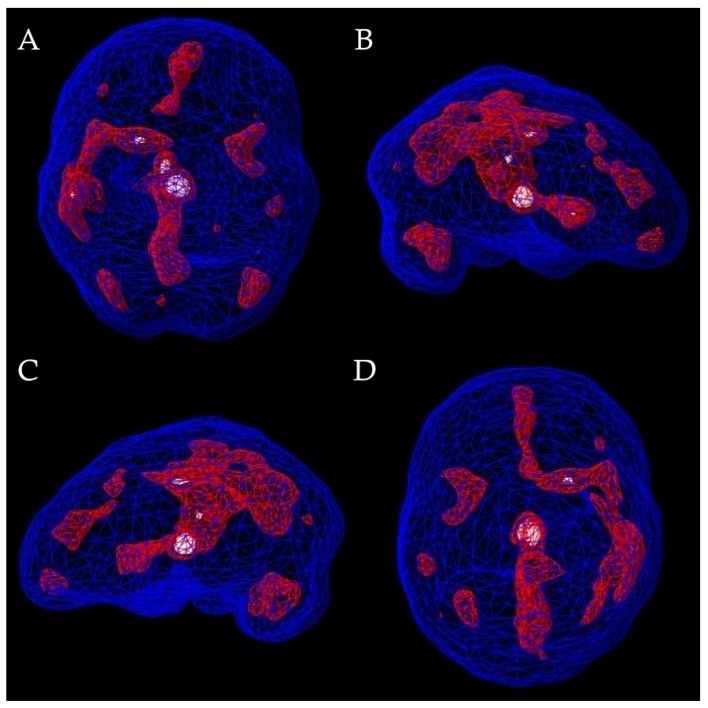
In October 2026, brain perfusion single-photon emission computed tomography (SPECT) was performed under resting and concentration conditions. Resting-state cortical surface renderings were interpreted as showing decreased relative tracer activity in the bilateral temporal lobes, moderate on the right and mild on the left, with additional mild bilateral inferior orbitofrontal reductions and very mild parietal and medial prefrontal differences (Figure 1).


Subcortical resting-state renderings were interpreted as showing moderately increased focal tracer activity in the medial thalamo-limbic region, mildly increased activity in the bilateral insular and basal ganglia regions, and decreased internal cerebellar tracer activity (Figure 2). These qualitative findings represented regional differences in relative tracer distribution and were considered nonspecific, without establishing a causal relationship to the reported neurocognitive disturbances.
In October 2026, quantitative electroencephalography (QEEG) was evaluated using a 19-electrode recording system based on the International 10–20 placement method, with a sampling rate of 256 Hz. Eyes-open and eyes-closed segments were subjected to spectral analysis and comparison with an age-referenced database. The QEEG interpretation described a posterior dominant rhythm of approximately 8.8–9 Hz and episodic 3–4 Hz sharp-and-slow-wave or notched rhythmic activity with low-amplitude spike-like morphology, predominantly in the left posterior temporal (P7/T5) and occipital (O1/O2) regions. Reported runs lasted approximately five to seven seconds. Low-resolution electromagnetic tomography (LORETA) source modeling estimated possible activity in the left mesial temporal/parahippocampal region, with additional modeled limbic and insular involvement. Possible enhancement during drowsiness or light sleep was proposed, but this modeling could not independently establish a deep anatomical source or mechanism.
A separate neurological EEG evaluation provided a formal clinical interpretation of an October 5, 2026 recording. The waking posterior dominant rhythm ranged from 7 to 9.5 Hz, with a predominance of slower frequencies for age. Very frequent 3.5–5 Hz occipital intermittent rhythmic delta activity (OIRDA), approximately 50–100 µV, was described, often with embedded low-amplitude sharp or spike-like waveforms. Posterior slow waves of youth were also reported. Drowsiness and stage N2 sleep were documented; N2 was identified by vertex transients, sleep spindles, and K-complexes. The interpretation characterized the EEG as abnormal for age because of background slowing and prominent OIRDA with embedded spike-like components. The findings were considered etiologically nonspecific, with possible increased seizure susceptibility raised as a clinical consideration. Representative awake, drowsy, and N2 waveforms were included in the source EEG report.
These QEEG and neurological EEG evaluations provided separate professional interpretations of electrophysiological findings, without establishing that two independent recordings had been acquired. Although the QEEG evaluation suggested possible sleep-related enhancement of abnormal activity, neither report supplied a quantified wake-versus-N2 comparison of discharge burden. The descriptions of posterior rhythmic and sharp/spike-like activity overlapped but were not demonstrated to represent identical waveforms on epoch-by-epoch review. No habitual episode of prolonged unresponsiveness or post-sleep recognition disturbance was documented with simultaneous electrographic correlation. Thus, neither a definite epileptic seizure, a specific epilepsy syndrome, nor a causal relationship between the reported waveforms and neurocognitive disturbances was established.
At the most recent documented evaluation in October 2026, caregiver-reported disturbances in autobiographical recognition and fluctuating academic and adaptive functioning persisted, particularly after sleep or intercurrent illness. No standardized longitudinal cognitive outcome measures or documented response to an intervention directed at the most recent EEG findings were available. Further neurological evaluation, including additional sleep-inclusive electrophysiological assessment and formal neurocognitive characterization when clinically indicated, could help clarify the presentation. The timing of subsequent follow-up and longer-term outcomes had not been established at the time of this report.
The most distinctive feature of this case was the caregiver-reported failure to recognize familiar people, surroundings, and aspects of personal identity after sleep, coupled with substantial fluctuations in access to previously acquired cognitive and adaptive abilities. The disturbance was not exclusively sleep-dependent, because deterioration also occurred during intercurrent illnesses without preceding sleep. These observations raise questions about memory consolidation, retrieval, and recognition, but they do not establish that previously stored information was erased. Recurrent prolonged unresponsiveness and the preceding gastrointestinal symptoms further complicated the presentation. Öztürk et al. reported an adolescent with abdominal pain, disorientation, and amnesia initially diagnosed as conversion disorder who subsequently demonstrated temporal EEG abnormalities during sleep and improved after treatment for abdominal epilepsy 7. In contrast, the present case involved longer and more heterogeneous episodes, without an independently confirmed seizure or documented antiseizure treatment response. The reports therefore illustrate overlapping clinical features rather than an established shared mechanism.
The potential relationship between sleep-associated electrophysiological activity and memory dysfunction warrants consideration. Certain childhood epilepsy syndromes feature neurocognitive regression associated with substantial activation of epileptiform discharges during non-rapid eye movement sleep 4. Burkholder et al. reported two adults with transient epileptic amnesia and frequent, predominantly sleep-related bitemporal seizures confirmed on prolonged video-EEG 8. Although these observations show that sleep-related epileptic activity can coexist with autobiographical memory impairment, they cannot be directly extrapolated to this pediatric presentation. The initial EEG did not attain N2 sleep, and the subsequent overnight study was reportedly unrevealing. More recent QEEG and separate neurological EEG interpretations described abnormal posterior rhythmic activity, while the neurological interpretation documented N2 sleep. Nevertheless, quantitative sleep-related potentiation, event-related EEG changes, and the epileptiform significance of the described waveforms remained unconfirmed. An epileptic contribution therefore remained a diagnostic possibility rather than a demonstrated explanation.
The differential diagnosis also included sleep-related focal seizures and epilepsy with spike-wave activation in sleep, particularly given the reported cognitive and adaptive regression 4. However, the available recordings did not demonstrate a characteristic quantified pattern of marked NREM spike-wave activation sufficient to establish a sleep-activated epileptic encephalopathy. Other considerations included disorders of sleep-wake arousal, migraine-related phenomena, and metabolic, autoimmune, infectious, or genetic conditions associated with acquired neurocognitive impairment. Routine biochemical findings and reported structural imaging did not identify a unifying cause, but disease-specific investigations were incompletely documented and these possibilities could not be comprehensively excluded. The prior diagnosis of FND remained part of the clinical history; because its original supporting signs and subsequent diagnostic reassessment were not available, the case did not permit retrospective confirmation or exclusion of FND, which may also coexist with neurological disease.
Cerebral perfusion SPECT was obtained as an adjunctive description of relative regional tracer distribution, not as a diagnostic test establishing epilepsy or another neurological mechanism. Gaggero et al. examined SPECT findings in 10 children with epilepsy characterized by continuous spike-wave activity during slow-wave sleep 9. Hypoperfusion corresponded to predominant EEG abnormalities in four patients, was discordant in two, and was not identified in four, illustrating the inconsistent anatomical correspondence of these modalities even in established epilepsy. In the present case, QEEG interpretation described predominantly left posterior temporal-occipital rhythmic activity, whereas resting SPECT showed bilateral temporal reductions in relative tracer activity, greater on the right, with additional cortical and subcortical differences. The investigations were performed under different conditions, without simultaneous EEG correlation or documented seizure-timed tracer administration. Accordingly, the SPECT observations remained descriptive and exploratory; they did not establish anatomical concordance, identify an epileptic focus, or explain the reported memory disturbances.
Several limitations restrict interpretation of this case. Rogeau et al. emphasized that epilepsy-related ictal and interictal perfusion SPECT depends on standardized protocols, radiotracer timing, and clinical-electrophysiological correlation 10; the present images were not acquired using a documented seizure-localization protocol. The electrophysiological reports did not include a quantified wake-versus-N2 burden or electrographic capture of habitual symptoms, and indirect QEEG source modeling did not establish a focal generator. The memory and functional disturbances were largely caregiver-reported, without available standardized serial neuropsychological testing to distinguish encoding, consolidation, retrieval, and recognition or to measure treatment outcomes. Historical imaging and diagnostic investigations were incompletely available for independent review, and targeted autoimmune, infectious, metabolic, and genetic test results were not available. Finally, the absence of a documented response to EEG-directed therapy and the lack of established longer-term follow-up precluded assessment of treatment-related change.
Despite these limitations, the case illustrates several practical diagnostic lessons. Profound post-sleep loss of recognition should be characterized through a detailed longitudinal history, including the duration of episodes, intervening sleep, intercurrent illness, and whether previously learned skills can subsequently be demonstrated. When clinically indicated, sleep-inclusive EEG with clearly documented sleep stages and simultaneous observation of representative symptoms may be more informative than an awake-only recording. Formal longitudinal assessment of memory and adaptive function could help distinguish apparent loss of stored information from impaired retrieval or inconsistent access. Finally, nonspecific EEG or perfusion abnormalities should be interpreted in context rather than treated as proof of a unifying diagnosis. The report's educational contribution lies in these diagnostic challenges while the underlying etiology remains unresolved.
This case describes an unusual pediatric presentation involving caregiver-reported sleep-associated disturbances in autobiographical recognition, recurrent prolonged unresponsiveness, and marked fluctuations in cognitive and adaptive functioning. Apparent memory loss after sleep does not necessarily establish permanent loss of stored information and may involve impaired retrieval or access to previously acquired knowledge. The case emphasizes careful longitudinal history-taking, consideration of sleep-related neurological disorders, and appropriately documented sleep-inclusive electrophysiological evaluation. Although EEG and perfusion SPECT identified findings of potential neurological interest, neither established a definitive diagnosis or causal mechanism. The absence of standardized longitudinal neuropsychological assessments, event-related EEG correlation, and complete specialized investigations limits interpretation. Further neurological evaluation may clarify the underlying condition, which remained unresolved at the reporting endpoint.
| [1] | Rasch B, Born J. About sleep's role in memory. Physiol Rev. 2013; 93(2): 681-766. | ||
| In article | View Article PubMed | ||
| [2] | Klinzing JG, Niethard N, Born J. Mechanisms of systems memory consolidation during sleep. Nat Neurosci. 2019;22(10):1598-1610. | ||
| In article | View Article PubMed | ||
| [3] | Tulving E. Episodic memory: from mind to brain. Annu Rev Psychol. 2002; 53: 1-25. | ||
| In article | View Article PubMed | ||
| [4] | Specchio N, Wirrell EC, Scheffer IE, et al. International League Against Epilepsy classification and definition of epilepsy syndromes with onset in childhood: position paper by the ILAE Task Force on Nosology and Definitions. Epilepsia. 2022; 63(6): 1398-1442. | ||
| In article | View Article PubMed | ||
| [5] | Peltola ME, Leitinger M, Halford JJ, et al. Routine and sleep EEG: minimum recording standards of the International Federation of Clinical Neurophysiology and the International League Against Epilepsy. Epilepsia. 2023; 64(3): 602-618. | ||
| In article | View Article PubMed | ||
| [6] | Singh J, Ebersole JS, Brinkmann BH. From theory to practical fundamentals of electroencephalographic source imaging in localizing the epileptogenic zone. Epilepsia. 2022; 63(10): 2476-2490. | ||
| In article | View Article PubMed | ||
| [7] | Öztürk Y, Göl G, Savcı U, Cansız MA, Tufan AE. Abdominal epilepsy mimicking conversion disorder: a case report. Psychiatry Clin Psychopharmacol. 2019; 29(4): 484-486. | ||
| In article | View Article | ||
| [8] | Burkholder DB, Jones AL, Jones DT, et al. Frequent sleep-related bitemporal focal seizures in transient epileptic amnesia syndrome: evidence from ictal video-EEG. Epilepsia Open. 2017; 2(2): 255-259. | ||
| In article | View Article PubMed | ||
| [9] | Gaggero R, Caputo M, Fiorio P, et al. SPECT and epilepsy with continuous spike waves during slow-wave sleep. Childs Nerv Syst. 1995; 11(3): 154-160. | ||
| In article | View Article PubMed | ||
| [10] | Rogeau A, Lilburn DML, Kaplar Z, et al. Identifying and troubleshooting the pitfalls of ictal/interictal brain perfusion SPECT studies. Nucl Med Commun. 2023; 44(11): 1053-1058. | ||
| In article | View Article PubMed | ||
Published with license by Science and Education Publishing, Copyright © 2026 Jay Gattis PsyD and Binh Pham DO MPH MS
 This work is licensed under a Creative Commons Attribution 4.0 International License. To view a copy of this license, visit
http://creativecommons.org/licenses/by/4.0/
| [1] | Rasch B, Born J. About sleep's role in memory. Physiol Rev. 2013; 93(2): 681-766. | ||
| In article | View Article PubMed | ||
| [2] | Klinzing JG, Niethard N, Born J. Mechanisms of systems memory consolidation during sleep. Nat Neurosci. 2019;22(10):1598-1610. | ||
| In article | View Article PubMed | ||
| [3] | Tulving E. Episodic memory: from mind to brain. Annu Rev Psychol. 2002; 53: 1-25. | ||
| In article | View Article PubMed | ||
| [4] | Specchio N, Wirrell EC, Scheffer IE, et al. International League Against Epilepsy classification and definition of epilepsy syndromes with onset in childhood: position paper by the ILAE Task Force on Nosology and Definitions. Epilepsia. 2022; 63(6): 1398-1442. | ||
| In article | View Article PubMed | ||
| [5] | Peltola ME, Leitinger M, Halford JJ, et al. Routine and sleep EEG: minimum recording standards of the International Federation of Clinical Neurophysiology and the International League Against Epilepsy. Epilepsia. 2023; 64(3): 602-618. | ||
| In article | View Article PubMed | ||
| [6] | Singh J, Ebersole JS, Brinkmann BH. From theory to practical fundamentals of electroencephalographic source imaging in localizing the epileptogenic zone. Epilepsia. 2022; 63(10): 2476-2490. | ||
| In article | View Article PubMed | ||
| [7] | Öztürk Y, Göl G, Savcı U, Cansız MA, Tufan AE. Abdominal epilepsy mimicking conversion disorder: a case report. Psychiatry Clin Psychopharmacol. 2019; 29(4): 484-486. | ||
| In article | View Article | ||
| [8] | Burkholder DB, Jones AL, Jones DT, et al. Frequent sleep-related bitemporal focal seizures in transient epileptic amnesia syndrome: evidence from ictal video-EEG. Epilepsia Open. 2017; 2(2): 255-259. | ||
| In article | View Article PubMed | ||
| [9] | Gaggero R, Caputo M, Fiorio P, et al. SPECT and epilepsy with continuous spike waves during slow-wave sleep. Childs Nerv Syst. 1995; 11(3): 154-160. | ||
| In article | View Article PubMed | ||
| [10] | Rogeau A, Lilburn DML, Kaplar Z, et al. Identifying and troubleshooting the pitfalls of ictal/interictal brain perfusion SPECT studies. Nucl Med Commun. 2023; 44(11): 1053-1058. | ||
| In article | View Article PubMed | ||