An adult man presented with several years of persistent fatigue, cognitive dysfunction, chronic pain, anxiety, depressive symptoms, and substantial functional impairment. He reported a prior diagnosis of Lyme disease and prolonged treatment, although documentation establishing the original diagnosis was unavailable. Subsequent infectious evaluation did not establish an active infectious etiology, and structural brain magnetic resonance imaging was reportedly unrevealing. Resting brain perfusion single-photon emission computed tomography demonstrated relatively decreased tracer activity involving bilateral temporal and occipital regions, the medial anterior prefrontal cortex, bilateral inferior orbitofrontal regions, and bilateral internal cerebellar regions, with relatively increased activity in the bilateral basal ganglia and thalami. These findings reflected qualitative interpretation of relative regional tracer distribution and were not used independently to establish diagnosis or etiology. Prior COVID-19 infection, sleep disturbance, chronic pain, psychiatric symptoms, stimulant exposure, caffeine use, and kratom use represented additional potential contributors. This case illustrates the descriptive role of brain perfusion SPECT in persistent cognitive and neuropsychiatric symptoms of uncertain and likely multifactorial origin.
Persistent fatigue, pain, cognitive complaints, and neuropsychiatric symptoms have been reported following treatment for Lyme disease, although such manifestations are nonspecific and do not by themselves establish persistent infection or treatment failure 1. Evidence regarding long-term cognitive and psychiatric effects remains inconsistent, with reported manifestations including difficulties with attention and memory, depression, anxiety, and sleep disturbance 2. In patients meeting criteria for post-treatment Lyme disease syndrome, subjective cognitive complaints may be prominent even when objective neuropsychological impairment is absent or relatively mild 3.
Fatigue and cognitive impairment are also well described following COVID-19, further complicating etiologic attribution when potentially overlapping postinfectious syndromes are present 4. Brain perfusion single-photon emission computed tomography (SPECT) depicts relative regional tracer distribution rather than providing absolute measurements of cerebral blood flow and requires interpretation within the broader clinical context 5. Cerebral perfusion differences have previously been described using SPECT in patients with objectively characterized Lyme encephalopathy; however, overlap with individuals without objective Lyme encephalopathy led investigators to conclude that SPECT could not independently establish the diagnosis or determine the presence of active central nervous system infection 6.
We describe an adult with persistent cognitive, neuropsychiatric, fatigue, and pain symptoms in the setting of a reported prior Lyme disease diagnosis, subsequent COVID-19 infection, and an inconclusive infectious evaluation. Resting brain perfusion SPECT demonstrated regional differences in relative tracer distribution. The imaging findings are presented descriptively as an adjunctive component of the clinical evaluation and are not interpreted as establishing a specific diagnosis or etiology.
An adult man presented for evaluation of several years of persistent fatigue, cognitive dysfunction, chronic pain, anxiety, depressive symptoms, and progressive functional impairment. He reported a prior diagnosis of Lyme disease for which he had undergone prolonged treatment, although documentation establishing the original diagnosis was not available for review. He also described persistent symptoms following COVID-19 infection. Despite previous medical evaluation and treatment, fatigue, cognitive complaints, pain, and neuropsychiatric symptoms remained substantially impairing.
Depressive symptoms included reduced motivation, anhedonia, low energy, impaired initiation, difficulty completing routine activities, and social withdrawal. He perceived his mood symptoms as closely related to the persistence and severity of his physical symptoms.
Anxiety had been present for many years and was described as pervasive, with prominent somatic manifestations including nausea, gastrointestinal discomfort, hyperventilation, shaking, transient visual disturbance, and episodes of detachment or dissociation. Some episodes reached panic intensity and resulted in marked functional impairment. Symptoms were more prominent during interpersonal and performance demands but also occurred without a clear external trigger.
He additionally described longstanding cognitive and executive difficulties characterized by impaired attention, poor concentration, disorganization, procrastination, slowed information processing, and difficulty completing tasks. He described persistent brain fog and difficulty processing otherwise routine information. These symptoms interfered with occupational, academic, social, and daily functioning.
Sleep was irregular, with a history of difficulty initiating and maintaining sleep followed more recently by prominent daytime sleepiness despite apparently adequate nocturnal sleep duration. Chronic musculoskeletal pain involving multiple regions periodically limited physical activity.
Relevant substance exposures were considered as potential contributors to the clinical presentation. Caffeine use was regular, while alcohol and tobacco use were infrequent. He reported limited prior cannabis exposure, occasional remote cocaine use, and more recent regular kratom use for pain and perceived improvement in concentration.
Previous psychiatric treatment had been limited. Brief trials of antidepressant and nonstimulant medications had not produced clear benefit. He had subsequently been prescribed an extended release stimulant for attention symptoms and reported partial improvement in concentration. Use was inconsistent because he associated the medication with episodic worsening of anxiety, physical discomfort, chills, and paranoid thoughts.
Previous structural brain magnetic resonance imaging was reportedly without significant abnormalities. Subsequent infectious testing did not establish a single active infectious explanation for the persistent symptoms. A Lyme disease antibody screen was negative. Epstein-Barr virus DNA was detected at a low level in whole blood, while Epstein-Barr virus early antigen D IgG was negative. Babesia and Bartonella serologies were negative. Spotted fever group rickettsial IgG was detected at a titer of 1:64, while IgM was negative. This isolated serologic finding was not considered sufficient by itself to establish acute rickettsial infection.
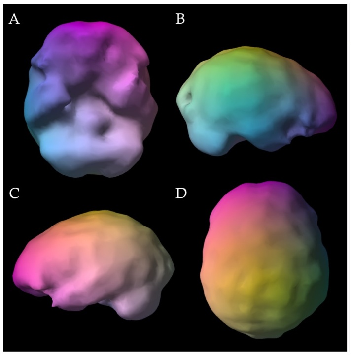
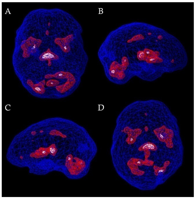
Brain perfusion single-photon emission computed tomography was obtained as an adjunctive component of the initial evaluation. Resting surface-rendered images demonstrated relatively decreased tracer activity involving the bilateral temporal lobes, bilateral occipital regions, medial anterior prefrontal cortex, bilateral inferior orbitofrontal regions, and bilateral internal cerebellar regions (Figure 1). Complementary subcortical renderings demonstrated relatively increased tracer activity involving the bilateral basal ganglia and thalami (Figure 2). The regional descriptions reflect the qualitative clinical SPECT interpretation based on relative regional tracer distribution; validated quantitative regional measurements were not available for retrospective analysis. The findings were not used independently to establish diagnosis or determine etiology.

The initial treatment approach emphasized diagnostic clarification and coordination of psychiatric and medical care rather than substantial modification of psychotropic treatment. Psychotherapy was recommended for anxiety, panic symptoms, depressive symptoms, and associated functional impairment. Behavioral recommendations focused on improving sleep regularity, moderating stimulant and caffeine exposure, maintaining physical activity as tolerated, and supporting general health behaviors. Continued medical evaluation was recommended because of persistent fatigue, cognitive dysfunction, chronic pain, daytime sleepiness, and uncertain contributions from previously identified infectious and postinfectious factors.
During subsequent follow-up, the patient continued to report fluctuating anxiety, mood symptoms, fatigue, cognitive difficulties, and disrupted sleep. Management remained focused on longitudinal assessment and coordination of psychiatric and medical care.

This case illustrates the difficulty of attributing persistent cognitive, neuropsychiatric, fatigue, and pain symptoms to a single cause when the historical diagnosis is uncertain and several potential contributors coexist. The patient reported a prior diagnosis of Lyme disease and prolonged treatment, but documentation establishing the original diagnosis was unavailable, and subsequent Lyme serology was negative. Persistent fatigue, pain, and cognitive complaints have been described after treatment for Lyme disease, but these symptoms are nonspecific and do not, in the absence of objective evidence, establish persistent infection or treatment failure 1. Accordingly, the prior Lyme diagnosis is best regarded as part of the clinical history rather than as an established explanation for the current presentation.
The subsequent infectious evaluation likewise did not identify a single active infectious explanation for the persistent symptoms. Lyme serology and Babesia and Bartonella testing were negative. Low-level Epstein-Barr virus DNA was detected, although its clinical significance in this setting was uncertain. Spotted fever group rickettsial IgG was detected at a titer of 1:64 with negative IgM, an isolated finding that did not establish acute rickettsial infection. Taken together, the available testing did not support attribution of the ongoing syndrome to a single active tick-borne or other infectious process. Evidence from randomized trials similarly does not support additional prolonged antibiotic treatment for nonspecific symptoms persisting after appropriately treated Lyme disease 7.
Etiologic attribution was further complicated by several plausible postinfectious and noninfectious contributors. The patient reported persistent symptoms following COVID-19, for which fatigue and cognitive impairment are well described 4. Sleep irregularity and excessive daytime sleepiness could also contribute to impaired concentration and reduced energy, while chronic pain, anxiety, and depressive symptoms may amplify cognitive complaints and functional impairment. Stimulant exposure, regular caffeine use, and more recent kratom use introduced additional potential influences. The relative contribution of these factors cannot be determined retrospectively, favoring a multifactorial rather than single-cause interpretation of the clinical presentation.
Brain perfusion SPECT provided descriptive functional imaging information but did not resolve this etiologic uncertainty. The resting study demonstrated relatively decreased tracer activity involving the bilateral temporal and occipital regions, medial anterior prefrontal cortex, bilateral inferior orbitofrontal regions, and bilateral internal cerebellar regions, together with relatively increased activity in the bilateral basal ganglia and thalami. These findings were based on qualitative interpretation of relative regional tracer distribution rather than validated quantitative regional measurements. Accordingly, the observed pattern should not be interpreted as diagnostic of a specific infectious, postinfectious, psychiatric, toxic, or other pathophysiologic process 5.
Prior SPECT studies in Lyme-associated neurologic syndromes provide useful context but do not establish a disease-specific perfusion pattern. In patients with objectively characterized Lyme encephalopathy, Logigian et al. reported reduced perfusion predominantly involving frontal cortical and subcortical regions and concluded that SPECT could not independently diagnose Lyme encephalopathy or establish active central nervous system infection 6. A later, broader cohort of patients clinically classified as having chronic Lyme disease reported perfusion deficits most frequently in frontal, temporal, and parietal regions 8. The present case showed partial anatomic overlap, particularly in temporal and prefrontal regions, but also differed with relative increases in the basal ganglia and thalami and additional occipital and cerebellar decreases. These similarities and differences do not establish a Lyme-associated imaging pattern, particularly because the historical Lyme diagnosis in this patient could not be independently verified.
Several limitations should be acknowledged. Historical documentation of the original Lyme disease diagnosis and treatment was incomplete, and contemporary evaluation did not establish active infection. Multiple concurrent factors could plausibly contribute to the fatigue, cognitive dysfunction, and neuropsychiatric symptoms. The SPECT findings were interpreted qualitatively without validated quantitative regional analysis, and no longitudinal imaging was available. Standardized neuropsychological testing was also unavailable to determine the extent to which subjective cognitive complaints corresponded to objectively measured impairment. These limitations preclude causal attribution between the clinical syndrome, prior infectious history, and observed relative perfusion findings.
This case documents persistent cognitive, neuropsychiatric, fatigue, and pain symptoms in an adult for whom no single etiology was established. Resting brain perfusion SPECT demonstrated qualitative differences in relative regional tracer distribution involving cortical, cerebellar, basal ganglia, and thalamic regions. These findings were adjunctive and should not be interpreted as diagnostic of Lyme disease, active infection, or another specific pathophysiologic process. The case underscores the importance of interpreting regional perfusion findings within the broader clinical context when multiple potential contributors coexist.
| [1] | Lantos PM, Rumbaugh J, Bockenstedt LK, et al. Clinical practice guidelines by the Infectious Diseases Society of America (IDSA), American Academy of Neurology (AAN), and American College of Rheumatology (ACR): 2020 guidelines for the prevention, diagnosis and treatment of Lyme disease. Clin Infect Dis. 2021; 72: e1-e48. | ||
| In article | |||
| [2] | Šegždaitė G, Aliukonytė O, Pociūtė K. Neuropsychiatric manifestations of Lyme disease: a literature review of psychiatric and cognitive impacts. Acta Med Litu. 2025; 32: 6-21. | ||
| In article | View Article PubMed | ||
| [3] | Touradji P, Aucott JN, Yang T, et al. Cognitive decline in post-treatment Lyme disease syndrome. Arch Clin Neuropsychol. 2019; 34: 455-465. | ||
| In article | View Article PubMed | ||
| [4] | Ceban F, Ling S, Lui LMW, et al. Fatigue and cognitive impairment in post-COVID-19 syndrome: a systematic review and meta-analysis. Brain Behav Immun. 2022; 101: 93-135. | ||
| In article | View Article PubMed | ||
| [5] | Kapucu OL, Nobili F, Varrone A, et al. EANM procedure guideline for brain perfusion SPECT using 99mTc-labelled radiopharmaceuticals, version 2. Eur J Nucl Med Mol Imaging. 2009; 36: 2093-2102. | ||
| In article | View Article PubMed | ||
| [6] | Logigian EL, Johnson KA, Kijewski MF, et al. Reversible cerebral hypoperfusion in Lyme encephalopathy. Neurology. 1997; 49: 1661-1670. | ||
| In article | View Article PubMed | ||
| [7] | Dersch R, Torbahn G, Rauer S. Treatment of post-treatment Lyme disease symptoms: a systematic review. Eur J Neurol. 2024; 31: e16293. | ||
| In article | View Article PubMed | ||
| [8] | Donta ST, Noto RB, Vento JA. SPECT brain imaging in chronic Lyme disease. Clin Nucl Med. 2012; 37: e219-e222. | ||
| In article | View Article PubMed | ||
Published with license by Science and Education Publishing, Copyright © 2026 Binh Pham DO MPH MS
 This work is licensed under a Creative Commons Attribution 4.0 International License. To view a copy of this license, visit
http://creativecommons.org/licenses/by/4.0/
| [1] | Lantos PM, Rumbaugh J, Bockenstedt LK, et al. Clinical practice guidelines by the Infectious Diseases Society of America (IDSA), American Academy of Neurology (AAN), and American College of Rheumatology (ACR): 2020 guidelines for the prevention, diagnosis and treatment of Lyme disease. Clin Infect Dis. 2021; 72: e1-e48. | ||
| In article | |||
| [2] | Šegždaitė G, Aliukonytė O, Pociūtė K. Neuropsychiatric manifestations of Lyme disease: a literature review of psychiatric and cognitive impacts. Acta Med Litu. 2025; 32: 6-21. | ||
| In article | View Article PubMed | ||
| [3] | Touradji P, Aucott JN, Yang T, et al. Cognitive decline in post-treatment Lyme disease syndrome. Arch Clin Neuropsychol. 2019; 34: 455-465. | ||
| In article | View Article PubMed | ||
| [4] | Ceban F, Ling S, Lui LMW, et al. Fatigue and cognitive impairment in post-COVID-19 syndrome: a systematic review and meta-analysis. Brain Behav Immun. 2022; 101: 93-135. | ||
| In article | View Article PubMed | ||
| [5] | Kapucu OL, Nobili F, Varrone A, et al. EANM procedure guideline for brain perfusion SPECT using 99mTc-labelled radiopharmaceuticals, version 2. Eur J Nucl Med Mol Imaging. 2009; 36: 2093-2102. | ||
| In article | View Article PubMed | ||
| [6] | Logigian EL, Johnson KA, Kijewski MF, et al. Reversible cerebral hypoperfusion in Lyme encephalopathy. Neurology. 1997; 49: 1661-1670. | ||
| In article | View Article PubMed | ||
| [7] | Dersch R, Torbahn G, Rauer S. Treatment of post-treatment Lyme disease symptoms: a systematic review. Eur J Neurol. 2024; 31: e16293. | ||
| In article | View Article PubMed | ||
| [8] | Donta ST, Noto RB, Vento JA. SPECT brain imaging in chronic Lyme disease. Clin Nucl Med. 2012; 37: e219-e222. | ||
| In article | View Article PubMed | ||