Fungal infections constitute an increasingly important global public health problem and are associated with substantial morbidity and mortality, particularly among immunocompromised and critically ill individuals. They range from highly prevalent superficial mycoses to invasive and disseminated infections with high case-fatality rates. Opportunistic pathogens, such as Aspergillus, Candida, Cryptococcus, Rhodotorula, Geotrichum, and Trichosporon cause diverse clinical syndromes, whereas endemic dimorphic fungi including Blastomyces, Coccidioides, Histoplasma, Paracoccidioides, and Talaromyces are strongly influenced by environmental and geographic exposure. Recent global estimates suggest that more than 6.5 million life-threatening invasive fungal infections occur annually, with approximately 3.8 million associated deaths. Climate change may alter fungal ecology, geographic distribution, and human exposure, while antifungal resistance increasingly compromises the effectiveness of the limited therapeutic options available. This review summarizes the epidemiology, major opportunistic and endemic mycoses, global morbidity and mortality, climate-related emergence, and antifungal resistance, and highlights the need for improved surveillance, laboratory diagnosis, antifungal stewardship, access to effective treatment, and coordinated One Health interventions.
Fungi are eukaryotic organisms that occur as yeasts, moulds, or thermally dimorphic forms and are widely distributed in environmental and host-associated niches 1. It is believed that around 5 million species of fungi are present in the world, out of which about 600 have been found to be implicated with various clinical disorders of humans as well as many species of animals 2. Fungal infections have been encountered in both sexes, all age groups, and can occur in all seasons, and in urban and rural settings 1. Human fungal diseases range from superficial and mucocutaneous infections to subcutaneous and invasive systemic mycoses. Superficial fungal infections are extremely common, whereas invasive fungal diseases are less frequent but are associated with substantial morbidity and mortality because diagnosis may be delayed and treatment options are limited 3. The burden is particularly high among individuals with impaired host defenses, including patients with malignancy, human immunodeficiency virus (HIV) infection, organ transplantation, neutropenia, diabetes mellitus, and those receiving corticosteroids, cytotoxic drugs, or other immunosuppressive therapies 1. It is important to mention that about 90% of invasive fungal infections have been described in immunocompromised subjects 2. Important opportunistic pathogens include Aspergillus, Candida, Cryptococcus, Geotrichum, and Trichosporon species 1, 4, 5.
Endemic fungal infections are associated with defined ecological niches and geographic regions and are commonly acquired from environmental reservoirs, particularly soil enriched with organic material or animal excreta 6. Inhalation of infectious propagules can cause disease even in immunocompetent individuals. Major endemic or thermally dimorphic pathogens include Blastomyces dermatitidis, Coccidioides immitis, Coccidioides posadasii, Histoplasma capsulatum, Paracoccidioides brasiliensis, Paracoccidioides lutzii, and Talaromyces marneffei; Sporothrix species also cause important implantation and zoonotic mycoses 7, 8. Contemporary estimates indicate that life-threatening fungal diseases impose a much larger global burden than previously recognized 9. Accordingly, this review delineates the epidemiology and public health importance of major opportunistic and endemic fungal infections, with particular emphasis on global burden, climate-related changes in fungal disease, and the emerging challenge of antifungal resistance.

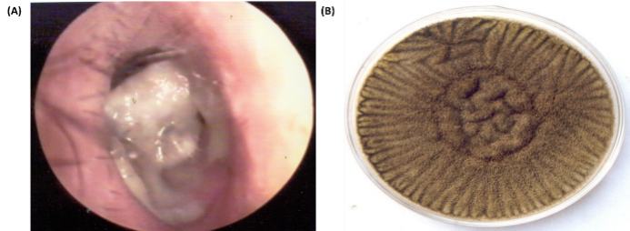
Aspergillosis is caused predominantly by Aspergillus fumigatus, although several other species may be implicated, including Aspergillus amstelodami, Aspergillus candidus, Aspergillus chevallieri, Aspergillus clavatus, Aspergillus deflectus, Aspergillus flavus, Aspergillus glaucus, Aspergillus nidulans, Aspergillus niger, Aspergillus ochraceous, Aspergillus restrictus, Aspergillus sydowii, Aspergillus tamari, Aspergillus terreus, Aspergillus udagawae, Aspergillus ustus, and Aspergillus versicolor 1, 10. The disease can occur in sporadic as well as in epidemic form. Nosocomial outbreaks of aspergillosis have been documented in the patients who were residing near the construction work 1. It is important to mention that the lungs were involved in about 75% of Aspergillus infection in HIV patients 1. Aspergillus species are ubiquitous in soil, air, water, and decaying plant material, and infection is usually acquired by inhalation of airborne conidia; traumatic inoculation can also produce localized disease. Aspergillus fumigatus is the species most frequently associated with invasive disease in humans and animals 11. Dave and Pal 12 additionally documented primary cutaneous aspergillosis caused by A. fumigatus in a 34-year-old immunocompetent poultry farmer following minor trauma while cleaning a poultry cage; direct microscopy and culture of a skin biopsy confirmed the infection, and environmental investigation recovered the organism from the patient's immediate surroundings. Aspergillus niger is an important cause of otomycosis, as illustrated in Figure 1.
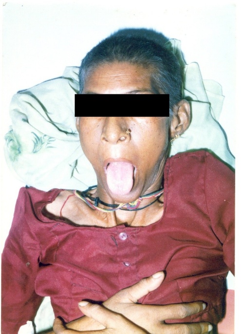
2.2. CandidiasisCandidiasis comprises mucosal, cutaneous, and invasive infections caused by Candida species 1. Candida commonly colonizes the oral cavity, gastrointestinal tract, genital tract, and skin without causing disease; however, disruption of host defenses or local microbial balance can permit pathogenic overgrowth. Oropharyngeal candidiasis (thrush) typically presents with white oral plaques, soreness, and dysphagia, whereas vulvovaginal candidiasis produces genital symptoms and is common among women 13. Mucosal candidiasis is especially frequent in patients with HIV infection or acquired immunodeficiency syndrome (AIDS), malignancy, or other forms of immunosuppression. A severe case of oropharyngeal candidiasis caused by Candida albicans is shown in Figure 2.

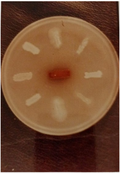
Candida albicans remains a major human pathogen, but non-albicans Candida species are also clinically important, particularly in invasive disease and in patients exposed to broad-spectrum antibiotics, corticosteroids, cytotoxic agents, indwelling devices, or prolonged hospitalization 4, 14. Of particular concern, Candida auris is a healthcare-associated pathogen capable of persistent colonization, nosocomial transmission, and resistance to multiple antifungal classes; the World Health Organization (WHO) includes C. auris among its critical-priority fungal pathogens 15, 16. Pal 17 was among the early investigators to report the isolation of Candida tropicalis from lung empyema in elderly, debilitated patient with diabetes, demonstrating the pathogenic potential of this opportunistic species. On Pal sunflower seed medium, Candida species produce white to cream colonies, whereas Cryptococcus neoformans develops a characteristic brown pigmentation, as shown in Figure 3.


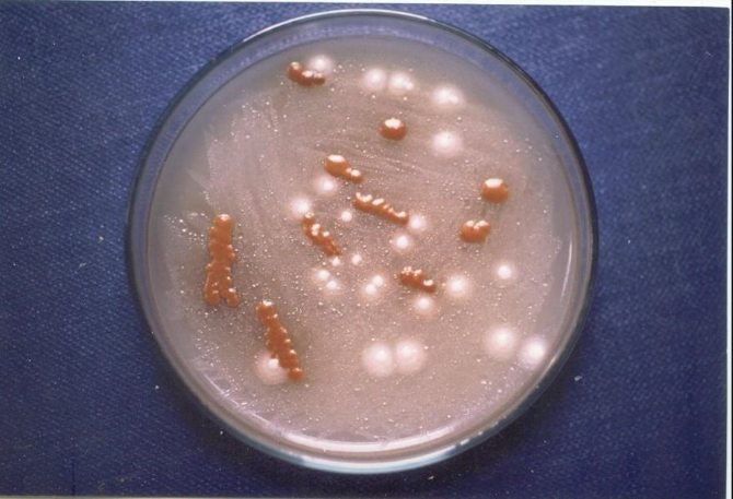
Cryptococcosis is an environmentally acquired mycosis caused mainly by Cryptococcus neoformans and Cryptococcus gattii 19. Infection most commonly follows inhalation of desiccated yeast cells or basidiospores from environmental reservoirs, and may remain confined to the lungs or disseminate, particularly to the central nervous system. It is reported that C. neoformans can survive for about 20 years in dry and old pigeon droppings that are not exposed to direct sunlight 20. The burden remains substantial: a recent global analysis estimated approximately 194,000 cases of cryptococcal meningitis and 147,000 associated deaths annually 9, replacing older estimates that were largely derived from specific HIV-associated populations. Dave and Pal 21 reported primary cutaneous cryptococcosis in an immunocompetent parrot keeper who sustained minor trauma while cleaning a cage; Cryptococcus neoformans was isolated from the skin lesion and from the immediate avian environment, supporting environmental acquisition. Clinical manifestations vary with the site of infection and may include cough, fever, dyspnea, headache, neurological symptoms, and cutaneous lesions 22. Pal sunflower seed medium developed by Pal in 1980 has been used in microbiology and public health laboratories for the rapid isolation and identification of C. neoformans from clinical as well as environmental specimens 1, 20. Cryptococcus neoformans imparts light to dark brown colored mucoid colonies on Pal sunflower seed medium. Figure 4 illustrates isolation of Cryptococcus neoformans from a clinical specimen from an immunocompromised patient on Pal sunflower seed medium.
2.4. MucormycosisMucormycosis is an emerging and re-emerging fungal infection reported globally, including in countries like India. It is caused by filamentous fungi from the genera Lichtheimia, Cunninghamella, Mucor, Rhizomucor, and Rhizopus. These fungi are widely present in the environment and can invade various organs of the body, such as the sinuses, lungs, brain, heart, kidneys, and joints. The infection is exogenous, with the respiratory tract being the primary route of entry for the fungi. Mucormycosis can manifest in both sporadic and epidemic forms, with most cases attributed to Rhizopus oryzae 24.
Mucormycosis presents in several clinical forms, including cutaneous, subcutaneous, rhinocerebral, gastrointestinal, pulmonary, and systemic mucormycosis 1, 25. Less common forms include pyelonephritis, endocarditis, and osteomyelitis. Clinical manifestations of mucormycosis can include fever, headache, shortness of breath, chest pain, coughing, hemoptysis, eyelid edema, pain and redness around the eyes and nose, blurred or double vision, protruding eyes, sudden loss of vision, nasal discharge, stuffy nose, diminished sense of smell, facial paresthesia, skin lesions, toothaches, and altered mental status 1, 25.
2.5. RhodotoruliosisRhodotoruliosis is an opportunistic and emerging mycotic disease affecting both humans and animals 1. It is caused by Rhodotorula, a basidiomycetous yeast that acts as a saprophyte in the environment. The disease can occur sporadically or in epidemic forms with exogenous sources of infection. The most frequently encountered Rhodotorula species include Rhodotorula glutinis, Rhodotorula mucilaginosa (formerly Rhodotorula rubra), and Rhodotorula minuta. These species can be isolated from a variety of sources, such as soil, air, plants, and household environments 1, 26.
In humans, Rhodotorula has been associated with infections such as fungemia, meningitis, ventriculitis, peritonitis, endocarditis, keratitis, endophthalmitis, hydrosalpinx, oral ulcers, and lymphadenitis. Systemic infection occurs in patients who are immunocompromised 1. In animals, it causes skin infections in chickens, sea lions, and cats; lung infections in sheep; epididymitis in dogs; and mastitis in cows and buffalo. Rhodotorula is an opportunistic pathogen that takes advantage of immunosuppressive conditions, indwelling devices, and antibiotic use 26.
2.6. GeotrichosisGeotrichosis is an uncommon opportunistic mycosis caused predominantly by Geotrichum candidum, a ubiquitous saprophytic, yeast-like filamentous fungus that can occur in soil, water, air, food products, and as a commensal of the human digestive, respiratory, and cutaneous microbiota 1. Disease is reported most often in individuals with impaired host defenses, including patients with malignancy, diabetes mellitus, HIV infection, leukosis, or organ transplantation, although clinically significant infection can also occur in immunocompetent hosts. Reported manifestations include oral, pulmonary, urinary, cutaneous, ocular, osteoarticular, and disseminated disease 27, 28, 29, 30.
Clinical diagnosis requires careful correlation because Geotrichum may be recovered as a colonizer or contaminant. Direct microscopy demonstrating hyaline septate hyphae and rectangular arthroconidia, together with repeated isolation from appropriate clinical specimens, supports etiologic significance. Dave and Pal 29 demonstrated G. candidum in oral lesions of an immunocompromised patient; however, Ramya and et al. 27 reported pulmonary geotrichosis in an immunocompetent patient. Urinary infection due to G. candidum was described in an elderly patient with lung cancer 28. A recent review of cutaneous geotrichosis noted that localized disease may follow trauma or burns, whereas disseminated cutaneous involvement occurs mainly in immunosuppressed patients; amphotericin B and voriconazole have been among the most frequently used systemic agents, although standardized clinical breakpoints for Geotrichum remain unavailable 30.
2.7. TrichosporonosisTrichosporonosis is an opportunistic mycosis caused by basidiomycetous yeast-like fungi of the genus Trichosporon, which are widely distributed in nature and may colonize human skin, the gastrointestinal tract, and the respiratory tract 1. Trichosporon asahii is the species most often associated with invasive disease, particularly in patients with hematological malignancies, neutropenia, organ transplantation, prolonged antibiotic exposure, or indwelling intravascular devices. Clinical presentations range from superficial white piedra to fungemia and disseminated infection with pulmonary or cutaneous involvement. Disseminated infection carries a grave prognosis, and hence, early diagnosis and prompt treatment are imperative 1. Trichosporon species are intrinsically resistant to echinocandins and may show reduced susceptibility to amphotericin B; triazoles, particularly voriconazole, are generally preferred for invasive trichosporonosis 31.
Blastomycosis is caused by the thermally dimorphic fungus Blastomyces dermatitidis, which thrives in moist, acidic soil 1. This species is the primary cause of infections in North America, especially around the Great Lakes, the St. Lawrence Seaway, and various south-central and southeastern states. Recently, other Blastomyces species have been identified as causing disease in different parts of the world 32.
Infection typically results from inhaling conidia from the mold phase of the fungus. Acute pulmonary infections can range from asymptomatic to typical community-acquired pneumonia, while chronic forms may present as mass-like lesions or cavitary pneumonia. In rare cases, pulmonary infections may progress to acute respiratory distress syndrome, which carries a high mortality rate. After an initial pulmonary infection, the yeast form of Blastomyces can spread hematogenously, often leading to cutaneous lesions. Additionally, it can involve the osteoarticular system, genitourinary tract, and central nervous system 32.
3.2. CoccidioidomycosisCoccidioidomycosis, commonly known as Valley Fever 1, is an invasive dimorphic fungal infection caused by Coccidioides immitis and Coccidioides posadasii. These fungi are found in the arid desert soils of the southwestern United States, as well as in parts of Mexico, Central America, and South America 33. Most Coccidioides infections are acquired by inhaling airborne arthroconidia. Occasionally, the infection can be acquired through primary cutaneous inoculation, leading to skin infections, or via organ transplantation from infected donor lungs, livers, or kidneys, resulting in disseminated disease 34, 35. Rare cases of neonatal transmission have also been reported, primarily due to the aspiration of infectious vaginal secretions during childbirth 36.
3.3. HistoplasmosisHistoplasmosis is an environmentally acquired systemic mycosis caused by the thermally dimorphic fungus Histoplasma capsulatum. Infection usually follows inhalation of airborne microconidia from contaminated soil and most commonly begins in the lungs 37. The majority of exposed immunocompetent individuals remain asymptomatic or develop a mild, self-limited respiratory illness; however, symptomatic pulmonary disease may present with fever, headache, non-productive cough, dyspnea, chest pain, and constitutional symptoms. In patients with impaired cell-mediated immunity or heavy exposure, infection may disseminate hematogenously and involve the skin, mucous membranes, lymphoreticular system, gastrointestinal tract, adrenal glands, or central nervous system. Because the clinical presentation can mimic tuberculosis and other systemic infections, laboratory confirmation is essential, particularly in endemic areas and among people living with HIV 6, 37.
3.4. ParacoccidioidomycosisParacoccidioidomycosis is a chronic systemic endemic mycosis caused principally by Paracoccidioides brasiliensis and Paracoccidioides lutzii 38. Infection is acquired mainly through inhalation of environmental propagules, producing a primary pulmonary infection that may remain subclinical or progress to chronic respiratory disease. Hematogenous or lymphatic dissemination can involve the oral and nasal mucosa, skin, lymph nodes, gastrointestinal tract, and other internal organs, where painful ulcerative lesions and systemic manifestations may occur 38, 39. Natural infection has also been documented in animals, including dogs and several wild species. Canine cases may present with weight loss, generalized lymphadenopathy, hepatosplenomegaly, and disseminated disease, supporting the ecological and One Health relevance of Paracoccidioides in endemic areas 40, 41.
3.5. Talaromycosis (formerly penicilliosis)Talaromycosis is an endemic systemic mycosis caused by Talaromyces marneffei, a thermally dimorphic fungus formerly known as Penicillium marneffei. The disease is particularly important among people with advanced HIV infection and other forms of immunosuppression in endemic regions of Southeast Asia and southern China 42. Talaromyces marneffei grows as a mould at approximately 25 °C and converts to a yeast-like form at 37 °C, a transition that is central to tissue invasion and pathogenesis 43. Phylogenetic and taxonomic studies transferred the species from Penicillium to Talaromyces; therefore, the current disease name talaromycosis should be used, with penicilliosis retained only as a historical synonym 44, 45.
Talaromycosis commonly presents as a disseminated febrile illness with weight loss, anemia, lymphadenopathy, respiratory symptoms, hepatosplenomegaly, and characteristic skin lesions, and it may clinically resemble histoplasmosis, cryptococcosis, or tuberculosis. Although infection is best recognized in humans, T. marneffei and related Talaromyces species have also been investigated in animals and environmental reservoirs, underscoring the importance of accurate species identification and ecological surveillance 46, 47.
Fungal diseases constitute a substantial but frequently underestimated cause of morbidity and mortality worldwide. Their clinical impact ranges from highly prevalent superficial and mucosal infections to life-threatening invasive and disseminated mycoses that may cause prolonged hospitalization, organ dysfunction, disability, chronic sequelae, and death. The burden is especially severe among patients with HIV/AIDS, malignancy, diabetes mellitus, neutropenia, organ transplantation, critical illness, and those receiving prolonged corticosteroid, antibiotic, cytotoxic, or other immunosuppressive therapies. Opportunistic fungi, such as Aspergillus, Candida, Cryptococcus, and Pneumocystis account for a major proportion of severe fungal disease 48.
More recent global estimates indicate that severe fungal disease is substantially more common than previously recognized. Denning 9 estimated approximately 6.5 million life-threatening invasive fungal infections annually and about 3.8 million associated deaths worldwide, of which roughly 2.5 million deaths were directly attributable to fungal disease. These estimates emphasize that invasive mycoses represent a major component of global infectious-disease mortality. The true burden is likely to remain underestimated because surveillance is limited, many infections are not diagnosed, access to mycology laboratories is uneven, and fungal disease may be recorded only as a complication of the underlying condition 9, 49. The principal current estimates are summarized in Table 1.
The consequences of fungal disease extend beyond mortality. Survivors may experience chronic respiratory impairment, neurological disability, recurrent infection, visual loss, or other long-term sequelae depending on the pathogen and site of infection. In resource-limited settings, delayed diagnosis, restricted access to sensitive laboratory tests, and limited availability or affordability of antifungal medicines further increase morbidity and the risk of poor outcomes. Strengthening surveillance, diagnostic capacity, and access to effective treatment is therefore essential for accurately defining and reducing the global burden of mycoses 49.
Climate change is increasingly recognized as an important environmental factor capable of influencing the ecology, geographic distribution, and epidemiology of fungal pathogens. Changes in temperature, rainfall, humidity, drought patterns, and the frequency of extreme weather events can alter fungal growth, sporulation, survival, dispersal and human exposure. Warming temperatures and changing precipitation patterns may also increase the suitability of previously non-endemic areas for some soil-associated fungi, while environmental disturbances such as dust storms, floods, wildfires, deforestation, and land-use change can increase aerosolization or redistribution of infectious fungal propagules 50. Cryptococcus gattii provides an important example of a fungal pathogen whose ecological range may be influenced by environmental and climatic conditions. Traditionally associated with tropical and subtropical regions, C. gattii has also caused infections and outbreaks in temperate regions. Global warming has been proposed as one factor contributing to its emergence in some geographical areas, while temperature, humidity, urbanization, deforestation, and other environmental changes may influence the survival and dispersal of Cryptococcus species 19, 23. Similar climate-related shifts are being considered for endemic mycoses such as coccidioidomycosis and histoplasmosis, where changes in soil conditions, rainfall, drought, and temperature can modify habitat suitability and airborne exposure 50.
Rising environmental temperatures may also impose selective pressure on fungi, potentially favoring organisms that can tolerate temperatures closer to those of the human body. Climate-related changes have consequently been discussed in relation to the emergence of new fungal threats and to changes in antifungal resistance. However, the relationship between climate change and the emergence of individual fungal pathogens is complex, and climate change should be considered one of several interacting ecological, biological, and socioeconomic drivers rather than the sole cause of fungal emergence. Continued environmental surveillance integrating human, animal, and ecosystem health is needed to better predict how changing climatic conditions will influence future patterns of fungal disease 50.
Antifungal resistance has emerged as a major challenge in the prevention and treatment of invasive fungal diseases. Therapeutic options are intrinsically limited because systemic therapy relies primarily on a small number of drug classes, including azoles, polyenes, echinocandins, and flucytosine. Resistance can delay effective therapy, prolong hospitalization, increase healthcare costs, and contribute to treatment failure and mortality. In response to the growing threat, the World Health Organization published the first Fungal Priority Pathogens List in 2022, identifying 19 fungal pathogens and prioritizing actions in surveillance, laboratory capacity, research and development, and public health intervention 15.
Resistance is particularly important among Candida and Aspergillus species. Candida auris is a healthcare-associated pathogen with a marked capacity for nosocomial transmission, environmental persistence, and resistance to multiple antifungal classes, whereas azole-resistant Aspergillus fumigatus increasingly complicates the management of invasive aspergillosis. Other Candida species may show intrinsic or acquired resistance to specific antifungal agents, making species-level identification clinically important. Molecular mechanisms include alteration or overexpression of drug targets, activation of efflux pumps, changes in membrane sterol pathways, stress-response adaptation, aneuploidy and other genomic changes, and biofilm-associated tolerance. Alterations involving ERG11 in Candida and cyp51A in A. fumigatus are well-described contributors to azole resistance 51.
Antifungal resistance may emerge during prolonged or repeated clinical therapy, but environmental selection is also a major concern. Agricultural use of azole fungicides can select azole-resistant environmental strains of A. fumigatus because agricultural and medical azoles act on related fungal sterol pathways. Resistant conidia can subsequently be inhaled by individuals with no prior exposure to medical azoles, illustrating the interconnected human, agricultural, veterinary, and environmental dimensions of antifungal resistance. A One Health response should therefore combine antifungal stewardship, species-level identification, susceptibility testing when indicated, infection prevention and control, environmental and clinical surveillance, and responsible antifungal use across sectors 49, 51.
An additional concern is the limited innovation and unequal access to fungal diagnostics and therapeutics. The WHO antifungal development landscape reported in 2025 that only four new antifungal drugs had received regulatory approval in the United States, European Union, or China during the preceding decade, while relatively few candidates had advanced to late-stage clinical development. Diagnostic gaps are also substantial, particularly in low- and middle-income countries, where delayed or unavailable species identification and susceptibility testing can promote inappropriate therapy. The 2026 WHO implementation blueprint consequently emphasizes coordinated national surveillance, strengthened mycology laboratory networks, equitable access to essential diagnostics and antifungals, stewardship, infection prevention, and sustained research investment as central components of the response to fungal disease and antifungal resistance 49, 52.
Fungal infections are important causes of morbidity and mortality across all age groups and in both developing and developed countries of the world. Opportunistic mycoses such as aspergillosis, candidiasis, cryptococcosis, mucormycosis, rhodotoruliosis, geotrichosis, and trichosporonosis are especially consequential in immunocompromised and critically ill patients, whereas endemic mycoses including blastomycosis, coccidioidomycosis, histoplasmosis, paracoccidioidomycosis, and talaromycosis remain important in regions where environmental exposure occurs. The increasing global burden, changing ecology of fungal pathogens, and emergence of antifungal resistance reinforce the need for timely diagnosis, effective treatment, surveillance, antifungal stewardship, and integrated One Health prevention strategies.
Based on these conclusions, the following recommendations are proposed:
• Promote veterinary care and monitoring for fungal infections in domestic and wildlife animals to prevent zoonotic transmission.
• Encourage the use of advanced molecular and serological methods to differentiate between fungal species and strains.
• Increase awareness and education about fungal infections, focusing on risk factors, symptoms, and preventive measures.
• Support research into new antifungal agents and treatment regimens to address resistant strains and improve patient outcomes.
• Implement strategies to reduce environmental exposure to fungal pathogens, particularly in areas where endemic fungi are prevalent.
• Further research work to elucidate the etiologic significance of opportunistic fungi in various clinical disorders of humans as well as animals may be rewarding.
The authors are highly indebted to Prof. Dr. R.K. Narayan for going through our manuscript. This paper is dedicated to all the scientists and academicians who conducted pioneering work in the field of fungal infections.
All authors made significant contributions to the preparation and revision of this review.
The authors have no competing interests.
No financial support was received from any organization.
WHO: World Health Organization;
HIV: Human immunodeficiency virus
AIDS: Acquired immunodeficiency syndrome
| [1] | Pal, M., Veterinary and Medical Mycology, Indian Council of Agricultural Research, New Delhi, India, 2007. | ||
| In article | |||
| [2] | Pal M, “Emerging role of saprobic fungi in human and animal health”, Journal of Mycopathological Research, 56 (3), i-ii, 2018. | ||
| In article | |||
| [3] | Brunet K, Alanio A, Lortholary O, Rammaert B, “Reactivation of dormant/latent fungal infection”, Journal of Infection, 77 (6), 463-468, 2018. | ||
| In article | View Article PubMed | ||
| [4] | Colombo AL, de Almeida Júnior JN, Slavin MA, Chen SC, Sorrell TC, “Candida and invasive mould diseases in non-neutropenic critically ill patients and patients with haematological cancer”, The Lancet Infectious Diseases, 17 (11), e344-e356, 2017. | ||
| In article | View Article PubMed | ||
| [5] | Lockhart SR, Guarner J, “Emerging and reemerging fungal infections”, Seminars in Diagnostic Pathology, 36 (3), 177-181, 2019. | ||
| In article | View Article PubMed | ||
| [6] | Rodríguez-Cerdeira C, Arenas R, Moreno-Coutiño G, Vásquez E, Fernández R, Chang P, “Systemic fungal infections in patients with human immunodeficiency virus”, Actas Dermo-Sifiliográficas (English Edition), 105 (1), 5-17, 2014. | ||
| In article | View Article | ||
| [7] | Brown GD, Denning DW, Gow NAR, Levitz SM, Netea MG, White TC, “Hidden killers: human fungal infections”, Science Translational Medicine, 4 (165), 165rv13, 2012. | ||
| In article | View Article PubMed | ||
| [8] | Limper AH, Adenis A, Le T, Harrison TS, “Fungal infections in HIV/AIDS”, The Lancet Infectious Diseases, 17 (11), e334-e343, 2017. | ||
| In article | View Article PubMed | ||
| [9] | Denning DW, “Global incidence and mortality of severe fungal disease”, The Lancet Infectious Diseases, 24 (7), e428-e438, 2024. | ||
| In article | View Article PubMed | ||
| [10] | Dave P, Mahendra R, Pal M, “Etiologic significance of Aspergillus terreus in primary cutaneous mycosis of an agricultural worker”, Molecular Microbiology Research, 5 (2), 1-4, 2015. | ||
| In article | View Article | ||
| [11] | Pal M, Dave P, “Aspergillosis: A sapromycotic zoonosis”, Intas Polivet, 7 (2), 421-428, 2006. | ||
| In article | |||
| [12] | Dave P, Pal M, “Cutaneous mycosis in a poultry farmer due to Aspergillus fumigatus”, International Journal of Cutaneous Disorders & Medicine, 4 (1), 180028, 2021. | ||
| In article | |||
| [13] | Vanani, A.R., Mahdavinia, M., Kalantari, H., Khoshnood, S. and Shirani, M., “Antifungal effect of Securigera securidaca L. vaginal gel on Candida species”, Current Medical Mycology, 5 (3), 31-35, 2019. | ||
| In article | |||
| [14] | Bertolini M, Dongari-Bagtzoglou A, “The relationship of Candida albicans with the oral bacterial microbiome in health and disease”, Advances in Experimental Medicine and Biology, 1197, 69-78, 2019. | ||
| In article | View Article PubMed | ||
| [15] | WHO, WHO fungal priority pathogens list to guide research, development and public health action, World Health Organization, Geneva, Switzerland, 2022. | ||
| In article | |||
| [16] | Pal M, Tariku F, Upadhyay D, Paula CR, Patil B, “Candida auris: an emerging life-threatening fungal pathogen of global public health concern”, Journal of Bacteriology and Mycology, 12 (2), 40-43, 2024. | ||
| In article | View Article | ||
| [17] | Pal M, “Isolation of Candida tropicalis from a case of empyema”, Current Science, 57(20), 1135-1136, 1988. | ||
| In article | |||
| [18] | Pal M, “Can Pal sunflower seed medium be considered as a simple inexpensive tool for the study of Candida species?”, EC Microbiology, 20 (9), 01-03, 2024. | ||
| In article | |||
| [19] | Pal M, “Cryptococcus gattii: An emerging global mycotic pathogen of humans and animals”, Journal of Mycopathological Research, 52 (1), 1-6, 2014. | ||
| In article | |||
| [20] | Pal M, “Cryptococcus neoformans and cryptococcosis: A contribution made by Prof. Dr. Mahendra Pal”, Journal of Bacteriology and Mycology: Open Access, 12, 25-29, 2024. | ||
| In article | View Article | ||
| [21] | Dave P, Pal M, “Primary cutaneous mycosis in an immunocompetent parrot keeper due to Cryptococcus neoformans”, Molecular Microbiology Research, 5 (4), 1-3, 2015. | ||
| In article | |||
| [22] | Pal M, Dave P, “Cryptococcosis: an emerging airborne mycosis of global concern”, Air and Water Borne Diseases, 5 (1), 1000127, 2016. | ||
| In article | View Article | ||
| [23] | Pal M, “Cryptococcosis and One Health Perspective”, Journal of Biology and Medicine: Open Access, 6 (1), 137, 2026. | ||
| In article | |||
| [24] | Pal M, “Mucormycosis in COVID-19 patients poses a challenge to public health”, Open Access Journal of Mycology and Mycological Sciences, 5 (1), 000158, 2022. | ||
| In article | View Article | ||
| [25] | Pal M, “Zygomycosis: A highly infectious emerging opportunistic fungal disease of public health concern”, Open Access Journal of Mycology and Mycological Sciences, 3 (1), 000122, 2020. | ||
| In article | View Article | ||
| [26] | Pal M, Rodrigues PC, da Silva Ruiz L, Gutama PK, “Rhodotoruliosis: An emerging opportunistic mycosis of humans and animals”, Open Access Journal of Mycology and Mycological Sciences, 4 (2), 000148, 2021. | ||
| In article | View Article | ||
| [27] | Ramya TG, Baby S, Geetha RK, “Pulmonary infection by Geotrichum candidum”, International Journal of Advances in Medicine, 1 (2), 171-172, 2014. | ||
| In article | |||
| [28] | Bilman FB, Yetik M, “Geotrichum candidum: a rare infection agent in urinary system: case report and review of the literature”, Journal of Clinical and Experimental Investigations, 8 (4), 127-129, 2017. | ||
| In article | View Article | ||
| [29] | Dave P, Pal M, “Etiologic role of Geotrichum candidum in oral lesions of an immunocompromised patient”, Madridge Journal of Vaccines, 1 (1), 24-26, 2017. | ||
| In article | View Article | ||
| [30] | Román-Montes CM, Sifuentes-Osornio J, Martínez-Gamboa A, “Cutaneous infections by Geotrichum spp”, Current Fungal Infection Reports, 18, 60-68, 2024. | ||
| In article | View Article | ||
| [31] | Mehta, V., Nayyar, C., Gulati, N., Singla, N., Rai, S. and Chandar, J., “A Comprehensive review of Trichosporon spp.: An Invasive and Emerging Fungus”, Cureus, 13 (8), e17345, August 2021. | ||
| In article | View Article | ||
| [32] | Linder KA, Kauffman CA, Miceli MH, “Blastomycosis: a review of mycological and clinical aspects”, Journal of Fungi, 9(1), 117, 2023. | ||
| In article | View Article PubMed | ||
| [33] | Crum NF, “Coccidioidomycosis: a contemporary review”, Infectious Diseases and Therapy, 11 (2), 713-742, 2022. | ||
| In article | View Article PubMed | ||
| [34] | Wright PW, Pappagianis D, Wilson M, Louro A, Moser SA, Komatsu K, Pappas PG, “Donor-related coccidioidomycosis in organ transplant recipients”, Clinical Infectious Diseases, 37 (9), 1265-1269, 2003. | ||
| In article | View Article PubMed | ||
| [35] | Nelson JK, Giraldeau G, Montoya JG, Deresinski S, Ho DY, Pham M, “Donor-derived Coccidioides immitis endocarditis and disseminated infection in the setting of solid organ transplantation”, Open Forum Infectious Diseases, 3 (3), ofw086, 2016. | ||
| In article | View Article PubMed | ||
| [36] | Charlton V, Ramsdell K, Sehring S, “Intrauterine transmission of coccidioidomycosis”, The Pediatric Infectious Disease Journal, 18 (6), 561-563, 1999. | ||
| In article | View Article PubMed | ||
| [37] | Pal M, “Histoplasmosis: An important mycosis of public health significance”, Open Access Journal of Mycology and Mycological Sciences, 4 (2), 000144, 2021. | ||
| In article | View Article | ||
| [38] | Bonifaz A, Vázquez-González D, Perusquía-Ortiz AM, “Endemic systemic mycoses: coccidioidomycosis, histoplasmosis, paracoccidioidomycosis and blastomycosis”, JDDG: Journal der Deutschen Dermatologischen Gesellschaft, 9 (9), 705-714, 2011. | ||
| In article | View Article PubMed | ||
| [39] | Ameen M, Talhari C, Talhari S, “Advances in paracoccidioidomycosis”, Clinical and Experimental Dermatology, 35 (6), 576-580, 2010. | ||
| In article | View Article PubMed | ||
| [40] | Ricci G, Mota FT, Wakamatsu A, Serafim RC, Borra RC, Franco M, “Canine paracoccidioidomycosis”, Medical Mycology, 42 (4), 379-383, 2004. | ||
| In article | View Article PubMed | ||
| [41] | de Farias MR, Condas LAZ, Ribeiro MG, Bosco SMG, Muro MD, Werner J, Theodoro RC, Bagagli E, Marques SA, Franco M, “Paracoccidioidomycosis in a dog: case report of generalized lymphadenomegaly”, Mycopathologia, 172 (2), 147-152, 2011. | ||
| In article | View Article PubMed | ||
| [42] | Vanittanakom N, Cooper CR Jr, Fisher MC, Sirisanthana T, “Penicillium marneffei infection and recent advances in the epidemiology and molecular biology aspects”, Clinical Microbiology Reviews, 19 (1), 95-110, 2006. | ||
| In article | View Article PubMed | ||
| [43] | Cooper CR, Vanittanakom N, “Insights into the pathogenicity of Penicillium marneffei”, Future Microbiology, 3 (1), 43-55, 2008. | ||
| In article | View Article PubMed | ||
| [44] | Samson RA, Yilmaz N, Houbraken J, Spierenburg H, Seifert KA, Peterson SW, Varga J, Frisvad JC, “Phylogeny and nomenclature of the genus Talaromyces and taxa accommodated in Penicillium subgenus Biverticillium”, Studies in Mycology, 70, 159-183, 2011. | ||
| In article | View Article PubMed | ||
| [45] | Yilmaz N, Visagie CM, Houbraken J, Frisvad JC, Samson RA, “Polyphasic taxonomy of the genus Talaromyces”, Studies in Mycology, 78, 175-341, 2014. | ||
| In article | View Article PubMed | ||
| [46] | Chaiwun B, Vanittanakom N, Jiviriyawat Y, Rojanasthien S, Thorner P, “Investigation of dogs as a reservoir of Penicillium marneffei in northern Thailand”, International Journal of Infectious Diseases, 15 (4), e236-e239, 2011. | ||
| In article | View Article PubMed | ||
| [47] | Tomlinson JK, Cooley AJ, Zhang S, Johnson ME, “Granulomatous lymphadenitis caused by Talaromyces helicus in a Labrador Retriever”, Veterinary Clinical Pathology, 40 (4), 553-557, 2011. | ||
| In article | View Article PubMed | ||
| [48] | Pal M, “Morbidity and mortality due to fungal infections”, Journal of Applied Microbiology and Biochemistry, 1(1), 1-3, 2017. | ||
| In article | |||
| [49] | WHO, Blueprint for strengthening responses to fungal disease and antifungal resistance: implementation guidance, World Health Organization, Geneva, Switzerland, 2026. | ||
| In article | |||
| [50] | Williams SL, Toda M, Chiller T, Brunkard JM, Litvintseva AP, “Effects of climate change on fungal infections”, PLoS Pathogens, 20(5), e1012219, 2024. | ||
| In article | View Article PubMed | ||
| [51] | Fisher MC, Alastruey-Izquierdo A, Berman J, Bicanic T, Bignell EM, Bowyer P, et al, “Tackling the emerging threat of antifungal resistance to human health”, Nature Reviews Microbiology, 20, 557-571, 2022. | ||
| In article | View Article PubMed | ||
| [52] | WHO, Antifungal agents in clinical and preclinical development: overview and analysis, World Health Organization, Geneva, Switzerland, 2025. | ||
| In article | |||
Published with license by Science and Education Publishing, Copyright © 2026 Mahendra Pal, Mahek Desai, Tesfaye Rebuma, Pratibha Dave, Claudete Rodrigues Paula, Neelima Ranjan and Oskar Nowak
 This work is licensed under a Creative Commons Attribution 4.0 International License. To view a copy of this license, visit
http://creativecommons.org/licenses/by/4.0/
This work is licensed under a Creative Commons Attribution 4.0 International License. To view a copy of this license, visit
http://creativecommons.org/licenses/by/4.0/
| [1] | Pal, M., Veterinary and Medical Mycology, Indian Council of Agricultural Research, New Delhi, India, 2007. | ||
| In article | |||
| [2] | Pal M, “Emerging role of saprobic fungi in human and animal health”, Journal of Mycopathological Research, 56 (3), i-ii, 2018. | ||
| In article | |||
| [3] | Brunet K, Alanio A, Lortholary O, Rammaert B, “Reactivation of dormant/latent fungal infection”, Journal of Infection, 77 (6), 463-468, 2018. | ||
| In article | View Article PubMed | ||
| [4] | Colombo AL, de Almeida Júnior JN, Slavin MA, Chen SC, Sorrell TC, “Candida and invasive mould diseases in non-neutropenic critically ill patients and patients with haematological cancer”, The Lancet Infectious Diseases, 17 (11), e344-e356, 2017. | ||
| In article | View Article PubMed | ||
| [5] | Lockhart SR, Guarner J, “Emerging and reemerging fungal infections”, Seminars in Diagnostic Pathology, 36 (3), 177-181, 2019. | ||
| In article | View Article PubMed | ||
| [6] | Rodríguez-Cerdeira C, Arenas R, Moreno-Coutiño G, Vásquez E, Fernández R, Chang P, “Systemic fungal infections in patients with human immunodeficiency virus”, Actas Dermo-Sifiliográficas (English Edition), 105 (1), 5-17, 2014. | ||
| In article | View Article | ||
| [7] | Brown GD, Denning DW, Gow NAR, Levitz SM, Netea MG, White TC, “Hidden killers: human fungal infections”, Science Translational Medicine, 4 (165), 165rv13, 2012. | ||
| In article | View Article PubMed | ||
| [8] | Limper AH, Adenis A, Le T, Harrison TS, “Fungal infections in HIV/AIDS”, The Lancet Infectious Diseases, 17 (11), e334-e343, 2017. | ||
| In article | View Article PubMed | ||
| [9] | Denning DW, “Global incidence and mortality of severe fungal disease”, The Lancet Infectious Diseases, 24 (7), e428-e438, 2024. | ||
| In article | View Article PubMed | ||
| [10] | Dave P, Mahendra R, Pal M, “Etiologic significance of Aspergillus terreus in primary cutaneous mycosis of an agricultural worker”, Molecular Microbiology Research, 5 (2), 1-4, 2015. | ||
| In article | View Article | ||
| [11] | Pal M, Dave P, “Aspergillosis: A sapromycotic zoonosis”, Intas Polivet, 7 (2), 421-428, 2006. | ||
| In article | |||
| [12] | Dave P, Pal M, “Cutaneous mycosis in a poultry farmer due to Aspergillus fumigatus”, International Journal of Cutaneous Disorders & Medicine, 4 (1), 180028, 2021. | ||
| In article | |||
| [13] | Vanani, A.R., Mahdavinia, M., Kalantari, H., Khoshnood, S. and Shirani, M., “Antifungal effect of Securigera securidaca L. vaginal gel on Candida species”, Current Medical Mycology, 5 (3), 31-35, 2019. | ||
| In article | |||
| [14] | Bertolini M, Dongari-Bagtzoglou A, “The relationship of Candida albicans with the oral bacterial microbiome in health and disease”, Advances in Experimental Medicine and Biology, 1197, 69-78, 2019. | ||
| In article | View Article PubMed | ||
| [15] | WHO, WHO fungal priority pathogens list to guide research, development and public health action, World Health Organization, Geneva, Switzerland, 2022. | ||
| In article | |||
| [16] | Pal M, Tariku F, Upadhyay D, Paula CR, Patil B, “Candida auris: an emerging life-threatening fungal pathogen of global public health concern”, Journal of Bacteriology and Mycology, 12 (2), 40-43, 2024. | ||
| In article | View Article | ||
| [17] | Pal M, “Isolation of Candida tropicalis from a case of empyema”, Current Science, 57(20), 1135-1136, 1988. | ||
| In article | |||
| [18] | Pal M, “Can Pal sunflower seed medium be considered as a simple inexpensive tool for the study of Candida species?”, EC Microbiology, 20 (9), 01-03, 2024. | ||
| In article | |||
| [19] | Pal M, “Cryptococcus gattii: An emerging global mycotic pathogen of humans and animals”, Journal of Mycopathological Research, 52 (1), 1-6, 2014. | ||
| In article | |||
| [20] | Pal M, “Cryptococcus neoformans and cryptococcosis: A contribution made by Prof. Dr. Mahendra Pal”, Journal of Bacteriology and Mycology: Open Access, 12, 25-29, 2024. | ||
| In article | View Article | ||
| [21] | Dave P, Pal M, “Primary cutaneous mycosis in an immunocompetent parrot keeper due to Cryptococcus neoformans”, Molecular Microbiology Research, 5 (4), 1-3, 2015. | ||
| In article | |||
| [22] | Pal M, Dave P, “Cryptococcosis: an emerging airborne mycosis of global concern”, Air and Water Borne Diseases, 5 (1), 1000127, 2016. | ||
| In article | View Article | ||
| [23] | Pal M, “Cryptococcosis and One Health Perspective”, Journal of Biology and Medicine: Open Access, 6 (1), 137, 2026. | ||
| In article | |||
| [24] | Pal M, “Mucormycosis in COVID-19 patients poses a challenge to public health”, Open Access Journal of Mycology and Mycological Sciences, 5 (1), 000158, 2022. | ||
| In article | View Article | ||
| [25] | Pal M, “Zygomycosis: A highly infectious emerging opportunistic fungal disease of public health concern”, Open Access Journal of Mycology and Mycological Sciences, 3 (1), 000122, 2020. | ||
| In article | View Article | ||
| [26] | Pal M, Rodrigues PC, da Silva Ruiz L, Gutama PK, “Rhodotoruliosis: An emerging opportunistic mycosis of humans and animals”, Open Access Journal of Mycology and Mycological Sciences, 4 (2), 000148, 2021. | ||
| In article | View Article | ||
| [27] | Ramya TG, Baby S, Geetha RK, “Pulmonary infection by Geotrichum candidum”, International Journal of Advances in Medicine, 1 (2), 171-172, 2014. | ||
| In article | |||
| [28] | Bilman FB, Yetik M, “Geotrichum candidum: a rare infection agent in urinary system: case report and review of the literature”, Journal of Clinical and Experimental Investigations, 8 (4), 127-129, 2017. | ||
| In article | View Article | ||
| [29] | Dave P, Pal M, “Etiologic role of Geotrichum candidum in oral lesions of an immunocompromised patient”, Madridge Journal of Vaccines, 1 (1), 24-26, 2017. | ||
| In article | View Article | ||
| [30] | Román-Montes CM, Sifuentes-Osornio J, Martínez-Gamboa A, “Cutaneous infections by Geotrichum spp”, Current Fungal Infection Reports, 18, 60-68, 2024. | ||
| In article | View Article | ||
| [31] | Mehta, V., Nayyar, C., Gulati, N., Singla, N., Rai, S. and Chandar, J., “A Comprehensive review of Trichosporon spp.: An Invasive and Emerging Fungus”, Cureus, 13 (8), e17345, August 2021. | ||
| In article | View Article | ||
| [32] | Linder KA, Kauffman CA, Miceli MH, “Blastomycosis: a review of mycological and clinical aspects”, Journal of Fungi, 9(1), 117, 2023. | ||
| In article | View Article PubMed | ||
| [33] | Crum NF, “Coccidioidomycosis: a contemporary review”, Infectious Diseases and Therapy, 11 (2), 713-742, 2022. | ||
| In article | View Article PubMed | ||
| [34] | Wright PW, Pappagianis D, Wilson M, Louro A, Moser SA, Komatsu K, Pappas PG, “Donor-related coccidioidomycosis in organ transplant recipients”, Clinical Infectious Diseases, 37 (9), 1265-1269, 2003. | ||
| In article | View Article PubMed | ||
| [35] | Nelson JK, Giraldeau G, Montoya JG, Deresinski S, Ho DY, Pham M, “Donor-derived Coccidioides immitis endocarditis and disseminated infection in the setting of solid organ transplantation”, Open Forum Infectious Diseases, 3 (3), ofw086, 2016. | ||
| In article | View Article PubMed | ||
| [36] | Charlton V, Ramsdell K, Sehring S, “Intrauterine transmission of coccidioidomycosis”, The Pediatric Infectious Disease Journal, 18 (6), 561-563, 1999. | ||
| In article | View Article PubMed | ||
| [37] | Pal M, “Histoplasmosis: An important mycosis of public health significance”, Open Access Journal of Mycology and Mycological Sciences, 4 (2), 000144, 2021. | ||
| In article | View Article | ||
| [38] | Bonifaz A, Vázquez-González D, Perusquía-Ortiz AM, “Endemic systemic mycoses: coccidioidomycosis, histoplasmosis, paracoccidioidomycosis and blastomycosis”, JDDG: Journal der Deutschen Dermatologischen Gesellschaft, 9 (9), 705-714, 2011. | ||
| In article | View Article PubMed | ||
| [39] | Ameen M, Talhari C, Talhari S, “Advances in paracoccidioidomycosis”, Clinical and Experimental Dermatology, 35 (6), 576-580, 2010. | ||
| In article | View Article PubMed | ||
| [40] | Ricci G, Mota FT, Wakamatsu A, Serafim RC, Borra RC, Franco M, “Canine paracoccidioidomycosis”, Medical Mycology, 42 (4), 379-383, 2004. | ||
| In article | View Article PubMed | ||
| [41] | de Farias MR, Condas LAZ, Ribeiro MG, Bosco SMG, Muro MD, Werner J, Theodoro RC, Bagagli E, Marques SA, Franco M, “Paracoccidioidomycosis in a dog: case report of generalized lymphadenomegaly”, Mycopathologia, 172 (2), 147-152, 2011. | ||
| In article | View Article PubMed | ||
| [42] | Vanittanakom N, Cooper CR Jr, Fisher MC, Sirisanthana T, “Penicillium marneffei infection and recent advances in the epidemiology and molecular biology aspects”, Clinical Microbiology Reviews, 19 (1), 95-110, 2006. | ||
| In article | View Article PubMed | ||
| [43] | Cooper CR, Vanittanakom N, “Insights into the pathogenicity of Penicillium marneffei”, Future Microbiology, 3 (1), 43-55, 2008. | ||
| In article | View Article PubMed | ||
| [44] | Samson RA, Yilmaz N, Houbraken J, Spierenburg H, Seifert KA, Peterson SW, Varga J, Frisvad JC, “Phylogeny and nomenclature of the genus Talaromyces and taxa accommodated in Penicillium subgenus Biverticillium”, Studies in Mycology, 70, 159-183, 2011. | ||
| In article | View Article PubMed | ||
| [45] | Yilmaz N, Visagie CM, Houbraken J, Frisvad JC, Samson RA, “Polyphasic taxonomy of the genus Talaromyces”, Studies in Mycology, 78, 175-341, 2014. | ||
| In article | View Article PubMed | ||
| [46] | Chaiwun B, Vanittanakom N, Jiviriyawat Y, Rojanasthien S, Thorner P, “Investigation of dogs as a reservoir of Penicillium marneffei in northern Thailand”, International Journal of Infectious Diseases, 15 (4), e236-e239, 2011. | ||
| In article | View Article PubMed | ||
| [47] | Tomlinson JK, Cooley AJ, Zhang S, Johnson ME, “Granulomatous lymphadenitis caused by Talaromyces helicus in a Labrador Retriever”, Veterinary Clinical Pathology, 40 (4), 553-557, 2011. | ||
| In article | View Article PubMed | ||
| [48] | Pal M, “Morbidity and mortality due to fungal infections”, Journal of Applied Microbiology and Biochemistry, 1(1), 1-3, 2017. | ||
| In article | |||
| [49] | WHO, Blueprint for strengthening responses to fungal disease and antifungal resistance: implementation guidance, World Health Organization, Geneva, Switzerland, 2026. | ||
| In article | |||
| [50] | Williams SL, Toda M, Chiller T, Brunkard JM, Litvintseva AP, “Effects of climate change on fungal infections”, PLoS Pathogens, 20(5), e1012219, 2024. | ||
| In article | View Article PubMed | ||
| [51] | Fisher MC, Alastruey-Izquierdo A, Berman J, Bicanic T, Bignell EM, Bowyer P, et al, “Tackling the emerging threat of antifungal resistance to human health”, Nature Reviews Microbiology, 20, 557-571, 2022. | ||
| In article | View Article PubMed | ||
| [52] | WHO, Antifungal agents in clinical and preclinical development: overview and analysis, World Health Organization, Geneva, Switzerland, 2025. | ||
| In article | |||